| 规格 | 价格 | 库存 | 数量 |
|---|---|---|---|
| 10 mM * 1 mL in DMSO |
|
||
| 2mg |
|
||
| 5mg |
|
||
| 10mg |
|
||
| 25mg |
|
||
| 50mg |
|
||
| 100mg |
|
||
| 250mg |
|
||
| 500mg |
|
||
| Other Sizes |
|
| 靶点 |
Selective inhibitor of farnesyl protein transferase (FTase) with the following inhibitory parameters:
- IC50 = 0.86 nM (recombinant human FTase), Ki = 0.4 nM (recombinant human FTase) [2] - High selectivity over geranylgeranyl protein transferase type I (GGTase-I): IC50 > 10 μM for GGTase-I, confirming no off-target inhibition of geranylgeranylation [1][2] |
|---|---|
| 体外研究 (In Vitro) |
Tipifarnib 的 ED50 为 4 nM,是 Cruzi 锥虫的强抑制剂。对于核纤层蛋白 B 肽和 K-RasB 肽,替比法尼的 IC50 分别为 0.86 nM 和 7.9 nM,替比法尼抑制这两种生物分子的分离的人法尼基转移酶 [2]。对于侵袭性前列腺癌 (PCa),替比法尼可减少血管生成和细胞增殖,同时诱导细胞凋亡 [3]。替比法尼(0.25 μM、1 μM;48 小时)显着减少 C4-2B 和 PC-3 细胞中外泌体的数量 [3]。 Tipifarnib (1 μM) 可显着抑制 C4-2B 细胞中的 Alix、nSMase2 和 Rab27a 蛋白数量 [3]。当应用于 C4-2B 和 PC-3 细胞时,替比法尼 (0.25 μM) 强烈抑制 p-ERK(Ras/Raf/ERK 信号通路的下游效应分子)的激活,但不会减少总 ERK [3]。暴露于替比法尼 (1.25-5 μM) 30 分钟会导致 U937 细胞内质网应激,进而导致细胞内钙稳态失衡 [4]。
癌细胞抗增殖活性: - 在H-Ras突变癌细胞(如SK-MES-1肺癌细胞、A549肺腺癌细胞)中,替匹法尼(Tipifarnib) 呈浓度依赖性强效抑制增殖: - SK-MES-1细胞:72小时MTT实验IC50=18 nM;100 nM 替匹法尼 使细胞活力降至对照组的28%; - A549细胞:72小时SRB实验IC50=35 nM;200 nM 替匹法尼 诱导G1期细胞周期阻滞(流式细胞术:G1期细胞占比从52%升至78%)。 - 在K-Ras或N-Ras突变细胞(如HCT116结肠癌细胞)中,抗增殖活性稍弱:IC50=85 nM(HCT116,72小时MTT)[2] - 抑制Ras法尼基化: - SK-MES-1细胞Western blot显示,100 nM 替匹法尼(Tipifarnib) 处理48小时后,法尼基化H-Ras蛋白减少70%(抗法尼基化Ras抗体检测),而总H-Ras蛋白水平无变化,证实其抑制FTase介导的Ras修饰[2] - 抑制外泌体生成与分泌: - 在MDA-MB-231乳腺癌细胞中,替匹法尼(Tipifarnib) (1 μM、10 μM)浓度依赖性抑制外泌体分泌: - 10 μM 替匹法尼 使CD63阳性外泌体数量减少45%(流式细胞术),外泌体相关蛋白TSG101水平降低42%(外泌体裂解液Western blot); - 机制上,其下调Rab27a(调控外泌体释放的GTP酶)的法尼基化:10 μM浓度下法尼基化Rab27a减少58%[3] |
| 体内研究 (In Vivo) |
在小鼠中,替比法尼(10 mg/kg;腹腔注射;单剂量)通过上调肝脏抗凋亡蛋白 Bcl-xL 抑制 GalN/LPS 引起的死亡率 [5]。
移植瘤模型抗肿瘤疗效: 1. SK-MES-1肺癌裸鼠移植瘤(n=6/组): - 肿瘤体积达100 mm³后,小鼠口服替匹法尼(Tipifarnib) 25 mg/kg每日两次(bid)和50 mg/kg bid,持续14天; - 肿瘤生长抑制率(TGI)分别为60%(25 mg/kg bid)和80%(50 mg/kg bid); - 最终肿瘤重量从溶剂组的1.2±0.3 g降至25 mg/kg组的0.5±0.1 g和50 mg/kg组的0.2±0.1 g; - 无显著体重下降(<5%)或死亡[2] 2. A549肺腺癌裸鼠移植瘤: - 50 mg/kg bid口服替匹法尼(Tipifarnib) 21天,TGI达75%,肿瘤组织中法尼基化H-Ras减少65%(Western blot)[2] |
| 酶活实验 |
FTase活性检测:
反应体系(50 μL)包含50 mM Tris-HCl(pH 7.5)、5 mM MgCl2、2 mM DTT、100 nM重组人FTase、200 nM生物素化CAAX肽(FTase底物)、100 nM [3H]-法尼基焦磷酸([3H]-FPP,放射性供体)及替匹法尼(Tipifarnib) (0.01~100 nM)。37°C孵育30分钟后,加入50 μL 20 mM EDTA终止反应。生物素化法尼基化肽通过链霉亲和素包被96孔板捕获,用含0.1% Tween-20的PBS洗涤3次,液体闪烁计数器检测结合放射性。与溶剂组比较计算抑制率,拟合曲线得IC50;通过双倒数作图法(改变[3H]-FPP浓度:25~200 nM)计算Ki[2] - GGTase-I选择性检测: 实验方案与FTase检测一致,仅以下参数调整: - 酶:重组人GGTase-I; - 供体底物:[3H]-香叶基香叶基焦磷酸([3H]-GGPP,100 nM); - 肽底物:适配GGTase-I的生物素化CAAX肽。 替匹法尼(Tipifarnib) 测试浓度高达10 μM,对GGTase-I的抑制率<5%,证实对FTase的高选择性[1] |
| 细胞实验 |
抗增殖实验(MTT法,文献[2]):
1. 细胞培养:SK-MES-1或A549细胞以5×10³细胞/孔接种于96孔板,在含10% FBS的RPMI 1640培养基中,37°C、5% CO2培养24小时[2] 2. 药物处理:加入替匹法尼(Tipifarnib) (0.1~1000 nM,溶于0.1% DMSO),溶剂对照组加入0.1% DMSO,继续培养72小时[2] 3. 活力检测:每孔加入10 μL MTT溶液(5 mg/mL),37°C孵育4小时;DMSO溶解甲瓒结晶后,酶标仪检测570 nm吸光度。细胞活力(%)=(处理组吸光度/对照组吸光度)×100%,拟合浓度-活力曲线得IC50[2] - Ras法尼基化Western blot: 1. SK-MES-1细胞(2×10⁵细胞/孔,6孔板)用替匹法尼(Tipifarnib) (10~200 nM)处理48小时[2] 2. 含蛋白酶抑制剂的RIPA缓冲液裂解细胞,4°C、12,000×g离心15分钟,BCA法测蛋白浓度[2] 3. 每泳道30 μg蛋白经12% SDS-PAGE分离后转移至PVDF膜,加入抗法尼基化H-Ras一抗(1:1000)和抗总H-Ras一抗(1:1000,内参)孵育;HRP标记二抗(1:5000)结合后ECL显影,ImageJ定量条带强度[2] - 外泌体分泌实验: 1. MDA-MB-231细胞(1×10⁶细胞/皿,10 cm培养皿)在无血清培养基中用替匹法尼(Tipifarnib) (1 μM、10 μM)处理48小时[3] 2. 收集细胞上清,300×g离心10分钟(去除细胞)、10,000×g离心30分钟(去除细胞碎片),随后4°C、100,000×g超速离心70分钟分离外泌体[3] 3. 外泌体沉淀用PBS重悬,流式细胞术(抗CD63-PE抗体染色)和Western blot(抗TSG101抗体)检测外泌体量,相对溶剂组计算分泌抑制率[3] |
| 动物实验 |
动物/疾病模型: GalN/LPS 攻击小鼠[5]
剂量: 10 mg/kg;同时用 GalN (400 mg/kg;腹腔注射) 和 LPS (32 g/kg) 进行攻击 给药途径: 腹腔注射;攻击前 60 分钟 实验结果: 保护原代肝细胞免受 GalN/肿瘤坏死因子诱导的细胞死亡。抑制 caspase 3 活化并上调抗凋亡蛋白。 裸鼠异种移植模型: 1. 动物和分组:雌性 BALB/c 裸鼠(6-8 周龄,18-22 g)随机分为 3 组(每组 n=6):载体对照组(0.5% CMC-Na)、Tipifarnib 25 mg/kg bid 组、Tipifarnib 50 mg/kg bid 组[2] 2. 肿瘤诱导:将 1×10⁶ 个 SK-MES-1 细胞(悬浮于 100 μL PBS:Matrigel = 1:1 混合液中)皮下注射到每只小鼠的右侧腹部。实验开始于肿瘤体积达到约 100 mm³ 时(第 0 天)[2] 3. 药物制备和给药:将替比法尼溶解于 0.5% 羧甲基纤维素钠 (CMC-Na) 溶液中制备混悬液。小鼠每日两次(8:00 和 20:00)灌胃给药,持续 14 天,给药剂量为 10 mL/kg [2] 4. 样本采集和检测: - 肿瘤体积:每 3 天使用游标卡尺测量一次,计算公式为 V = (长度 × 宽度²) / 2。 - 体重:每 3 天记录一次,以监测毒性。 - 肿瘤重量:第 14 天,处死小鼠,解剖并称量肿瘤。肿瘤生长抑制率(TGI)= [1 – (治疗组肿瘤重量 / 对照组肿瘤重量)] × 100%。 - 肿瘤组织分析:取部分肿瘤组织进行裂解,用于Western blot检测法尼基化H-Ras [2] |
| 药代性质 (ADME/PK) |
口服吸收:
- 在大鼠中,口服Tipifarnib(10 mg/kg)的口服生物利用度(F)为35%,达到最大浓度的时间(Tmax)为1.5小时,最大血浆浓度(Cmax)为89 ng/mL [1] - 分布: - 在携带SK-MES-1异种移植瘤的裸鼠中,口服50 mg/kg Tipifarnib 2小时后,肿瘤组织浓度是血浆浓度的2.3倍(肿瘤:156 ng/g;血浆:68 ng/mL)[2] - 消除: - 在大鼠中,Tipifarnib的消除半衰期(t1/2)为4.2小时(静脉注射剂量:5 mg/kg)。 72小时内,粪便排泄量占总剂量的65%,尿液排泄量占12%[1] |
| 毒性/毒理 (Toxicokinetics/TK) |
体外细胞毒性选择性:
- 在正常人肺成纤维细胞 (MRC-5) 中,Tipifarnib 显示出较低的细胞毒性:IC50 = 500 nM (72 小时 MTT 法),比在 SK-MES-1 癌细胞 (18 nM) 中高约 28 倍,表明其具有选择性抗肿瘤活性 [2] - 体内安全性: - 在接受 Tipifarnib 治疗的裸鼠中(剂量高达 50 mg/kg,每日两次,持续 14 天): - 血清肝功能指标(ALT、AST)或肾功能指标(BUN、肌酐)均无显著变化; - 外周血白细胞计数保持在正常范围内(无骨髓抑制); - 体重减轻 <5%(无严重毒性)[2] - 血浆蛋白结合率: - Tipifarnib 显示出较高的血浆蛋白结合率 (>97%)采用平衡透析法(37°C,pH 7.4)测定人、大鼠和小鼠血浆[1] |
| 参考文献 |
|
| 其他信息 |
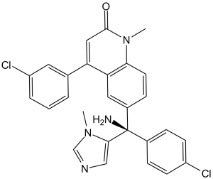
替比法尼是一种喹诺酮类化合物,其结构为1-甲基喹诺林-2-酮,在4位和6位分别连接有3-氯苯基和氨基(4-氯苯基)(1-甲基咪唑-5-基)甲基(R-异构体)。它是一种抗肿瘤药物,属于EC 2.5.1.58(蛋白质法尼基转移酶)抑制剂和细胞凋亡诱导剂。它是一种喹诺酮类化合物,属于单氯苯类、咪唑类和伯氨基化合物。
替比法尼(R-115777)是一种正在研究用于治疗急性髓系白血病(AML)和其他类型癌症的药物。它属于法尼基转移酶抑制剂类药物。它也被称为扎内斯特拉(Zarnestra)。 2005年6月,FDA向Zarnestra发出了不予批准函。 Tipifarnib是一种非肽类喹诺酮,具有潜在的抗肿瘤活性。Tipifarnib可与法尼基蛋白转移酶结合并抑制该酶的活性,该酶参与蛋白质加工(法尼基化)以进行信号转导。通过抑制蛋白质的法尼基化,该药物可阻止Ras癌基因的激活,抑制细胞生长,诱导细胞凋亡,并抑制血管生成。 (NCI04) 药物适应症 已研究用于治疗结直肠癌、白血病(髓系)、胰腺癌和实体瘤。 治疗头颈部上皮恶性肿瘤 作用机制 法尼基转移酶抑制剂 (FTI) 是一类实验性抗癌药物,其靶向蛋白质法尼基转移酶,下游效应是阻止 Ras 蛋白的正常功能,Ras 蛋白通常在癌症中异常活跃。RAS 翻译后经历四个修饰步骤:异戊二烯化、蛋白水解、甲基化和棕榈酰化。异戊二烯化涉及法尼基转移酶 (FTase) 将法尼基从法尼基焦磷酸 (FPP) 转移到前体 RAS 蛋白上。此外,一种相关的酶——牻牛儿基牻牛儿基转移酶I (GGTase I) 能够将牻牛儿基牻牛儿基转移至K-和N-RAS。法尼基是RAS与细胞膜连接所必需的。若不与细胞膜连接,RAS就无法传递来自膜受体的信号(Reuter等人,2000)。 药效学 R115777是一种非肽类法尼基转移酶抑制剂,能够抑制人胰腺腺癌细胞系的生长。这种生长抑制与信号转导和转录激活因子 3 (STAT3) 和细胞外信号调节激酶 (ERK) 的磷酸化水平的调节有关。 Tipifarnib (R115777) 是一种合成的、口服的、选择性的法尼基蛋白转移酶 (FTase) 抑制剂,是首个用于治疗 Ras 突变癌症的 FTase 抑制剂 [1][2] - 其核心机制:FTase 催化 Ras 蛋白(H-Ras、K-Ras、N-Ras)的法尼基化,这是一种翻译后修饰,是 Ras 定位到细胞膜和激活致癌信号通路(例如 MAPK/ERK 通路)所必需的。 Tipifarnib通过抑制FTase,阻断Ras的法尼基化和膜定位,从而抑制Ras介导的癌细胞增殖和存活[1][2]。Tipifarnib选择性抑制外泌体生物合成(通过抑制Rab27a法尼基化)提示其具有额外的抗转移作用,因为外泌体能够促进癌细胞侵袭、血管生成和免疫逃逸。这拓展了其在Ras突变型癌症之外的潜在治疗应用[3]。临床前研究证实,Tipifarnib在H-Ras突变型肿瘤中具有强大的抗肿瘤活性,且口服生物利用度良好,毒性可控,支持其在Ras驱动型恶性肿瘤中的临床开发[1][2]。 |
| 分子式 |
C27H22CL2N4O
|
|---|---|
| 分子量 |
489.4
|
| 精确质量 |
488.117
|
| CAS号 |
192185-72-1
|
| 相关CAS号 |
Tipifarnib (S enantiomer);192185-71-0
|
| PubChem CID |
159324
|
| 外观&性状 |
White to light yellow solid powder
|
| 密度 |
1.3±0.1 g/cm3
|
| 沸点 |
681.7±55.0 °C at 760 mmHg
|
| 熔点 |
211-213ºC (dec.)
|
| 闪点 |
366.1±31.5 °C
|
| 蒸汽压 |
0.0±2.1 mmHg at 25°C
|
| 折射率 |
1.672
|
| LogP |
4.94
|
| tPSA |
65.84
|
| 氢键供体(HBD)数目 |
1
|
| 氢键受体(HBA)数目 |
3
|
| 可旋转键数目(RBC) |
4
|
| 重原子数目 |
34
|
| 分子复杂度/Complexity |
785
|
| 定义原子立体中心数目 |
1
|
| SMILES |
CN1C=NC=C1[C@@](C2=CC=C(C=C2)Cl)(C3=CC4=C(C=C3)N(C(=O)C=C4C5=CC(=CC=C5)Cl)C)N
|
| InChi Key |
PLHJCIYEEKOWNM-HHHXNRCGSA-N
|
| InChi Code |
InChI=1S/C27H22Cl2N4O/c1-32-16-31-15-25(32)27(30,18-6-9-20(28)10-7-18)19-8-11-24-23(13-19)22(14-26(34)33(24)2)17-4-3-5-21(29)12-17/h3-16H,30H2,1-2H3/t27-/m1/s1
|
| 化学名 |
6-[(R)-amino-(4-chlorophenyl)-(3-methylimidazol-4-yl)methyl]-4-(3-chlorophenyl)-1-methylquinolin-2-one
|
| 别名 |
R115777; R 115777; R-115777; LX81; NSC702818; D03720; trade name Zarnestra
|
| HS Tariff Code |
2934.99.9001
|
| 存储方式 |
Powder -20°C 3 years 4°C 2 years In solvent -80°C 6 months -20°C 1 month |
| 运输条件 |
Room temperature (This product is stable at ambient temperature for a few days during ordinary shipping and time spent in Customs)
|
| 溶解度 (体外实验) |
|
|||
|---|---|---|---|---|
| 溶解度 (体内实验) |
配方 1 中的溶解度: ≥ 1.43 mg/mL (2.92 mM) (饱和度未知) in 10% DMSO + 40% PEG300 + 5% Tween80 + 45% Saline (这些助溶剂从左到右依次添加,逐一添加), 澄清溶液。
例如,若需制备1 mL的工作液,可将100 μL 14.3 mg/mL澄清DMSO储备液加入400 μL PEG300中,混匀;然后向上述溶液中加入50 μL Tween-80,混匀;加入450 μL生理盐水定容至1 mL。 *生理盐水的制备:将 0.9 g 氯化钠溶解在 100 mL ddH₂O中,得到澄清溶液。 配方 2 中的溶解度: 1.43 mg/mL (2.92 mM) in 10% DMSO + 90% (20% SBE-β-CD in Saline) (这些助溶剂从左到右依次添加,逐一添加), 悬浊液; 超声助溶。 例如,若需制备1 mL的工作液,可将 100 μL 14.3mg/mL澄清的DMSO储备液加入到900μL 20%SBE-β-CD生理盐水中,混匀。 *20% SBE-β-CD 生理盐水溶液的制备(4°C,1 周):将 2 g SBE-β-CD 溶解于 10 mL 生理盐水中,得到澄清溶液。 View More
配方 3 中的溶解度: ≥ 1.43 mg/mL (2.92 mM) (饱和度未知) in 10% DMSO + 90% Corn Oil (这些助溶剂从左到右依次添加,逐一添加), 澄清溶液。 配方 4 中的溶解度: 15% Captisol+citrate vehicle: 5 mg/mL 配方 5 中的溶解度: 10 mg/mL (20.43 mM) in 20% HP-β-CD/10 mM citrate pH 2.0 (这些助溶剂从左到右依次添加,逐一添加), 澄清溶液; 超声助溶. 1、请先配制澄清的储备液(如:用DMSO配置50 或 100 mg/mL母液(储备液)); 2、取适量母液,按从左到右的顺序依次添加助溶剂,澄清后再加入下一助溶剂。以 下列配方为例说明 (注意此配方只用于说明,并不一定代表此产品 的实际溶解配方): 10% DMSO → 40% PEG300 → 5% Tween-80 → 45% ddH2O (或 saline); 假设最终工作液的体积为 1 mL, 浓度为5 mg/mL: 取 100 μL 50 mg/mL 的澄清 DMSO 储备液加到 400 μL PEG300 中,混合均匀/澄清;向上述体系中加入50 μL Tween-80,混合均匀/澄清;然后继续加入450 μL ddH2O (或 saline)定容至 1 mL; 3、溶剂前显示的百分比是指该溶剂在最终溶液/工作液中的体积所占比例; 4、 如产品在配制过程中出现沉淀/析出,可通过加热(≤50℃)或超声的方式助溶; 5、为保证最佳实验结果,工作液请现配现用! 6、如不确定怎么将母液配置成体内动物实验的工作液,请查看说明书或联系我们; 7、 以上所有助溶剂都可在 Invivochem.cn网站购买。 |
| 制备储备液 | 1 mg | 5 mg | 10 mg | |
| 1 mM | 2.0433 mL | 10.2166 mL | 20.4332 mL | |
| 5 mM | 0.4087 mL | 2.0433 mL | 4.0866 mL | |
| 10 mM | 0.2043 mL | 1.0217 mL | 2.0433 mL |
1、根据实验需要选择合适的溶剂配制储备液 (母液):对于大多数产品,InvivoChem推荐用DMSO配置母液 (比如:5、10、20mM或者10、20、50 mg/mL浓度),个别水溶性高的产品可直接溶于水。产品在DMSO 、水或其他溶剂中的具体溶解度详见上”溶解度 (体外)”部分;
2、如果您找不到您想要的溶解度信息,或者很难将产品溶解在溶液中,请联系我们;
3、建议使用下列计算器进行相关计算(摩尔浓度计算器、稀释计算器、分子量计算器、重组计算器等);
4、母液配好之后,将其分装到常规用量,并储存在-20°C或-80°C,尽量减少反复冻融循环。
计算结果:
工作液浓度: mg/mL;
DMSO母液配制方法: mg 药物溶于 μL DMSO溶液(母液浓度 mg/mL)。如该浓度超过该批次药物DMSO溶解度,请首先与我们联系。
体内配方配制方法:取 μL DMSO母液,加入 μL PEG300,混匀澄清后加入μL Tween 80,混匀澄清后加入 μL ddH2O,混匀澄清。
(1) 请确保溶液澄清之后,再加入下一种溶剂 (助溶剂) 。可利用涡旋、超声或水浴加热等方法助溶;
(2) 一定要按顺序加入溶剂 (助溶剂) 。
| NCT Number | Recruitment | interventions | Conditions | Sponsor/Collaborators | Start Date | Phases |
| NCT04865159 | Terminated | Drug: Tipifarnib | Advanced Solid Tumor | Kura Oncology, Inc. | May 6, 2021 | Phase 1 |
| NCT02807272 | Completed | Drug: Tipifarnib | Leukemia, Myelomonocytic, Chronic | Kura Oncology, Inc. | October 2016 | Phase 2 |
| NCT03496766 | Terminated Has Results | Drug: Tipifarnib | Non Small Cell Lung Cancer | Spanish Lung Cancer Group | May 7, 2018 | Phase 2 |
| NCT02210858 | Completed | Other: Laboratory Biomarker Analysis Drug: Tipifarnib |
Accelerated Phase of Disease Atypical Chronic Myeloid Leukemia, BCR-ABL1 Negative |
National Cancer Institute (NCI) | May 2000 | Phase 1 Phase 2 |
|
|---|
|
|
|
|---|
|
InvivoChem的所有产品仅用于作科学研究,不面向患者销售
Copyright 2020 InvivoChem LLC | All Rights Reserved 粤ICP备20063088号-1


 COA
COA


 463611831
