| 规格 | 价格 | 库存 | 数量 |
|---|---|---|---|
| 10g |
|
||
| 25g |
|
||
| 50g |
|
||
| 100g |
|
||
| 200g |
|
||
| Other Sizes |
|
| 靶点 |
Secondary bile acid metabolite; Endogenous Metabolite
|
|---|---|
| 体外研究 (In Vitro) |
通过 FXR 介导的 ACE2 调节,Ursodeoxycholic acid/熊去氧胆酸(10 μM;24 小时)可降低不同细胞类型中的 SARS-CoV-2 感染,并降低初级气道和受损类器官中的 ACE2 和 SHP 水平 [4]。
FXR在体外调节病毒感染[4] 我们的结果表明,用临床批准的药物Ursodeoxycholic acid/熊去氧胆酸或非处方药ZGG抑制FXR信号传导,可以降低多种细胞类型中ACE2的表达,Ursodeoxycholic acid/熊去氧胆酸是原发性胆汁性胆管炎(PBC)的一线治疗药物。为了考虑这一发现与新冠肺炎的相关性,我们研究了FXR介导的ACE2下调是否可以降低体外对SARS-CoV-2感染的易感性。为此,我们将胆囊胆管细胞、气道和肠道类器官暴露于生理水平的CDCA,以模拟体内存在的FXR激活的基线水平,并在UDCA或ZGG存在或不存在的情况下,用从患者鼻咽拭子中分离出的严重急性呼吸系统综合征冠状病毒2型感染它们(图1e、f)。用UDCA或ZGG抑制FXR信号传导可减少所有三种类器官中的病毒感染(图1e、f和扩展数据图5a)。然后,我们研究了观察到的病毒感染减少是否是FXR介导的ACE2下调的直接结果。首先,我们发现,使用shRNAs敲除FXR会降低ACE2的表达,并抑制胆管细胞类器官中的病毒感染,而与CDCA或UDCA或ZGG的存在无关(扩展数据图4d)。因此,在敲除后,用UDCA或ZGG治疗对病毒感染没有影响(扩展数据图4d)。接下来,为了确定ACE2的调节是否是UDCA和ZGG减少严重急性呼吸系统综合征冠状病毒2感染的唯一机制,我们用UDCA或ZGG处理了HEK293T细胞,这些细胞经过基因工程改造,能够独立于FXR过表达ACE2(扩展数据图6a,b),然后用严重急性呼吸系综合征冠状病毒-2感染它们。正如预期的那样,在没有ACE2调节的情况下,UDCA和ZGG不影响病毒复制(扩展数据图6c)。这些结果共同证实,UDCA和ZGG通过FXR介导的ACE2调节,在体外降低了多种细胞类型对严重急性呼吸系统综合征冠状病毒2型感染的易感性。 为了进一步探讨次级胆汁酸抑制肝脏FATP的生理和药理学意义,我们测试了Ursodeoxycholic acid/熊去氧胆酸/UDCA对原代肝细胞摄取LCFA的影响。使用基于FACS的LCFA摄取测定法,允许对活细胞进行门控,我们发现UDCA而不是TUDCA抑制了原代人肝细胞对LCFA的摄取(见图5)。UDCA还抑制了C57Bl/6动物原代小鼠肝细胞对LCFA的摄取,没有任何可检测到的细胞毒性作用(图5A右)。重要的是,这种作用完全依赖于FATP5,因为UDCA未能抑制FATP5缺失动物原代肝细胞对LCFA的摄取(图5A)。正如我们稳定的细胞系结果所预测的那样,次级胆汁酸DCA显著抑制了肝细胞对LCFA的摄取(图5B),而LCA对原代小鼠肝细胞对LCMA的摄取没有抑制作用(图5D)。此外,DCA介导的抑制与毒性无关(图5B右),重要的是,主要依赖于FATP5,因为这种作用在FATP5缺失的肝细胞中被消除(图5B)。我们使用14C标记的油酸盐证实DCA抑制了原代肝细胞对脂肪酸的摄取(补充图6)。随后的检测侧重于原代肝细胞对放射性标记代谢物的摄取,并证明DCA可以抑制多种脂肪酸的摄取,而不影响2-脱氧-D-[3H]葡萄糖/葡萄糖混合物的摄取(图5C)[5]。 |
| 体内研究 (In Vivo) |
在 C57BL/6J 野生型小鼠中,熊去氧胆酸(50、150 和 450 mg/kg;途径);每天一次,持续 21 天——导致体重减轻 [1]。在小鼠和仓鼠中,熊去氧胆酸(1% w/w 或 416 mg/kg;端口;7 天)可降低 ACE2 表达 [4]。在仓鼠中,熊去氧胆酸(416 mg/kg;侧壁;7 天)可有效减少 SARS-CoV-2 感染 [4]。
熊去氧胆酸(市售熊二醇)是一种天然胆汁酸,用于治疗各种肝脏和胃肠道疾病。Ursodiol可以调节胆汁酸库,这有可能改变肠道微生物群落结构。反过来,肠道微生物群落可以调节胆汁酸库,从而突出了肠道微生物群胆汁酸宿主轴的相互联系。尽管存在这些相互作用,但尚不清楚外源性施用熊二醇是否以及如何塑造传统小鼠的肠道微生物群落结构和胆汁酸库。本研究旨在描述熊二醇如何改变传统小鼠的胃肠道生态系统。C57BL/6J野生型小鼠经口灌胃给予三种剂量的熊二醇(50、150或450mg/kg/天)中的一种,持续21天。检查了肠道微生物群和胆汁酸的变化,包括粪便、回肠和盲肠内容物。血清中胆汁酸也进行了测定。用低剂量和高剂量熊二醇治疗的小鼠体重明显减轻。与预处理相比,回肠和盲肠内容物中的微生物群落结构和胆汁酸池发生了变化,在熊二醇治疗21天后,粪便中的微生物群结构和胆汁酸池也发生了纵向变化。在回肠和盲肠内容物中,Lachnospiraceae家族的成员对观察到的变化做出了重大贡献。这项研究首次全面阐述了外源性熊二醇如何塑造传统小鼠的健康胃肠道生态系统。进一步研究这些变化如何反过来改变宿主的生理反应非常重要。[1] 关于原发性胆汁性肝硬化(PBC)患者的死亡率和恶性肿瘤风险,以及使用Ursodeoxycholic acid/熊去氧胆酸是否会降低这种风险,存在争议。为了调查这个问题,我们从英国全科医学研究数据库中确定了930名PBC患者和9202名对照受试者。我们将常规熊去氧胆酸分为6个或更多处方的治疗和少于6个处方的非常规治疗。我们发现,与普通人群相比,PBC队列的死亡率增加了2.7倍[调整后的风险比(HR)为2.69;95%置信区间为2.35-3.09]。在服用常规熊去氧胆酸(43%)的患者中,死亡率增加了2.2倍(HR,2.19;95%CI,1.66-2.87),在未接受治疗的患者中增加了2.7倍(HR;2.69;95%CI2.18-3.33)。熊去氧胆酸治疗组的疾病不那么严重,这并不能解释死亡率的明显降低。经熊去氧胆酸治疗的患者患原发性肝癌的风险增加了3倍(HR,3.17;95%CI,0.64-15.62),而未经治疗的患者则增加了8倍(HR(7.77);95%CI(1.30-46.65))。 结论:我们发现,与普通人群相比,PBC患者的死亡率增加了3倍,定期使用熊去氧胆酸治疗后,死亡率有所降低。然而,熊去氧胆酸的观察效果没有统计学意义。[2] 众所周知,胆汁酸在吸收疏水性营养物质方面作为洗涤剂和在调节代谢方面作为信号分子发挥着重要作用。我们测试了天然胆汁酸干扰蛋白质介导的肝脏长链游离脂肪酸(LCFA)摄取的新假设。为此,将表达脂肪酸转运蛋白的稳定细胞系以及来自小鼠和人类肝脏的原代肝细胞与原代和继发胆汁酸一起孵育,以确定其对LCFA摄取率的影响。我们确定Ursodeoxycholic acid/熊去氧胆酸(UDCA)和脱氧胆酸(DCA)是肝脏特异性脂肪酸转运蛋白5(FATP5)的两种最有效的抑制剂。UDCA和DCA都能够以FATP5依赖的方式抑制原代肝细胞对LCFA的摄取。随后,在体内用这些次生胆汁酸处理小鼠,以评估其抑制饮食诱导的肝甘油三酯积聚的能力。通过注射或作为高脂肪饮食的一部分在体内施用DCA显著抑制了肝脏脂肪酸的摄取,并将肝脏甘油三酯降低了50%以上。 结论:这些数据表明,特定胆汁酸,特别是次级胆汁酸DCA,在调节肝脏LCFA摄取中起着新的作用。这些结果阐明了一种以前未被重视的方法,即特定的胆汁酸,如UDCA和DCA,可以影响肝脏甘油三酯代谢,并可能导致对抗肥胖相关脂肪肝疾病的新方法[5]。 |
| 酶活实验 |
FXR活性的调节[4]
CDCA、ZGG和UDCA/熊去氧胆酸购自xxx,并按照制造商的说明重新配制。为了调节FXR活性,将类器官与终浓度为10μM的CDCA或10μM CDCA与10μM UDCA或ZGG联合孵育。 |
| 细胞实验 |
ChIP[4]
每个ChIP大约使用6×10⁶个细胞,在收集前2小时,将细胞与含有100μM CDCA、UDCA/熊去氧胆酸或ZGG的新鲜培养基一起孵育。根据制造商的说明,使用True Micro ChIP试剂盒进行ChIP。简而言之,在预清除后,将裂解物与FXR抗体(补充表1)或非免疫IgG一起孵育过夜。完成ChIP,并使用MicroChip DiaPure柱纯化免疫沉淀的DNA。如前所述,使用ΔΔCt方法通过qPCR对样本进行分析51(引物序列见补充表3)。众所周知的FXR靶基因OSTα(也称为SLC51A;参考文献54)上FXRE侧翼的引物用作阳性对照,而ACE2启动子上远离FXRE的位点侧翼的引物则用作阴性对照。结果标准化为非免疫IgG-ChIP对照观察到的富集。 萤光素酶记者[4] 使用人类基因组DNA作为模板扩增ACE2基因和SHP基因(也称为NR0B2)中含有FXRE IR-1的两个不同片段,并将其插入pGL3启动子荧光素酶载体上。ACE2和SHP IR-1突变体是使用定点突变方法产生的。所用引物的序列见补充表4。使用TransIT-293转染试剂将这些基因报告构建体与市售的FXR表达质粒共转染到HEK293细胞中。转染后24小时,细胞在新鲜培养基中用50μM的CDCA、UDCA/Ursodeoxycholic acid/熊去氧胆酸和ZGG处理8小时 h.用GLO萤光素酶报告物测定系统测定萤光素酶活性,并将值归一化到空的pGL3载体上。 细胞毒性和存活率[4] 用0.1μM–100μM的CDCA、UDCA/熊去氧胆酸或ZGG处理初级类器官,并使用台盼蓝和Countess II细胞计数器计数活细胞的百分比。在SpectraMax M2上使用SoftMax Pro 5.4.4,使用基于刃天青的PrestoBlue测定法测量用10μM CDCA、UDCA或ZGG处理的初级类器官的细胞存活率。 严重急性呼吸系统综合征冠状病毒2型复制的萤光素酶报告基因[4] 如前所述,在病毒复制过程中产生了用于检测严重急性呼吸系统综合征冠状病毒2型蛋白酶活性的萤光素酶报告基因28。简而言之,将稳定表达ACE2、肾荧光素酶(Rluc)和严重急性呼吸系综合征冠状病毒-2木瓜蛋白酶样蛋白酶可激活的圆形置换萤火虫萤光素酶(FFluc)的HEK293T报告细胞接种在平底96孔板中。第二天早上,用指定剂量的CDCA、UDCA/熊去氧胆酸和ZGG处理细胞,并以0.01的MOI感染严重急性呼吸系统综合征冠状病毒2型。将严重急性呼吸综合征冠状病毒2型RdRp抑制剂瑞德西韦和阻断严重急性呼吸系统综合征冠状病毒-2刺突与ACE2(REGN-COV2)相互作用的中和抗体混合物作为阳性对照。24小时后,细胞在用PBS和1%NP-40 1:1稀释的双Glo萤光素酶缓冲液中裂解。然后将裂解物转移到不透明的96孔板上,根据制造商的说明,使用Dual-Glo试剂盒测量病毒复制,以FFluc/Rluc活性的比率进行定量。FFluc/Rluc比值表示为最大值的分数,然后使用GraphPad Prism中的Sigmoid、4PL、X is log(浓度)函数进行分析。 |
| 动物实验 |
动物/疾病模型: 5周龄C57BL/6J野生型小鼠(雄性和雌性)[1]
剂量: 50、150和450 mg/kg,溶于玉米油中 给药途径: 灌胃(po);每日一次,持续21天 实验结果: 50 mg/kg组和450 mg/kg组的小鼠在一周内体重持续显著下降。50 mg/kg组的体重下降持续整个实验过程。450 mg/kg组的体重下降最初出现在熊去氧胆酸给药的第一周和第三周。150 mg/kg组的体重与未治疗组小鼠相比无显著差异。 动物/疾病模型: FVB/N 小鼠和叙利亚金仓鼠 [4] 剂量: 小鼠 1% w/w,仓鼠 416 mg/kg。 给药途径: 饲料或灌胃,持续 7 天。 实验结果: ACE2 表达降低。 动物/疾病模型:叙利亚金仓鼠,SARS-CoV-2 感染模型 [4] 剂量:416 mg/kg 给药途径:po(灌胃),7 天 实验结果:9 只哨兵动物中有 6 只阻止了 SARS-CoV-2 的传播(33% 感染 vs. 67% 未感染)。 熊去氧胆酸给药实验和样本采集 [1] 将 5 周龄 C57BL/6J 野生型小鼠(雄性和雌性)分组,分别给予三种不同剂量(50、150 和 450 mg/kg,溶于玉米油)的熊去氧胆酸,每日一次。通过灌胃法连续21天给予小鼠熊去氧胆酸。选择这些不同的剂量进行概念验证实验,旨在使肠道内熊去氧胆酸浓度达到足以改变艰难梭菌体内生命周期的水平。三个不同剂量组的灌胃总量保持一致,以控制玉米油的给药量。熊去氧胆酸的剂量根据小鼠的当前体重每周调整一次。进行了两项独立的实验,每组治疗组共纳入8只小鼠(雌性/雄性)。实验期间每天称量小鼠体重。每天收集两次粪便,速冻后储存于-80°C直至进一步分析。在开始任何治疗之前,对一组小鼠进行解剖(预处理组)。该预处理组作为小鼠接受熊去氧胆酸治疗前的肠道菌群和胆汁酸代谢组的基线。另设一组对照组小鼠,每日进行与治疗组相似的操作,但不给予熊去氧胆酸(未治疗对照组)。所有熊去氧胆酸治疗组小鼠和未治疗对照组小鼠均于第21天进行尸检。收集回肠和盲肠的胃肠道内容物及组织,液氮速冻后保存于-80℃直至进一步分析。从胆囊抽取血清和胆汁,液氮速冻后保存于-80℃直至进一步分析。 小鼠[4] 小鼠饲养于12小时光照/12小时黑暗循环环境中,湿度为45-65%,温度为20-24℃。使用年龄匹配的雌性小鼠。小鼠随机分配至治疗组和对照组。治疗组小鼠饲喂添加了1% (w/w) 熊去氧胆酸 (UDCA) 和1% (w/w) 胆酸的饲料,而对照组小鼠饲喂添加了1% (w/w) 胆酸的饲料58。胆酸用于激活FXR,并研究UDCA对FXR激活的影响19。小鼠自由采食7天。数据分析采用盲法,分析人员不知晓实验分组情况。 仓鼠[4] 金叙利亚仓鼠购自Janvier Labs。仓鼠饲养于12小时光照/12小时黑暗循环环境中,湿度为45-65%,温度为20-24℃。使用年龄相仿、体重在80克至100克之间的雄性仓鼠。仓鼠随机分配至治疗组和对照组。治疗组仓鼠每日通过灌胃给予熊去氧胆酸(UDCA,416 mg/kg),而对照组仓鼠仅给予赋形剂。仓鼠自由采食,治疗持续7天,以达到与服用UDCA的患者相似的血药浓度29(扩展数据图9a)。[4] 为了测试熊去氧胆酸/UDCA对SARS-CoV-2感染的疗效,将1 × 102噬斑形成单位(PFU)的病毒溶于100 µl PBS中,通过鼻内途径直接接种于一只仓鼠。每只感染的仓鼠被放置在传播笼的一侧。笼子用透气隔板隔开,使感染的仓鼠可以与另一侧饲养的先前已治疗的未感染仓鼠共同饲养,从而使我们能够研究病毒通过气溶胶传播的感染情况。每天从所有仓鼠身上采集拭子,通过qPCR检测病毒N基因来监测感染情况。感染后第4天,对仓鼠实施安乐死,并采集肺和鼻甲用于病毒感染定量分析。该实验重复3次,共使用9只UDCA组仓鼠和6只载体对照组仓鼠。数据分析采用盲法,分析人员对实验分组情况不知情。 动物实验[5] FATP5基因敲除小鼠的构建方法已在之前文献中描述。在进行高脂饮食实验(60%热量来自脂肪)之前,小鼠饲喂标准饲料。所有实验均在单独饲养的动物身上进行。在胆汁酸注射实验中,小鼠背部右侧髋部上方皮下注射3.2mg/kg的DCA或LCA(溶于20μl DMSO),每日一次,持续7周,同时喂食高脂饮食。在胆汁酸喂养实验中,将5mg/g的熊去氧胆酸(UDCA)、0.5mg/g的DCA或0.5mg/g的LCA混合于高脂食物中。小鼠喂食添加胆汁酸的高脂饮食7周。每周记录一次小鼠的食物摄入量和体重。在此期间,小鼠可自由摄取水和高脂食物。为了采集组织和血浆,小鼠在处死前禁食4小时。采用二氧化碳窒息法处死小鼠后,检查皮下注射部位,确认无胆汁酸沉淀积聚。取出肝脏和其他器官,进行裂解,并分别使用BCA蛋白测定试剂盒和Infinity TAG测定试剂盒测定器官裂解液中的蛋白质和TAG浓度。所有操作均已获得加州大学伯克利分校动物护理与使用委员会(ACUC)的批准。 |
| 药代性质 (ADME/PK) |
吸收、分布和排泄
正常情况下,内源性熊去氧胆酸仅占人体总胆汁酸池的一小部分(约5%)。口服后,大部分熊去氧胆酸通过被动扩散吸收,但吸收并不完全。吸收后,在无肝病的情况下,熊去氧胆酸经肝脏提取约50%。随着肝病严重程度的增加,提取率降低。长期服用熊去氧胆酸后,它成为胆汁和血浆中的主要胆汁酸。在13至15 mg/kg/天的长期剂量下,熊去氧胆酸占胆汁和血浆胆汁酸的30-50%。 熊去氧胆酸主要经粪便排泄。肾脏排泄是次要的排泄途径。治疗后,尿排泄量增加,但除严重胆汁淤积性肝病外,仍低于1%。 熊去氧胆酸 (UDCA) 的分布容积尚未确定;然而,由于 UDCA 主要分布于胆囊和小肠的胆汁中,因此预计其分布容积较小。 代谢/代谢物 给药后,熊去氧胆酸 (UDCA) 进入门静脉并进入肝脏,在肝脏中与甘氨酸或牛磺酸结合。UDCA 也进入胆汁。甘氨酸或牛磺酸结合物通过被动和主动机制在小肠中被吸收。这些结合物也可在回肠中被肠道酶去结合,生成游离的 UDCA,后者可在肝脏中被重新吸收并重新结合。未被吸收的熊去氧胆酸(UDCA)进入结肠,在肠道细菌的作用下发生7-脱羟基化,生成石胆酸。部分UDCA通过7-氧代中间体异构化为鹅去氧胆酸。鹅去氧胆酸也发生7-脱羟基化,生成石胆酸。这些代谢产物溶解度低,主要随粪便排出。少量石胆酸被重吸收,在肝脏中与甘氨酸或牛磺酸结合,并在3位进行硫酸化。生成的硫酸化石胆酸结合物经胆汁排出,最终随粪便排出体外。 生物半衰期 估计半衰期为3.5至5.8天。 |
| 毒性/毒理 (Toxicokinetics/TK) |
肝毒性
在多项针对不同疾病的临床试验中,熊去氧胆酸并未被发现会导致血清酶升高、原发性肝病恶化或出现临床上明显的肝损伤。然而,有少数报道称,晚期肝病和肝硬化患者在开始服用熊去氧胆酸后出现临床失代偿,但其原因尚不清楚。至少有一例患者在重新服用熊去氧胆酸后出现黄疸复发。因此,熊去氧胆酸对多种肝病具有有益作用,并且尚未有确凿证据表明其与非肝硬化患者出现临床上明显的急性肝损伤有关。有研究担心熊去氧胆酸可能对晚期肝病患者(Child-Pugh B级和C级)有害,此类患者可能不应服用熊去氧胆酸。 可能性评分:D(可能是既往肝病急性失代偿的罕见原因)。 妊娠期和哺乳期影响 ◉ 哺乳期用药概述 熊去氧胆酸天然存在于母乳中。由于外源性给药后母乳中熊去氧胆酸(熊去氧胆酸)含量较低,婴儿摄入量很小,预计不会对母乳喂养的婴儿造成任何不良影响。熊去氧胆酸已用于新生儿,安全有效地治疗新生儿黄疸。无需采取特殊预防措施。 ◉ 对母乳喂养婴儿的影响 一名母乳喂养(喂养程度未说明)的婴儿在出生后的前6个月发育正常,其母亲每日服用750至1000毫克熊去氧胆酸。 七名妇女在临近分娩和产后每日服用14毫克/公斤熊去氧胆酸。她们报告称,在产后早期,其母乳喂养的婴儿未出现不良反应。 据报道,一名患有原发性胆汁性肝硬化的母亲每日三次口服250毫克熊去氧胆酸,并正常母乳喂养了她的婴儿,但未说明母乳喂养的程度和持续时间。 一名患有原发性胆汁性肝硬化的妇女在产后3周出现严重瘙痒和血清胆汁酸升高。熊去氧胆酸起始剂量为每日 500 毫克(7.5 毫克/公斤),并在接下来的 8 周内逐渐增加至每日 1500 毫克(25 毫克/公斤)。她母乳喂养(喂养程度未说明)的婴儿精神运动发育正常,且未观察到明显的副作用。 对土耳其安卡拉一家医院诊断为原发性胆汁性肝硬化的孕妇病历进行回顾性分析发现,有 8 名患者在产后服用熊去氧胆酸,剂量为每日 13-15 毫克/公斤。“大多数”患者对婴儿进行了母乳喂养(喂养程度未说明)。未报告婴儿出现副作用。 一名妇女每天给 8 天大的早产儿哺乳 10 次,每次约 15 分钟。该婴儿于妊娠 34 周时通过剖腹产出生,体重 3600 克。她被诊断患有胆汁淤积症、1型糖尿病和甲状腺功能减退症。她接受了熊去氧胆酸(每日500毫克)、来得时胰岛素和门冬胰岛素以及左甲状腺素钠的治疗。她还服用头孢呋辛、氟比洛芬以及对乙酰氨基酚、丙吡芬酮和咖啡因的复方制剂。这位母亲共服用熊去氧胆酸12天,头孢呋辛和复方镇痛药10天,氟比洛芬15天。在熊去氧胆酸治疗期间未观察到不良反应。 20名哺乳期妇女因胆汁淤积症服用熊去氧胆酸,每日剂量为500至1500毫克或13至15毫克/公斤,具体剂量取决于病情。产后3天停用熊去氧胆酸。根据新生儿早期产后标准临床检查,未观察到任何新生儿出现明显副作用;在常规儿科检查的1年随访中,也未观察到产后发育恶化。 ◉ 对哺乳和母乳的影响 截至修订日期,未找到相关的已发表信息。 蛋白质结合 在健康个体中,非结合型熊去氧胆酸与血浆蛋白的结合率至少为70%。目前尚无关于结合型熊去氧胆酸蛋白质结合的信息。 毒性概述 熊去氧胆酸已被证实具有潜在的毒性分子特性。熊去氧胆酸会分解成有毒的石胆酸。熊去氧胆酸在小肠吸收后,会在肝脏进行结合。结合后,熊去氧胆酸不会在肝脏或肠黏膜中进一步分解。它可被氧化或还原,生成7-酮基石胆酸或石胆酸。石胆酸对肝细胞有毒性,甚至在硫酸化受损者中可导致肝功能衰竭。它还会导致节段性胆管损伤、肝细胞衰竭和死亡。 |
| 参考文献 | |
| 其他信息 |
药效学
熊去氧胆酸 (UDCA) 是一种次级胆汁酸,具有细胞保护、免疫调节和利胆作用。它能降低胆汁脂质中的胆固醇含量。UDCA 抑制肠道对胆固醇的吸收和胆固醇向胆汁的分泌,从而降低胆汁胆固醇饱和度。UDCA 增加胆汁酸流量并促进胆汁酸分泌。 熊去氧胆酸是一种胆汁酸,存在于熊科动物的胆汁中,以牛磺酸结合物的形式存在。在治疗上,它能抑制胆固醇的合成和吸收,并有助于溶解胆结石。它既是人体的代谢产物,也是小鼠的代谢产物。它是一种胆汁酸,属于二羟基-5β-胆烷酸,也是一种 C24 甾体。它是熊去氧胆酸的共轭酸。 熊去氧胆酸(UDCA),又称熊二醇,是一种天然存在的胆汁酸,在人体胆汁酸池中含量较少。UDCA 已被用于治疗肝病数十年:其在传统医学中的应用可追溯到一百多年前。UDCA 最初是从中国黑熊的胆汁中发现的,它是由初级胆汁酸——鹅去氧胆酸——发生 7β-差向异构化而形成的。由于其亲水性,UDCA 的毒性低于胆酸或鹅去氧胆酸。熊去氧胆酸(UDCA)于1987年首次获得美国食品药品监督管理局(FDA)批准用于溶解胆结石,并于1996年获批用于治疗原发性胆汁性肝硬化。UDCA的作用机制是置换胆汁酸池中疏水性或毒性较大的胆汁酸。 熊去氧胆酸是一种胆汁酸。 熊去氧胆酸(又称熊二醇)是一种天然存在的胆汁酸,用于溶解胆固醇结石,并治疗包括原发性胆汁性肝硬化在内的胆汁淤积性肝病。熊去氧胆酸与治疗期间罕见的短暂性轻度血清转氨酶升高以及罕见的黄疸和既往肝硬化患者肝病恶化有关。 据报道,海狸鼠体内含有熊去氧胆酸,相关数据可查。LOTUS——天然产物数据库。熊去氧胆酸是熊去氧胆酸的合成衍生物,熊去氧胆酸是一种由肝脏产生并分泌储存在胆囊中的胆汁酸。熊去氧胆酸也由中国黑熊肝脏产生,几个世纪以来一直用于治疗肝病。该药物通过抑制肝脏胆固醇生成和降低胆汁胆固醇来溶解或预防胆固醇结石。熊去氧胆酸还能减少肠道对胆固醇的吸收。 熊去氧胆酸是一种小分子药物,目前临床试验阶段最高为IV期(涵盖所有适应症),于1987年首次获批,用于治疗原发性胆汁性肝硬化和胆汁性肝硬化,并有25项在研适应症。 熊去氧胆酸是鹅去氧胆酸的差向异构体。它是一种哺乳动物胆汁酸,最初在熊体内发现,显然是鹅去氧胆酸盐的前体或产物。服用熊去氧胆酸可改变胆汁成分,并可能溶解胆结石。它被用作利胆剂和利胆药。 熊去氧胆酸(商品名为熊去氧胆酸)是一种天然存在的胆汁酸,用于治疗多种肝脏和胃肠道疾病。熊去氧胆酸可以调节胆汁酸池,从而有可能改变肠道菌群的结构。反过来,肠道微生物群可以调节胆汁酸池,从而凸显了肠道菌群-胆汁酸-宿主轴的相互关联性。尽管存在这些相互作用,但外源性熊去氧胆酸是否以及如何影响常规小鼠的肠道微生物群结构和胆汁酸池仍不清楚。本研究旨在阐明熊去氧胆酸如何改变常规小鼠的胃肠道生态系统。C57BL/6J野生型小鼠分别接受三种剂量的熊去氧胆酸(50、150或450 mg/kg/天)灌胃,持续21天。检测了肠道菌群和胆汁酸的变化,包括粪便、回肠和盲肠内容物。同时检测了血清中的胆汁酸水平。低剂量和高剂量熊去氧胆酸处理的小鼠均出现显著的体重下降。与治疗前相比,回肠和盲肠内容物中的微生物群落结构和胆汁酸池发生了改变;在为期21天的熊去氧胆酸治疗后,粪便中也观察到了类似的纵向变化。在回肠和盲肠内容物中,毛螺菌科(Lachnospiraceae)的成员对观察到的变化均有显著贡献。本研究首次全面阐述了外源性熊去氧胆酸如何塑造常规小鼠健康的胃肠道生态系统。进一步研究这些变化如何反过来影响宿主的生理反应至关重要。[1] 熊去氧胆酸是目前唯一获批用于治疗慢性胆汁淤积性肝病的药物。它具有细胞保护、抗凋亡、膜稳定、抗氧化和免疫调节作用。长期服用熊去氧胆酸可延长原发性胆汁性肝硬化(PBC)患者的生存期并延缓肝移植。有证据表明,熊去氧胆酸甚至可能阻止原发性胆汁性胆管炎(PBC)组织学分期的进展。它对原发性硬化性胆管炎、妊娠期肝内胆汁淤积症、囊性纤维化相关肝病、慢性移植物抗宿主病、全肠外营养相关性胆汁淤积症以及各种儿童胆汁淤积性肝病也具有有益作用。本综述讨论了目前关于熊去氧胆酸的作用机制及其在治疗各种肝病中的作用的认识。[3] 通过调节病毒宿主受体(例如血管紧张素转化酶2 (ACE2))1来预防SARS-CoV-2感染,可能代表一种新的COVID-19化学预防方法,可作为疫苗接种的补充2,3。然而,控制ACE2表达的机制仍不清楚。本文证明,法尼醇X受体(FXR)是多种受COVID-19影响的组织(包括胃肠道和呼吸系统)中ACE2转录的直接调控因子。我们随后使用非处方化合物Z-古古甾酮和已过专利期的药物熊去氧胆酸(UDCA)来降低FXR信号传导,并下调人肺、胆管细胞和肠道类器官以及小鼠和仓鼠相应组织中的ACE2表达。我们发现,UDCA介导的ACE2下调可降低体外、体内以及离体灌注的人肺和肝脏对SARS-CoV-2感染的易感性。此外,我们还发现UDCA可降低人鼻上皮细胞中ACE2的表达。最后,我们利用回顾性登记数据,确定了UDCA治疗与SARS-CoV-2感染后良好临床结局之间的相关性,并在一个独立的肝移植受者验证队列中证实了这些发现。总之,我们证明FXR在控制ACE2表达中发挥作用,并提供了证据表明,调节该通路可能有助于降低SARS-CoV-2感染,为未来的临床试验铺平了道路。[4] 已知胆汁酸在疏水性营养物质的吸收中作为去污剂发挥重要作用,并在代谢调节中作为信号分子发挥作用。我们检验了一个新的假设,即天然存在的胆汁酸会干扰蛋白质介导的肝脏长链游离脂肪酸(LCFA)的摄取。为此,我们将表达脂肪酸转运蛋白的稳定细胞系以及来自小鼠和人肝脏的原代肝细胞与初级和次级胆汁酸一起孵育,以确定它们对LCFA摄取率的影响。我们发现熊去氧胆酸 (UDCA) 和脱氧胆酸 (DCA) 是肝脏特异性脂肪酸转运蛋白 5 (FATP5) 的两种最强效抑制剂。UDCA 和 DCA 均能以 FATP5 依赖的方式抑制原代肝细胞对长链脂肪酸 (LCFA) 的摄取。随后,我们用这些次级胆汁酸在体内治疗小鼠,以评估它们抑制饮食诱导的肝脏甘油三酯积累的能力。体内注射 DCA 或将其作为高脂饮食的一部分,均能显著抑制肝脏脂肪酸的摄取,并使肝脏甘油三酯水平降低 50% 以上。结论:这些数据表明,特定胆汁酸,特别是次级胆汁酸 DCA,在调节肝脏 LCFA 摄取方面发挥着新的作用。研究结果揭示了熊去氧胆酸 (UDCA) 和脱氧胆酸 (DCA) 等特定胆汁酸影响肝脏甘油三酯代谢的一种此前未被重视的方式,并可能为对抗肥胖相关脂肪肝疾病提供新的方法。[5] |
| 分子式 |
C24H40O4
|
|---|---|
| 分子量 |
392.5720
|
| 精确质量 |
392.292
|
| 元素分析 |
C, 73.43; H, 10.27; O, 16.30
|
| CAS号 |
128-13-2
|
| 相关CAS号 |
Ursodeoxycholic acid sodium;2898-95-5
|
| PubChem CID |
31401
|
| 外观&性状 |
White to off-white solid powder
|
| 密度 |
1.1±0.1 g/cm3
|
| 沸点 |
547.1±25.0 °C at 760 mmHg
|
| 熔点 |
203-206 ºC
|
| 闪点 |
298.8±19.7 °C
|
| 蒸汽压 |
0.0±3.3 mmHg at 25°C
|
| 折射率 |
1.543
|
| LogP |
4.66
|
| tPSA |
77.76
|
| 氢键供体(HBD)数目 |
3
|
| 氢键受体(HBA)数目 |
4
|
| 可旋转键数目(RBC) |
4
|
| 重原子数目 |
28
|
| 分子复杂度/Complexity |
605
|
| 定义原子立体中心数目 |
10
|
| SMILES |
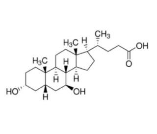
C[C@H](CCC(=O)O)[C@H]1CC[C@@H]2[C@@]1(CC[C@H]3[C@H]2[C@H](C[C@H]4[C@@]3(CC[C@H](C4)O)C)O)C
|
| InChi Key |
RUDATBOHQWOJDD-UZVSRGJWSA-N
|
| InChi Code |
InChI=1S/C24H40O4/c1-14(4-7-21(27)28)17-5-6-18-22-19(9-11-24(17,18)3)23(2)10-8-16(25)12-15(23)13-20(22)26/h14-20,22,25-26H,4-13H2,1-3H3,(H,27,28)/t14-,15+,16-,17-,18+,19+,20+,22+,23+,24-/m1/s1
|
| 化学名 |
(4R)-4-[(3R,5S,7S,8R,9S,10S,13R,14S,17R)-3,7-dihydroxy-10,13-dimethyl-2,3,4,5,6,7,8,9,11,12,14,15,16,17-tetradecahydro-1H-cyclopenta[a]phenanthren-17-yl]pentanoic acid
|
| 别名 |
URSODEOXYCHOLIC ACID; ursodiol; 128-13-2; Actigall; UDCA; Ursodesoxycholic acid; Urso Forte; Litursol;
|
| HS Tariff Code |
2934.99.9001
|
| 存储方式 |
Powder -20°C 3 years 4°C 2 years In solvent -80°C 6 months -20°C 1 month |
| 运输条件 |
Room temperature (This product is stable at ambient temperature for a few days during ordinary shipping and time spent in Customs)
|
| 溶解度 (体外实验) |
DMSO : ≥ 100 mg/mL (~254.73 mM)
H2O : ~1 mg/mL (~2.55 mM) |
|---|---|
| 溶解度 (体内实验) |
配方 1 中的溶解度: ≥ 2.08 mg/mL (5.30 mM) (饱和度未知) in 10% DMSO + 40% PEG300 + 5% Tween80 + 45% Saline (这些助溶剂从左到右依次添加,逐一添加), 澄清溶液。
例如,若需制备1 mL的工作液,可将100 μL 20.8 mg/mL澄清DMSO储备液加入400 μL PEG300中,混匀;然后向上述溶液中加入50 μL Tween-80,混匀;加入450 μL生理盐水定容至1 mL。 *生理盐水的制备:将 0.9 g 氯化钠溶解在 100 mL ddH₂O中,得到澄清溶液。 配方 2 中的溶解度: ≥ 2.08 mg/mL (5.30 mM) (饱和度未知) in 10% DMSO + 90% (20% SBE-β-CD in Saline) (这些助溶剂从左到右依次添加,逐一添加), 澄清溶液。 例如,若需制备1 mL的工作液,可将 100 μL 20.8 mg/mL澄清DMSO储备液加入900 μL 20% SBE-β-CD生理盐水溶液中,混匀。 *20% SBE-β-CD 生理盐水溶液的制备(4°C,1 周):将 2 g SBE-β-CD 溶解于 10 mL 生理盐水中,得到澄清溶液。 View More
配方 3 中的溶解度: ≥ 2.08 mg/mL (5.30 mM) (饱和度未知) in 10% DMSO + 90% Corn Oil (这些助溶剂从左到右依次添加,逐一添加), 澄清溶液。 1、请先配制澄清的储备液(如:用DMSO配置50 或 100 mg/mL母液(储备液)); 2、取适量母液,按从左到右的顺序依次添加助溶剂,澄清后再加入下一助溶剂。以 下列配方为例说明 (注意此配方只用于说明,并不一定代表此产品 的实际溶解配方): 10% DMSO → 40% PEG300 → 5% Tween-80 → 45% ddH2O (或 saline); 假设最终工作液的体积为 1 mL, 浓度为5 mg/mL: 取 100 μL 50 mg/mL 的澄清 DMSO 储备液加到 400 μL PEG300 中,混合均匀/澄清;向上述体系中加入50 μL Tween-80,混合均匀/澄清;然后继续加入450 μL ddH2O (或 saline)定容至 1 mL; 3、溶剂前显示的百分比是指该溶剂在最终溶液/工作液中的体积所占比例; 4、 如产品在配制过程中出现沉淀/析出,可通过加热(≤50℃)或超声的方式助溶; 5、为保证最佳实验结果,工作液请现配现用! 6、如不确定怎么将母液配置成体内动物实验的工作液,请查看说明书或联系我们; 7、 以上所有助溶剂都可在 Invivochem.cn网站购买。 |
| 制备储备液 | 1 mg | 5 mg | 10 mg | |
| 1 mM | 2.5473 mL | 12.7366 mL | 25.4732 mL | |
| 5 mM | 0.5095 mL | 2.5473 mL | 5.0946 mL | |
| 10 mM | 0.2547 mL | 1.2737 mL | 2.5473 mL |
1、根据实验需要选择合适的溶剂配制储备液 (母液):对于大多数产品,InvivoChem推荐用DMSO配置母液 (比如:5、10、20mM或者10、20、50 mg/mL浓度),个别水溶性高的产品可直接溶于水。产品在DMSO 、水或其他溶剂中的具体溶解度详见上”溶解度 (体外)”部分;
2、如果您找不到您想要的溶解度信息,或者很难将产品溶解在溶液中,请联系我们;
3、建议使用下列计算器进行相关计算(摩尔浓度计算器、稀释计算器、分子量计算器、重组计算器等);
4、母液配好之后,将其分装到常规用量,并储存在-20°C或-80°C,尽量减少反复冻融循环。
计算结果:
工作液浓度: mg/mL;
DMSO母液配制方法: mg 药物溶于 μL DMSO溶液(母液浓度 mg/mL)。如该浓度超过该批次药物DMSO溶解度,请首先与我们联系。
体内配方配制方法:取 μL DMSO母液,加入 μL PEG300,混匀澄清后加入μL Tween 80,混匀澄清后加入 μL ddH2O,混匀澄清。
(1) 请确保溶液澄清之后,再加入下一种溶剂 (助溶剂) 。可利用涡旋、超声或水浴加热等方法助溶;
(2) 一定要按顺序加入溶剂 (助溶剂) 。
Fenofibrate in Patients with Primary Biliary Cholangitis (PBC)
CTID: NCT06365424
Phase: Phase 2/Phase 3 Status: Recruiting
Date: 2024-09-19
InvivoChem的所有产品仅用于作科学研究,不面向患者销售
Copyright 2020 InvivoChem LLC | All Rights Reserved 粤ICP备20063088号-1


 463611831
