| 规格 | 价格 | 库存 | 数量 |
|---|---|---|---|
| 10 mM * 1 mL in DMSO |
|
||
| 1mg |
|
||
| 5mg |
|
||
| 10mg |
|
||
| 25mg |
|
||
| 50mg |
|
||
| 100mg |
|
||
| 250mg |
|
||
| 500mg |
|
||
| Other Sizes |
|

| 靶点 |
Aurora A (Ki = 0.6 nM); Aurora B (Ki = 18 nM); Aurora C (Ki = 4.6 nM)
From [1] (Aurora kinase-focused assays): - Tozasertib (VX680; MK0457) is a potent, pan-Aurora kinase inhibitor with high selectivity for Aurora A, Aurora B, and Aurora C; - IC50 values for recombinant human Aurora kinases: Aurora A = 0.6 nM, Aurora B = 18 nM, Aurora C = 6 nM (≥30/10-fold selectivity for Aurora A over Aurora B/C); - Weak inhibition of non-Aurora kinases: IC50 for CDK1 = 200 nM, IC50 for PLK1 = 350 nM (≥333/583-fold selectivity over Aurora A) [1] - From [2] (ABL2-focused binding assays): - Inhibits ABL-related gene 2 (ABL2) kinase activity; - Ki for recombinant human ABL2 = 38 nM; IC50 for ABL2 kinase activity = 120 nM; - No significant inhibition of ABL1 (IC50 > 500 nM) [2] |
|---|---|
| 体外研究 (In Vitro) |
当用 ABL 或 FLT-3(突变型和野生型)激酶转染的 BaF3 细胞暴露于 tozasertib 时,除了相当的细胞毒性(IC50 约为 300 nM)外,细胞还表现出 G2/M 停滞、核内复制和凋亡。抑制表型类似于 AUR B。tozasertib 具有时间依赖性抑制 CAL-62 的生长。在 tozasertib 治疗 14 天后,8305C 的集落数量和大小显着减少约 70%,CAL-62、8505C 和 BHT-101 显着减少 90%。用 tozasertib 处理不同的 ATC 细胞系,可降低生长,IC50 范围为 25 至 150 nM。 Tozasertib 显着降低了许多细胞系在软琼脂中建立集落的能力。 Caspase-3 活性分析表明,Tozasertib 会导致多种细胞类型凋亡。暴露于tozasertib 12小时后,CAL-62细胞积累了DNA含量低于4N的细胞。延时成像显示,Tozasertib 处理的 CAL-62 细胞退出中期而不增殖。此外,使用 tozasertib 会消除组蛋白 H3 磷酸化 [2]。在患者来源的样本中,tozasertib 显着抑制带有 T315I 突变的 BCR-Abl [3]。
实体癌细胞抗增殖与促凋亡活性(来自[1]): - 在人癌细胞系(HCT116:结肠癌;HeLa:宫颈癌;MCF-7:乳腺癌)中: 1. Tozasertib (0.01–100 nM)剂量依赖性抑制增殖:IC50 = 1.2 nM(HCT116)、0.8 nM(HeLa)、1.5 nM(MCF-7)(72小时MTT法); 2. 10 nM诱导G2/M周期阻滞:HCT116细胞G2/M期比例从溶剂组12%升至70%(PI染色,流式细胞术); 3. 20 nM诱导凋亡:HCT116细胞Annexin V阳性比例52% vs 溶剂组6%(流式细胞术); 4. 蛋白质印迹法:10 nM使p-Aurora A(Thr288)减少95%、p-Aurora B(Thr232)减少90%,上调cleaved caspase-3 4.0倍[1] - 间变性甲状腺癌细胞(ATC)活性(来自[3]): - 在人ATC细胞系(CAL-62、8505C、SW1736)中: 1. Tozasertib (0.1–50 nM)抑制增殖:IC50 = 2.3 nM(CAL-62)、1.8 nM(8505C)、2.5 nM(SW1736)(72小时CCK-8法); 2. 5 nM使p-Aurora A/B减少85%(蛋白质印迹法),抑制CAL-62细胞克隆形成75%(14天甲基纤维素实验); 3. 15 nM诱导凋亡:8505C细胞Annexin V阳性比例45% vs 溶剂组7%[3] - ABL2激酶抑制活性(来自[2]): - 在重组人ABL2激酶实验中: 1. Tozasertib (0.1–500 nM)剂量依赖性抑制ABL2活性:IC50 = 120 nM; 2. 对ABL1介导的STAT5磷酸化无显著影响(K562细胞蛋白质印迹法,IC50 > 500 nM)[2] |
| 体内研究 (In Vivo) |
VX-680 使人类 AML (HL-60) 异种移植模型中的肿瘤大小显着减小。在 mude 小鼠中,每天两次腹腔注射 (bidip) 75 mg/kg VX-680 治疗 13 天,平均肿瘤体积减少了 98%。肿瘤生长减少是剂量依赖性的,并且在 12.5 mg/kg bid 的剂量下显着,VX-680 具有良好的耐受性,仅在最高剂量时观察到体重小幅下降。 VX-680 还在胰腺和结肠异种移植模型中引发肿瘤消退。当静脉注射给携带 HCT116 肿瘤的 mude 大鼠时,VX-680 也显示出有效的抗肿瘤活性。更高剂量的 VX-680 (2 mg/kg/h) 可提高疗效,平均肿瘤体积减少 56%。
HCT116结肠癌异种移植疗效(来自[1]): - 6–8周龄雌性裸鼠(n=6/组)皮下接种HCT116细胞(5×10⁶个,第0天): 1. 处理组: - 溶剂组:5% DMSO + 95%生理盐水(腹腔注射,IP,每日1次,第7–20天); - Tozasertib 10 mg/kg组:IP,每日1次,第7–20天; - Tozasertib 25 mg/kg组:IP,每日1次,第7–20天; 2. 疗效(第21天): - 25 mg/kg实现90%肿瘤生长抑制率(TGI):治疗组肿瘤体积180 mm³ vs 溶剂组1800 mm³; - 肿瘤裂解液:p-Aurora A/B减少90%(蛋白质印迹法); - IHC检测:有丝分裂指数(p-组蛋白H3阳性细胞)从溶剂组25%降至治疗组3%[1] |
| 酶活实验 |
ABL2激酶结构域的蛋白表达与纯化[2]
用Sf9细胞培养获得的杆状病毒感染悬浮培养的Trichoplusia ni (Hi5)细胞,以2 × 10⁶个/mL的密度表达该蛋白。感染后48 h,离心收获细胞,细胞微球保存于- 80℃。细胞重悬于由5mm咪唑、500 mM NaCl、50 mM Hepes、pH 7.4、5%甘油、0.5 mM三(2-羧基乙基)膦(TCEP)组成的缓冲液中,补充完整的蛋白酶抑制剂混合物,超声裂解。裂解液在45000g下,在4℃下离心1 h。过滤后的上清液上载于镍螯合树脂上。洗涤后,用上述缓冲液加50 ~ 300 mM咪唑洗脱蛋白,洗脱液混合。用TEV蛋白酶在4°C下处理过夜,去除六组氨酸标签。将消化后的ABL2浓缩至2.5 mL体积,上载于Superdex75凝胶过滤柱(hilload 16/60, GE Healthcare),在10 mM Hepes、pH 8.0、300 mM NaCl和0.5 mM TCEP中平衡。[2] 通过电喷雾电离飞行时间质谱法对该蛋白进行了鉴定。在去除六组氨酸标签之前,与计算质量33 502相比,观察到的质量为33 414 Da;质量的差异很可能是由于去除了n端蛋氨酸,然后乙酰化。去除六组氨酸标签后,观察到的质量为30 980 Da,与计算质量完全吻合。 结晶与数据收集[2] ABL2:伊马替尼复合物(PDB代码3GVU,表2)2)在4°C下从ABL2:伊马替尼(4 mg/mL含有1 mM伊马替尼的蛋白质)和储层溶液(20% PEG3350, 0.1 M柠檬酸盐,pH 5.5)的1:1比例滴入200 nL滴液中结晶。然后晶体在含20% (v/v) PEG300的储层溶液中冷冻,并在液氮中速冻。在瑞士光源(SLS)下,在X10SA光束线上采集了100 K下的x射线衍射数据。 Aurora A/B激酶活性实验(放射性法,来自[1]): 1. 将纯化人Aurora A(0.1 μg/mL)或Aurora B(0.2 μg/mL)与组蛋白H3(1 μg/mL,底物)、[γ-³²P]ATP(5 μCi,10 μM)在激酶缓冲液(50 mM Tris-HCl pH 7.5、10 mM MgCl₂、1 mM DTT)中30°C孵育15分钟。 2. 加入系列浓度Tozasertib (0.01–500 nM),继续孵育30分钟。 3. 将反应液点样于P81磷酸纤维素纸,用1%磷酸洗涤3次以去除未结合的ATP。 4. 液体闪烁计数仪检测放射性,通过四参数逻辑回归计算IC50[1] - ABL2激酶活性实验(基于HTRF,来自[2]): 1. 将纯化人ABL2激酶域(0.2 μg/mL)与生物素化STAT5肽(含Tyr694基序,1 μg/mL)、ATP(10 μM)在实验缓冲液(50 mM HEPES pH 7.4、5 mM MgCl₂、0.1 mM Na₃VO₄)中37°C孵育20分钟。 2. 加入系列浓度Tozasertib (0.1–500 nM),继续孵育30分钟。 3. 用20 mM EDTA终止反应,加入抗磷酸化STAT5穴状化合物抗体和链霉亲和素-铕偶联物。 4. 检测时间分辨荧光(激发光340 nm,发射光665 nm/620 nm比值),用1:1结合模型计算Ki[2] |
| 细胞实验 |
免疫印迹[3]
细胞在RIPA缓冲液(50 mM Tris-HCl (pH 7.4), 1% NP-40, 0.5%脱氧胆酸钠,150 mM氯化钠,1 mM EDTA, 1×蛋白酶抑制剂鸡尾酒集III)中裂解,超声,然后在15 000 g下离心20分钟。用Bradford法测定蛋白浓度。50 μg细胞蛋白提取物等分液在12.5%聚丙烯酰胺凝胶上电泳,并转移到硝化纤维素膜上。然后用TBST (50 mM Tris-HCl (pH 7.4), 150 mM NaCl, 0.05% twee -20)洗涤膜,用5%低脂牛奶在TBST中饱和,然后在4°C下与抗极光a(1:500),极光b(1:500),极光C(1:500)或肌动蛋白(1:100)抗体在TBS-T中孵育过夜。洗涤后,将膜与合适的山根过氧化物酶偶联抗小鼠或兔IgG二抗(1:20 000)在TBST中孵育,并使用化学发光超级信号试剂盒 进行开发。使用Bio-Rad 670型扫描密度计的Molecular Analyst PC软件,通过扫描密度测定法定量极光激酶和肌动蛋白免疫反应带。计算不同的极光激酶/肌动蛋白比值,得到Tozasertib (VX680;MK0457)处理的细胞与对照细胞中的细胞相比归一化,并报告为变异的倍数。 扩散试验[3] CAL-62细胞在不含(二甲基亚砜,DMSO)或500 nM Tozasertib (VX680;MK0457)的不同时间段(1-5天)。 用不同浓度的Aurora抑制剂(5-500 nM)处理不同的ATC细胞4天,观察Tozasertib (VX680;MK0457)对细胞增殖的影响。在孵育结束前,用30 mM BrdU脉冲标记细胞2小时。根据制造商的说明,使用细胞增殖ELISA试剂盒(罗氏应用科学)通过比色免疫分析法分析BrdU掺入。Tozasertib (VX680;MK0457)处理的细胞与对照细胞中观察到的细胞进行比较,表达为与对照相比的变异倍数。 流式细胞仪分析[3]< br > CAL-62细胞在DMSO缺失或250 nM Tozasertib (VX680;MK0457)持续6、12或72小时。在一些实验中,细胞在收获前20分钟用30 mM BrdU处理。用橡皮警棍刮擦,在PBS中收集细胞,并将其固定在冰冷的乙醇中。使用EPICS Elite流式细胞仪分析细胞样本的DNA含量(碘化丙啶)和/或BrdU含量(FITC),如前所述(30)。 软琼脂的菌落形成[3] 首先加入3 ml含0.4%软琼脂的完整培养基,制备直径为3.5 cm的培养皿。将贴壁ATC细胞培养物胰蛋白酶化并离心,获得150000个活细胞/ml的单细胞悬液。将悬浮液与含0.4%软琼脂的完全培养基按1:2的比例混合,然后分成两份,其中一份添加Tozasertib (VX680;MK0457) 250 nM,另一个与载具(DMSO)。将这些悬浮液涂于培养皿中,1 ml/皿,在37°C和5% CO2下孵育。处理过的和未处理过的板在电镀后几小时拍照,以排除细胞聚集物的存在(数据未显示),并在2周后再次拍照。利用MetaVue软件 测量菌落大小,对直径大于50 μm的菌落进行评分。 Caspase-3试验[3] 不同的ATC细胞在不含DMSO或存在250 nM Tozasertib (VX680;MK0457) 72 h。处理后,用PBS冲洗细胞,在PBS中刮取。然后使用caspase-3/CPP32荧光测定试剂盒评估细胞的caspase-3活性。 延时分析[3] CAL-62细胞在DMSO缺失或250 nM Tozasertib (VX680;MK0457),在配备孵育室的徕卡DM-IRBE显微镜下,在37°C和5% CO2条件下观察24 h。使用MetaVue软件每5分钟拍摄一次细胞图片。 免疫荧光(如果)[3] 玻璃片上培养的CAL-62细胞用250 nM Tozasertib (VX680;MK0457)或Vehicle(DMSO) 6小时。细胞在冷甲醇中固定2分钟,洗涤,在室温下用3% BSA在PBS中预孵育1小时。PBS洗涤三次后,细胞与抗tacc3抗体(1:100)、抗aurora - a抗体(1:200)、抗aurora - b抗体(1:200)、抗aurora - c抗体(1:200)、抗p -组蛋白H3抗体(1:100)、抗γ-微管蛋白抗体(1:200)、抗β-微管蛋白抗体(1:200)室温孵育2小时。 HCT116细胞增殖与周期实验(来自[1]): 1. HCT116细胞(5×10³细胞/孔)接种于96孔板,37°C(5% CO₂)过夜孵育。 2. 加入系列浓度Tozasertib (0.01/0.1/1/10/100 nM),培养72小时。 3. 每孔加入MTT试剂(5 mg/mL,10 μL),孵育4小时;DMSO溶解甲臜结晶,检测570 nm吸光度计算IC50。 4. 周期分析:HCT116细胞(1×10⁶细胞/mL)用10 nM Tozasertib 处理24小时,70%乙醇固定,PI(50 μg/mL)+ RNase A(100 μg/mL)染色,流式细胞术分析[1] - ATC细胞增殖与凋亡实验(来自[3]): 1. CAL-62/8505C细胞(5×10³细胞/孔)接种于96孔板,37°C(5% CO₂)过夜孵育。 2. 加入系列浓度Tozasertib (0.1/0.5/1/5/10/50 nM),培养72小时;每孔加入CCK-8试剂(10 μL),检测450 nm吸光度计算IC50。 3. 凋亡检测:8505C细胞(1×10⁵细胞/mL)用15 nM Tozasertib 处理48小时,Annexin V-FITC/PI染色,流式细胞术分析[3] |
| 动物实验 |
溶于 50% PEG300 的 50 mM 磷酸盐缓冲液中;剂量为 50 和 75 mg/kg;腹腔注射。用于携带 HL-60 白血病细胞的雌性无胸腺 NCr-nu 小鼠。Aurora 激酶在有丝分裂过程中对染色体分离和胞质分裂的调控至关重要。这些激酶的异常表达和活性在多种人类肿瘤中均有发生,并导致非整倍体和肿瘤发生。本文报道了一种高效且选择性的 Aurora 激酶小分子抑制剂 VX-680 的发现,该抑制剂可阻断多种人类肿瘤细胞的细胞周期进程并诱导其凋亡。该化合物在多种体内异种移植模型中均能显著抑制肿瘤生长,并在耐受剂量下使白血病、结肠癌和胰腺癌肿瘤消退。我们的数据表明,Aurora激酶抑制剂为治疗多种人类恶性肿瘤提供了一种新方法。[1]HCT116结肠癌异种移植模型(引自[1]):1. 动物:雌性裸鼠(6-8周龄,18-20克,每组n=6)。2. 异种移植瘤建立:第0天:将5×10⁶个HCT116细胞(100 μL 1:1 PBS-基质胶)皮下注射到右侧腹部。3. 开始治疗:第7天(肿瘤体积约100 mm³)。4. 治疗组:- 对照组:5% DMSO + 95%生理盐水,腹腔注射(IP),每日一次,第7-20天。 - 托扎替尼 10 mg/kg:溶于 5% DMSO + 95% 生理盐水,腹腔注射,每日一次,第 7-20 天。- 托扎替尼 25 mg/kg:溶剂/给药途径与 10 mg/kg 相同,第 7-20 天。5. 监测和取样:- 每 3 天测量一次肿瘤体积(长×宽²/2);每周监测体重。- 第 21 天:处死小鼠,收集肿瘤组织进行蛋白质印迹(p-Aurora A/B)和免疫组化(磷酸化组蛋白 H3)分析[1]
|
| 药代性质 (ADME/PK) |
代谢/代谢物
托扎替尼已知的代谢物包括Unii-1S9W4WJ8WL和Unii-wuu5ros9AF。 血浆蛋白结合率(引自[1]):- 人血浆:98%(平衡透析,37°C,4小时);- 小鼠血浆:97% [1] - 小鼠半衰期(引自[1]):- 腹腔注射托扎替尼25 mg/kg的雌性裸鼠(每组n=3):末端半衰期(t1/2)= 3.2小时;- 血浆峰浓度(Cmax)= 8.5 μg/mL,达峰时间(Tmax)= 0.5小时 [1] |
| 毒性/毒理 (Toxicokinetics/TK) |
异种移植小鼠体内安全性(来自[1]):- 小鼠接受托扎替尼治疗,剂量高达25 mg/kg(腹腔注射,14天):- 体重变化≤5%(与载体组相比);- 无明显临床毒性(嗜睡、腹泻);- 血清ALT/AST/肌酐在正常范围内(第21天)[1]
- 体外正常细胞安全性(来自[3]):- 人正常甲状腺滤泡细胞(Nthy-ori 3-1)用托扎替尼(≤50 nM)处理72小时:- 细胞活力>90%(CCK-8检测); |
| 参考文献 |
|
| 其他信息 |
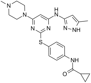
N-[4-[[4-(4-甲基-1-哌嗪基)-6-[(5-甲基-1H-吡唑-3-基)氨基]-2-嘧啶基]硫代]苯基]环丙烷甲酰胺是一种N-芳基哌嗪类化合物。
另见:托扎替尼乳酸盐(注释已移至)。 ABL2(也称为ARG(ABL相关基因))与研究较为深入的Abelson激酶cABL密切相关。ABL2参与人类肿瘤疾病的发生,并在实体瘤中表达失调。致癌基因易位发生在急性白血病中。目前尚未有关于ABL2结构信息的报道。为了阐明抑制剂相互作用的结构决定因素,我们解析了ABL2与抗癌药物伊马替尼的共晶体结构。有趣的是,伊马替尼不仅与非活性激酶的ATP结合位点相互作用,而且还与调节性肉豆蔻酸结合位点结合。因此,这种结构可能作为开发变构ABL抑制剂的工具。此外,我们解析了ABL2与VX-680以及ATP模拟I型抑制剂的复合物结构,揭示了DFG基序在活性和非活性构象之间的有趣中间位置,这也可能作为未来抑制剂设计的模板。[2] 间变性甲状腺癌(ATC)是一种侵袭性肿瘤,其细胞周期失调导致细胞不受控制地增殖和基因组不稳定。它们对化疗药物和放射疗法均无反应,大多数患者在确诊后几个月内死亡。在本研究中,我们评估了VX-680(一种参与染色体分离和胞质分裂多个环节调控的Aurora丝氨酸/苏氨酸激酶抑制剂)对ATC细胞的体外作用。我们检测了VX-680对ATC衍生细胞系CAL-62、8305C、8505C和BHT-101的增殖、凋亡、软琼脂克隆形成、细胞周期和倍性的影响。VX-680处理不同ATC细胞后,细胞增殖呈时间和剂量依赖性抑制,IC50值介于25至150 nM之间。VX-680显著抑制了不同细胞系在软琼脂中形成克隆的能力。caspase-3活性分析表明,VX-680诱导了不同细胞系的凋亡。 CAL-62 细胞暴露于 VX-680 12 小时后,DNA 含量 ≥ 4N 的细胞数量显著增加。延时分析表明,VX-680 处理的 CAL-62 细胞在未分裂的情况下退出有丝分裂中期。此外,VX-680 处理后组蛋白 H3 磷酸化水平降低。总之,我们的数据表明 VX-680 能有效抑制不同 ATC 衍生细胞系的生长,值得进一步研究以挖掘其在 ATC 治疗中的潜在治疗价值。[3] 作用机制(引自 [1,2,3]):1. 抑制 Aurora 激酶:托扎替尼与 Aurora A/B/C 的 ATP 结合口袋竞争性结合,抑制其激酶活性,阻断纺锤体组装和染色体分离,导致 G2/M 期阻滞和细胞凋亡 [1,3]; 2. 针对 ABL2:抑制 ABL2 介导的 STAT5 磷酸化,但效力低于 Aurora 激酶 [2] - 治疗潜力(引自 [1,3]): - 在实体瘤(结肠癌、宫颈癌、乳腺癌)中显示出临床前疗效 [1]; - 具有治疗难治性甲状腺间变性癌的潜力 [3] - 药物类别(引自 [1]):托扎替尼属于吡唑并嘧啶类激酶抑制剂,针对泛 Aurora 激酶抑制进行了优化 [1] |
| 分子式 |
C23H28N8OS
|
|
|---|---|---|
| 分子量 |
464.59
|
|
| 精确质量 |
464.21
|
|
| 元素分析 |
C, 59.46; H, 6.07; N, 24.12; O, 3.44; S, 6.90
|
|
| CAS号 |
639089-54-6
|
|
| 相关CAS号 |
|
|
| PubChem CID |
5494449
|
|
| 外观&性状 |
White to off-white solid powder
|
|
| 密度 |
1.4±0.1 g/cm3
|
|
| 折射率 |
1.708
|
|
| LogP |
1.18
|
|
| tPSA |
127.37
|
|
| 氢键供体(HBD)数目 |
3
|
|
| 氢键受体(HBA)数目 |
8
|
|
| 可旋转键数目(RBC) |
7
|
|
| 重原子数目 |
33
|
|
| 分子复杂度/Complexity |
650
|
|
| 定义原子立体中心数目 |
0
|
|
| SMILES |
CC1=CC(=NN1)NC2=CC(=NC(=N2)SC3=CC=C(C=C3)NC(=O)C4CC4)N5CCN(CC5)C
|
|
| InChi Key |
GCIKSSRWRFVXBI-UHFFFAOYSA-N
|
|
| InChi Code |
InChI=1S/C23H28N8OS/c1-15-13-20(29-28-15)25-19-14-21(31-11-9-30(2)10-12-31)27-23(26-19)33-18-7-5-17(6-8-18)24-22(32)16-3-4-16/h5-8,13-14,16H,3-4,9-12H2,1-2H3,(H,24,32)(H2,25,26,27,28,29)
|
|
| 化学名 |
(N-[4({4-(4-methylpiperazin-1-yl)-6-[(3-methyl-1H-pyrazol-5 -yl)amino]pyrimidin-2-yl}thio)phenyl]cyclopropanecarboxamide)
|
|
| 别名 |
Tozasertib, MK-0457; VX-680; MK 0457; MK-0457; VX680; VX 680; MK0457; VE 465; VE465; VE-465
|
|
| HS Tariff Code |
2934.99.9001
|
|
| 存储方式 |
Powder -20°C 3 years 4°C 2 years In solvent -80°C 6 months -20°C 1 month 注意: 本产品在运输和储存过程中需避光。 |
|
| 运输条件 |
Room temperature (This product is stable at ambient temperature for a few days during ordinary shipping and time spent in Customs)
|
| 溶解度 (体外实验) |
|
|||
|---|---|---|---|---|
| 溶解度 (体内实验) |
配方 1 中的溶解度: ≥ 2.08 mg/mL (4.48 mM) (饱和度未知) in 10% DMSO + 40% PEG300 + 5% Tween80 + 45% Saline (这些助溶剂从左到右依次添加,逐一添加), 澄清溶液。
例如,若需制备1 mL的工作液,可将100 μL 20.8 mg/mL澄清DMSO储备液加入400 μL PEG300中,混匀;然后向上述溶液中加入50 μL Tween-80,混匀;加入450 μL生理盐水定容至1 mL。 *生理盐水的制备:将 0.9 g 氯化钠溶解在 100 mL ddH₂O中,得到澄清溶液。 配方 2 中的溶解度: ≥ 2.08 mg/mL (4.48 mM) (饱和度未知) in 10% DMSO + 90% (20% SBE-β-CD in Saline) (这些助溶剂从左到右依次添加,逐一添加), 澄清溶液。 例如,若需制备1 mL的工作液,可将 100 μL 20.8 mg/mL澄清DMSO储备液加入900 μL 20% SBE-β-CD生理盐水溶液中,混匀。 *20% SBE-β-CD 生理盐水溶液的制备(4°C,1 周):将 2 g SBE-β-CD 溶解于 10 mL 生理盐水中,得到澄清溶液。 View More
配方 3 中的溶解度: ≥ 2.08 mg/mL (4.48 mM) (饱和度未知) in 10% DMSO + 90% Corn Oil (这些助溶剂从左到右依次添加,逐一添加), 澄清溶液。 配方 4 中的溶解度: 30% PEG400+0.5% Tween80+5% propylene glycol:30mg/mL 1、请先配制澄清的储备液(如:用DMSO配置50 或 100 mg/mL母液(储备液)); 2、取适量母液,按从左到右的顺序依次添加助溶剂,澄清后再加入下一助溶剂。以 下列配方为例说明 (注意此配方只用于说明,并不一定代表此产品 的实际溶解配方): 10% DMSO → 40% PEG300 → 5% Tween-80 → 45% ddH2O (或 saline); 假设最终工作液的体积为 1 mL, 浓度为5 mg/mL: 取 100 μL 50 mg/mL 的澄清 DMSO 储备液加到 400 μL PEG300 中,混合均匀/澄清;向上述体系中加入50 μL Tween-80,混合均匀/澄清;然后继续加入450 μL ddH2O (或 saline)定容至 1 mL; 3、溶剂前显示的百分比是指该溶剂在最终溶液/工作液中的体积所占比例; 4、 如产品在配制过程中出现沉淀/析出,可通过加热(≤50℃)或超声的方式助溶; 5、为保证最佳实验结果,工作液请现配现用! 6、如不确定怎么将母液配置成体内动物实验的工作液,请查看说明书或联系我们; 7、 以上所有助溶剂都可在 Invivochem.cn网站购买。 |
| 制备储备液 | 1 mg | 5 mg | 10 mg | |
| 1 mM | 2.1524 mL | 10.7622 mL | 21.5244 mL | |
| 5 mM | 0.4305 mL | 2.1524 mL | 4.3049 mL | |
| 10 mM | 0.2152 mL | 1.0762 mL | 2.1524 mL |
1、根据实验需要选择合适的溶剂配制储备液 (母液):对于大多数产品,InvivoChem推荐用DMSO配置母液 (比如:5、10、20mM或者10、20、50 mg/mL浓度),个别水溶性高的产品可直接溶于水。产品在DMSO 、水或其他溶剂中的具体溶解度详见上”溶解度 (体外)”部分;
2、如果您找不到您想要的溶解度信息,或者很难将产品溶解在溶液中,请联系我们;
3、建议使用下列计算器进行相关计算(摩尔浓度计算器、稀释计算器、分子量计算器、重组计算器等);
4、母液配好之后,将其分装到常规用量,并储存在-20°C或-80°C,尽量减少反复冻融循环。
计算结果:
工作液浓度: mg/mL;
DMSO母液配制方法: mg 药物溶于 μL DMSO溶液(母液浓度 mg/mL)。如该浓度超过该批次药物DMSO溶解度,请首先与我们联系。
体内配方配制方法:取 μL DMSO母液,加入 μL PEG300,混匀澄清后加入μL Tween 80,混匀澄清后加入 μL ddH2O,混匀澄清。
(1) 请确保溶液澄清之后,再加入下一种溶剂 (助溶剂) 。可利用涡旋、超声或水浴加热等方法助溶;
(2) 一定要按顺序加入溶剂 (助溶剂) 。
| NCT Number | Recruitment | interventions | Conditions | Sponsor/Collaborators | Start Date | Phases |
| NCT02532868 | Terminated | Drug: MK-0457 | Cancer | Merck Sharp & Dohme LLC | May 2005 | Phase 1 |
| NCT00099346 | Terminated | Drug: MK0457, VX-680 (Aurora Kinase Inhibitor) |
Colorectal Cancer Advanced Solid Tumors |
Merck Sharp & Dohme LLC | January 2005 | Phase 1 |
ABL2 bound to a type I inhibitor2. (A) ABL2:2, showing the compound bound to the ATP binding site, and the ordered activation loop. Compound2is shown in yellow.J Med Chem.2011 Apr 14;54(7):2359-67. |
Myristate binding pocket of ABL2. (A) Surface of the myristate binding pocket of ABL2, with imatinib shown as a yellow ball-and-stick representation.J Med Chem.2011 Apr 14;54(7):2359-67. |
Comparison of ABL2:imatinib and ABL2:1with ABL1:imatinib and ABL1:1.J Med Chem.2011 Apr 14;54(7):2359-67. |
InvivoChem的所有产品仅用于作科学研究,不面向患者销售
Copyright 2020 InvivoChem LLC | All Rights Reserved 粤ICP备20063088号-1


 COA
COA
")
")
")
 463611831
