| 规格 | 价格 | 库存 | 数量 |
|---|---|---|---|
| 1mg |
|
||
| 5mg |
|
||
| 10mg |
|
||
| 25mg |
|
||
| 50mg |
|
||
| 100mg |
|
||
| 250mg |
|
||
| 500mg |
|
||
| Other Sizes |
|

| 靶点 |
TNKS2: 2 nM (IC50)
TNKS1: 5 nM (IC50) ARTD2: 479 nM (IC50) ARTD1: 5500 nM (IC50) XAV-939 specifically targets tankyrase 1 (TNKS1) and tankyrase 2 (TNKS2) (TNKS1 IC50 = 5 nM; TNKS2 IC50 = 9 nM) [1] XAV-939 does not significantly inhibit other poly(ADP-ribose) polymerases (PARPs: IC50 > 100 μM for PARP1, PARP2) [1] |
|---|---|
| 体外研究 (In Vitro) |
XAV-939 针对 TNKS1 和 TNKS2 的 IC50 值分别为 5 nM 和 2 nM[1]。 XAV-939 (0.3–30 μM) 持续三天或十天可改善 hBMSC 的成骨细胞发育 [2]。通过引起 SH3BP2 积累,XAV-939 (3 μM) 刺激 hMSC 的成骨细胞分化 [2]。在成骨细胞发育过程中,XAV-939(3 μM;10 天)会增加 hBMSC 细胞中 OPG 的表达并降低 RANKL 的表达 [2]。 XAV-939 刺激 SFRP3 和 SFRP4 的产生,同时抑制 Wnt/β-连环蛋白信号传导 [3]。
在重组TNKS1/TNKS2酶活性实验中,XAV-939 剂量依赖性抑制ADP核糖基化活性,IC50分别为5 nM(TNKS1)和9 nM(TNKS2),进而稳定AXIN蛋白并抑制Wnt/β-连环蛋白(β-catenin)信号通路 [1] - 在人间充质干细胞(hMSCs)中,XAV-939(1 μM)培养21天后促进成骨分化:第7天碱性磷酸酶(ALP)活性提高2.4倍,第21天矿化结节形成增加65%(茜素红S染色),成骨标志物(Runx2、ALP、骨钙素(OCN))的mRNA水平分别上调2.1倍、1.8倍和2.3倍 [2] - 在经历机械应力(10%周期性拉伸)的人颞下颌关节(TMJ)软骨细胞中,XAV-939(2 μM)抑制Wnt/β-catenin激活:核内β-catenin水平降低70%,分泌型卷曲相关蛋白(sFRP1、sFRP3)的mRNA表达分别上调1.9倍和2.2倍。它还下调分解代谢酶(MMP13降低62%;ADAMTS5降低58%),减少细胞外基质降解 [3] - 在从骨关节炎(OA)模型中分离的小鼠关节软骨细胞中,XAV-939(1 μM)抑制β-catenin核转位(降低65%),在mRNA水平下调OA相关基因(MMP13、Col10a1、Runx2)55-70%,同时上调合成代谢基因(Col2a1、聚集蛋白聚糖)1.8倍和1.6倍 [4] - 在正常hMSCs和软骨细胞中,XAV-939 在浓度高达20 μM时毒性较低(细胞活力较对照组>85%)[2][3] |
| 体内研究 (In Vivo) |
在体内,XAV-939可恢复机械应力引起的软骨退变 [3]。XAV-939改善OA严重程度,减少体内软骨退变和滑膜炎。[4]
为了评估Wnt活性的消融是否可以改善膝关节OA的严重程度,10周大的雄性小鼠接受了DMM手术。手术后3周,小鼠关节内注射生理盐水对照或小分子Wnt抑制剂XAV-939,每10天注射一次。术后10周,即最后一次注射后10天采集膝关节(图1A)。我们使用Wnt信号标记物β-catenin抗体和间质/成纤维细胞标记物periostin对关节切片进行免疫荧光,评估典型Wnt/β-catenin信号的时间和空间变化。损伤前,极少染色;然而,损伤后,β-catenin在膝关节,特别是滑膜中强烈上调(图1B),并在XAV-939治疗后减弱(图1C)。数据显示滑膜中典型Wnt信号的显著和动态增加,并且增强的染色明显与骨膜蛋白共定位。同型对照显示最小信号[4]。 体内实验结果显示,iovd诱导的TMJ机械应力破坏下颌骨生长,诱导TMJ软骨发生oa样变化,增加oa相关细胞因子表达。此外,iOVD激活Wnt/β-catenin信号通路,抑制Sfrp1、Sfrp3和Sfrp4在髁突软骨中的表达。此外,体外研究表明,应激破坏体内平衡,激活Wnt/β-catenin信号,抑制软骨细胞中SFRP3和SFRP4的表达。XAV-939抑制Wnt/β-catenin信号通路可促进SFRP3和SFRP4的表达,并在体内和体外挽救机械应力诱导的软骨变性。 结论:本研究提示机械应力在体内和体外均可降低SFRPs的表达,并通过Wnt/β-catenin信号通路促进TMJOA。抑制Wnt/β-catenin信号传导可促进SFRPs的表达,尤其是SFRP3和SFRP4的表达,并挽救机械应力诱导的软骨变性。Wnt/β-catenin信号和SFRPs可能是TMJOA的潜在治疗靶点。 关键词:机械应力;分泌卷曲相关蛋白(SFRPs);颞下颌关节骨关节炎(TMJOA);Wnt /β连环蛋白[3]。 在半月板失稳(DMM)诱导的小鼠OA模型中,腹腔注射 XAV-939(10 mg/kg/天,持续8周)显著改善关节损伤。OARSI组织学评分从溶媒组的8.2降至3.5,关节软骨厚度增加45%,MMP13阳性软骨细胞减少60%。它还抑制软骨下骨硬化(骨体积分数降低38%)[4] - 在咬合干扰诱导的大鼠TMJ OA模型中,关节内注射 XAV-939(5 μM/10 μL/关节,每周1次,持续4周)减少软骨侵蚀(侵蚀面积减少55%)和滑膜炎(滑膜厚度减少48%)。它恢复TMJ软骨中sFRP3的表达,抑制Wnt/β-catenin信号(核β-catenin阳性细胞减少62%)[3] |
| 酶活实验 |
抑制剂的筛选和抑制剂效力的测量[1]
通过搜索商业上可获得的化合物库来鉴定仅具有一个取代的黄酮衍生物,并通过Molport从不同的供应商处购买。将化合物储存在−20°C的二甲基亚砜中,并在TNKS1测定缓冲液中稀释,然后将其加入反应混合物中。在10μM和1μM下对化合物进行重复测试。在该筛选中使用化合物对照以排除化合物荧光和猝灭的影响。基于两点初始筛选,测量IC50值低于10μM的抑制剂的抑制能力。使用半对数稀释液测量IC50值,并对TNKS1进行一式四份的三次单独反应。调节孵育时间,使得底物转化率在筛选的情况下大于50%,在IC50测量的情况下小于30%。使用Graphpad Prism(Windows版本5.0)使用四个参数拟合剂量-反应曲线。 抑制剂的选择性[1] 使用上述同质活性测定法,还针对其他六个人类ARTD家族成员对鉴定的最佳坦激酶抑制剂进行了分析。基于先前进行的优化,每种酶的培养时间和条件各不相同。在分析测定中,底物NAD+浓度为250或500nM。为了获得稳健的信号,调节孵育时间,使得底物转化率在每种情况下都超过50%。为了有效地评估化合物的选择性,在1μM下对它们进行了表征。DMSO、化合物和蛋白质对照与所有酶一起使用,以排除或校正DMSO、自发荧光和荧光猝灭的影响。 TNKS1/TNKS2 ADP核糖基化实验:将纯化的重组人TNKS1或TNKS2与多聚ADP核糖聚合酶底物(组蛋白H1)和 XAV-939(0.1 nM-100 nM)在实验缓冲液(50 mM Tris-HCl,pH 7.5,10 mM MgCl₂,1 mM DTT,0.2 mM NAD⁺)中于37°C孵育45分钟。使用多聚ADP核糖特异性抗体通过Western blot检测ADP核糖基化底物,从剂量-效应曲线计算IC50值 [1] - PARP选择性实验:在与TNKS实验相同的缓冲液和底物(组蛋白H1)中,将 XAV-939(100 μM)对PARP1和PARP2进行筛选。通过Western blot的光密度分析量化ADP核糖基化活性,未观察到对PARP1或PARP2的显著抑制(活性降低>50%)[1] |
| 细胞实验 |
细胞活力测定[2]
细胞类型: hMSC-TERT 细胞系 测试浓度: 0.3、3 和 30 μM 孵育时间:3天 实验结果:证明0.3和3μM剂量对第1、2、3天的增殖没有显着影响,但在当天抑制hMSCs细胞增殖3 剂量为 30 μM。 细胞凋亡分析[2] 细胞类型: hMSC-TERT 细胞系 测试浓度: 3 μM 孵育时间: 3 天 实验结果: 在 XAV-939 处理的 hBMSC RT-PCR 中显示细胞死亡(凋亡和坏死)的微小百分比[2] 细胞类型: hMSC-TERT 细胞系 测试浓度: 3 µM 孵育时间: 10 天 <实验结果:成骨细胞相关基因标记物的基因表达上调,包括:ALP、COL1A1、RUNX2 和 OC。 hMSC成骨分化实验:人间充质干细胞以2×10⁴个/孔接种到6孔板中,在成骨培养基中培养。加入 XAV-939(0.1-10 μM),培养21天。第7天比色法检测ALP活性,第21天茜素红S染色矿化结节,第14天qPCR分析成骨标志物(Runx2、ALP、OCN)的mRNA水平 [2] - TMJ软骨细胞机械应力实验:原代人TMJ软骨细胞以1×10⁵个/孔接种到胶原包被的6孔板中,经历10%周期性拉伸(0.5 Hz)24小时。应力施加前1小时,用 XAV-939(0.5-5 μM)预处理细胞。qPCR检测sFRP1/3和分解代谢酶(MMP13、ADAMTS5)的mRNA水平,免疫荧光分析核β-catenin [3] - OA软骨细胞Wnt信号实验:从小鼠DMM诱导OA的膝关节中分离关节软骨细胞,以5×10³个/孔接种到96孔板中。用 XAV-939(0.1-5 μM)处理细胞24小时。免疫细胞化学检测β-catenin核转位,qPCR分析合成/分解代谢基因表达 [4] - 细胞活力实验:hMSCs和TMJ软骨细胞以3×10³个/孔接种到96孔板中,用 XAV-939(0.1-50 μM)处理72小时。CCK-8法评估细胞活力 [2][3] |
| 动物实验 |
2.5 mg/kg;腹腔注射
小鼠模型 所有小鼠均购自杰克逊实验室。10周龄雄性C57BL/6J小鼠接受DMM手术以诱导OA,方法如前所述。术后3周,每隔10天进行5次关节内注射XAV-939(0.4 mM)或生理盐水,总注射量为5 μl。术后10周收集对照组(n = 8)和Wnt抑制剂组(n = 8)小鼠的膝关节。[4] 研究人员使用改良的增加垂直咬合高度(iOVD)错颌畸形体内模型和分离的软骨细胞经受机械应力的体外模型,研究了机械应力诱导的颞下颌关节骨关节炎(TMJOA)的进展。本研究通过体外药物添加和体内关节内注射XAV-939,探讨了Wnt/β-catenin信号通路抑制对机械应力诱导的颞下颌关节骨关节炎(TMJOA)的影响。采用锥形束计算机断层扫描(CBCT)分析、组织化学和免疫组织化学(IHC)染色、定量实时PCR(qRT-PCR)、蛋白质印迹(WB)和免疫荧光(IF)染色评估TMJOA的进展、Wnt/β-catenin信号通路和SFRPs。[3] 小鼠DMM诱导OA模型:8周龄C57BL/6小鼠接受DMM手术以诱导OA。术后一周,将XAV-939溶解于DMSO中,并用生理盐水稀释(最终DMSO浓度≤5%),以10 mg/kg/天的剂量腹腔注射,持续8周。对照组注射DMSO/生理盐水混合物。处死小鼠后,收集膝关节进行组织学分析(番红O-固绿染色)和OARSI评分[4] - 咬合干扰诱导的大鼠颞下颌关节骨关节炎模型:对6周龄Sprague-Dawley大鼠进行咬合干扰以诱导颞下颌关节骨关节炎。同时,将XAV-939溶解于无菌PBS中,配制成5 μM的浓度,每周向颞下颌关节内注射10 μL,持续4周。对照组注射PBS。收集颞下颌关节组织进行组织学检查(苏木精-伊红染色和番红O染色)和β-catenin免疫组化染色[3] |
| 毒性/毒理 (Toxicokinetics/TK) |
体外实验表明,XAV-939 对正常间充质干细胞和软骨细胞的毒性较低(hMSCs IC50 > 20 μM;颞下颌关节软骨细胞 IC50 > 25 μM)[2][3]
- 体内研究表明,在测试剂量(10 mg/kg 腹腔注射,5 μM 关节内注射)下,XAV-939 不会引起小鼠和大鼠显著的体重减轻(<5% vs. 基线)或明显的死亡[3][4] - 与载体对照组相比,XAV-939 治疗组动物的肝功能(ALT、AST)或肾功能(肌酐、BUN)均未观察到显著变化[4] |
| 参考文献 |
|
| 其他信息 |
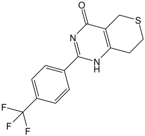
XAV939 是一种硫吡喃并嘧啶类化合物,其 7,8-二氢-5H-硫吡喃并[4,3-d]嘧啶骨架的 C-4 位被羟基取代,C-2 位被对位三氟甲基苯基取代。它是一种硫吡喃并嘧啶类化合物,属于三氟甲基苯类化合物。
硫吡喃并嘧啶类化合物是 ADP-核糖基转移酶,在多种细胞通路中发挥关键作用,包括细胞增殖的调控,因此是治疗癌症的潜在药物靶点。黄酮类化合物已被证明能够抑制硫吡喃并嘧啶类化合物,本文报道了我们发现的更有效、更具选择性的黄酮衍生物。我们使用市售的单取代黄酮类化合物进行构效关系研究,并分析了 18 个先导化合物的共晶结构,以解释其效力和选择性。研究人员还对活性最强的抑制剂进行了细胞实验,结果表明它们能有效拮抗Wnt信号通路。为了评估其选择性,研究人员进一步测试了这些抑制剂对一系列同源人ADP-核糖基转移酶的抑制作用。其中,活性最强的化合物22(MN-64)对tankyrase 1的抑制效力为6 nM,并表现出同工酶选择性和Wnt信号通路抑制作用。这项工作为合理开发黄酮类化合物作为tankyrase抑制剂奠定了基础,并指导了其他结构相关抑制剂的开发。[1] Tankyrase是聚(ADP-核糖)聚合酶超家族的成员,参与多种细胞和分子过程。Tankyrase抑制可负调控Wnt信号通路。因此,Tankyrase抑制剂已被广泛研究用于治疗与Wnt信号通路激活相关的临床疾病,例如癌症和纤维化疾病。此外,近期有报道称,Tankyrase抑制剂可通过SH3结构域结合蛋白2(一种骨代谢必需的衔接蛋白)的积累来上调成骨作用。本研究探讨了Tankyrase抑制剂对人骨骼(间充质)干细胞(hMSCs)成骨分化的影响。Tankyrase抑制剂XAV-939是在小分子功能筛选库中发现的。碱性磷酸酶活性和茜素红染色分别作为成骨分化和体外矿化基质形成的标志物。采用安捷伦微阵列平台进行全基因组表达谱分析。结果表明,Tankyrase抑制剂XAV-939可增强hBMSCs的成骨分化,表现为ALP活性增强、体外矿化基质形成增加以及成骨相关基因表达上调。对经 XAV-939 处理的细胞进行全基因组表达谱分析,发现与载体处理的对照细胞相比,有 847 个 mRNA 转录本上调,614 个 mRNA 转录本下调。分析结果还表明,多种信号通路可能发生改变,包括 TGFβ、胰岛素信号通路、黏着斑、雌激素代谢、氧化应激、RANK-RANKL(核因子 κB 受体激活因子配体)信号通路、维生素 D 合成、IL-6 以及细胞因子和炎症反应。进一步的生物信息学分析(采用 Ingenuity Pathway Analysis)发现,经 XAV-939 处理的细胞中,与 TNF、NFκB 和 STAT 信号通路相关的功能类别和网络显著富集。我们发现了一种Tankyrase抑制剂(XAV-939)能够有效增强人骨髓间充质干细胞(hBMSC)的成骨分化,这可能成为治疗骨形成减少相关疾病的一种有效疗法。[2]骨关节炎(OA)是一种累及软骨和滑膜的退行性关节疾病。在OA中被激活的经典Wnt/β-catenin信号通路正逐渐被认为是组织修复和纤维化的重要调节因子。本研究旨在探讨Wnt信号通路对滑膜成纤维细胞和关节软骨细胞的影响,以及Wnt信号通路抑制对OA疾病严重程度的治疗效果。小鼠接受了内侧半月板不稳定手术,并通过关节内注射Wnt/β-catenin信号通路小分子抑制剂XAV-939进行治疗。 Wnt/β-catenin信号通路在小鼠滑膜成纤维细胞以及骨关节炎(OA)来源的人滑膜成纤维细胞中均高度激活。XAV-939可减轻OA的严重程度,并减少体内软骨退变和滑膜炎。使用作用机制不同的两种小分子抑制剂XAV-939和C113抑制Wnt信号通路,可减弱体外滑膜成纤维细胞的增殖和I型胶原合成,但对人OA来源的软骨细胞增殖无影响。然而,Wnt信号通路的调节可增加COL2A1和PRG4的转录水平,而这两种基因在OA软骨细胞中表达下调。总之,在创伤性骨关节炎模型中,Wnt抑制剂通过促进软骨细胞的抗分解代谢作用和滑膜成纤维细胞的抗纤维化作用,降低了疾病严重程度,可能是一类有前景的骨关节炎治疗药物。[4] XAV-939是一种强效、选择性的TNKS1/2小分子抑制剂[1]。其作用机制包括与TNKS1/2的催化结构域结合,抑制其ADP核糖基化活性,稳定AXIN蛋白,并促进β-catenin降解,从而抑制经典的Wnt/β-catenin信号通路[1][3][4]。体外实验表明,XAV-939能够促进间充质干细胞的成骨分化,并保护软骨细胞免受分解代谢损伤;体内实验也证实了其在小鼠骨关节炎和大鼠颞下颌关节损伤中的治疗效果。 OA模型[2][3][4] - 它被广泛用作研究发育生物学、干细胞分化和骨关节炎发病机制中Wnt/β-catenin信号通路的工具化合物[1][2][3][4] - XAV-939在骨关节炎和其他Wnt/β-catenin信号通路相关的肌肉骨骼疾病中具有潜在的治疗应用价值[3][4] |
| 分子式 |
C14H11F3N2OS
|
|
|---|---|---|
| 分子量 |
312.31
|
|
| 精确质量 |
312.054
|
|
| 元素分析 |
C, 53.84; H, 3.55; F, 18.25; N, 8.97; O, 5.12; S, 10.27
|
|
| CAS号 |
284028-89-3
|
|
| 相关CAS号 |
|
|
| PubChem CID |
135418940
|
|
| 外观&性状 |
White to off-white solid powder
|
|
| 密度 |
1.5±0.1 g/cm3
|
|
| 沸点 |
429.3ºC at 760 mmHg
|
|
| 闪点 |
213.4ºC
|
|
| 折射率 |
1.634
|
|
| LogP |
2.98
|
|
| tPSA |
71.31
|
|
| 氢键供体(HBD)数目 |
1
|
|
| 氢键受体(HBA)数目 |
6
|
|
| 可旋转键数目(RBC) |
1
|
|
| 重原子数目 |
21
|
|
| 分子复杂度/Complexity |
505
|
|
| 定义原子立体中心数目 |
0
|
|
| SMILES |
S1C([H])([H])C2C(N([H])C(C3C([H])=C([H])C(C(F)(F)F)=C([H])C=3[H])=NC=2C([H])([H])C1([H])[H])=O
|
|
| InChi Key |
KLGQSVMIPOVQAX-UHFFFAOYSA-N
|
|
| InChi Code |
InChI=1S/C14H11F3N2OS/c15-14(16,17)9-3-1-8(2-4-9)12-18-11-5-6-21-7-10(11)13(20)19-12/h1-4H,5-7H2,(H,18,19,20)
|
|
| 化学名 |
2-(4-(trifluoromethyl)phenyl)-7,8-dihydro-3H-thiopyrano[4,3-d]pyrimidin-4(5H)-one
|
|
| 别名 |
|
|
| HS Tariff Code |
2934.99.9001
|
|
| 存储方式 |
Powder -20°C 3 years 4°C 2 years In solvent -80°C 6 months -20°C 1 month |
|
| 运输条件 |
Room temperature (This product is stable at ambient temperature for a few days during ordinary shipping and time spent in Customs)
|
| 溶解度 (体外实验) |
|
|||
|---|---|---|---|---|
| 溶解度 (体内实验) |
配方 1 中的溶解度: 1.56 mg/mL (5.00 mM) in 10% DMSO + 40% PEG300 + 5% Tween80 + 45% Saline (这些助溶剂从左到右依次添加,逐一添加), 悬浮液;超声助溶。
例如,若需制备1 mL的工作液,可将100 μL 15.6 mg/mL澄清DMSO储备液加入400 μL PEG300中,混匀;然后向上述溶液中加入50 μL Tween-80,混匀;加入450 μL生理盐水定容至1 mL。 *生理盐水的制备:将 0.9 g 氯化钠溶解在 100 mL ddH₂O中,得到澄清溶液。 配方 2 中的溶解度: 1.56 mg/mL (5.00 mM) in 10% DMSO + 90% (20% SBE-β-CD in Saline) (这些助溶剂从左到右依次添加,逐一添加), 悬浊液; 超声助溶。 例如,若需制备1 mL的工作液,可将 100 μL 15.6 mg/mL 澄清 DMSO 储备液加入 900 μL 20% SBE-β-CD 生理盐水溶液中,混匀。 *20% SBE-β-CD 生理盐水溶液的制备(4°C,1 周):将 2 g SBE-β-CD 溶解于 10 mL 生理盐水中,得到澄清溶液。 View More
配方 3 中的溶解度: 1.56 mg/mL (5.00 mM) in 10% DMSO + 90% Corn Oil (这些助溶剂从左到右依次添加,逐一添加), 澄清溶液; 超声助溶. 配方 4 中的溶解度: 配方 1 中的溶解度: ~1.6 mg/mL (5.0 mM) in 10% DMSO + 40% PEG300 + 5% Tween80 + + 45% Saline (这些助溶剂从左到右依次添加,逐一添加), 悬浮液。 例如,若需制备1 mL的工作液,您可以取100 μL 16 mg/mL DMSO 储备液加入到400 μL PEG300中,混匀;然后再向上述溶液中加入50 μL Tween 80,混匀;最后向上述溶液中加入450 μL 生理盐水,混匀。 生理盐水的配制:将0.9 g氯化钠溶解于100 mL ddH ₂ O中,得到澄清溶液。 配方 2 中的溶解度: ~1.6 mg/mL(5.0 mM)在10% DMSO + 90% (20% SBE-β-CD in saline)中(这些助溶剂从左到右依次添加,逐一添加),得到悬浮溶液。 例如,如果要制备1 mL工作液,则可以取100 μL 16 mg/mL DMSO储备液并添加到900 μL 20% SBE-β-CD 生理盐水溶液,混匀。 *20% SBE-β-CD 生理盐水溶液的制备(4°C,1 周):将 2 g SBE-β-CD 溶解于 10 mL 生理盐水中,得到澄清溶液。 配方 3 中的溶解度: ~1.6 mg/mL (5.0 mM) in 10% DMSO + 90% Corn oil (这些助溶剂从左到右依次添加,逐一添加), 澄清溶液。 例如,如果要制备1 mL工作液,则可以取100 μL 16 mg/mL DMSO储备液并添加到900 μL 玉米油,混合均匀。 配方 4 中的溶解度: ~5 mg/mL (16.0 mM) in 50% PEG300 + 50% Saline (这些助溶剂从左到右依次添加,逐一添加), 悬浮液。 配方 5 中的溶解度: ~30 mg/mL (96 mM) in 30% PEG 400+0.5% Tween 80+5% Propylene glycol (这些助溶剂从左到右依次添加,逐一添加). 配方 6 中的溶解度: 5 mg/mL (16.01 mM) in 50% PEG300 50% Saline (这些助溶剂从左到右依次添加,逐一添加), 悬浊液; 超声助溶。 *生理盐水的制备:将 0.9 g 氯化钠溶解在 100 mL ddH₂O中,得到澄清溶液。 1、请先配制澄清的储备液(如:用DMSO配置50 或 100 mg/mL母液(储备液)); 2、取适量母液,按从左到右的顺序依次添加助溶剂,澄清后再加入下一助溶剂。以 下列配方为例说明 (注意此配方只用于说明,并不一定代表此产品 的实际溶解配方): 10% DMSO → 40% PEG300 → 5% Tween-80 → 45% ddH2O (或 saline); 假设最终工作液的体积为 1 mL, 浓度为5 mg/mL: 取 100 μL 50 mg/mL 的澄清 DMSO 储备液加到 400 μL PEG300 中,混合均匀/澄清;向上述体系中加入50 μL Tween-80,混合均匀/澄清;然后继续加入450 μL ddH2O (或 saline)定容至 1 mL; 3、溶剂前显示的百分比是指该溶剂在最终溶液/工作液中的体积所占比例; 4、 如产品在配制过程中出现沉淀/析出,可通过加热(≤50℃)或超声的方式助溶; 5、为保证最佳实验结果,工作液请现配现用! 6、如不确定怎么将母液配置成体内动物实验的工作液,请查看说明书或联系我们; 7、 以上所有助溶剂都可在 Invivochem.cn网站购买。 |
| 制备储备液 | 1 mg | 5 mg | 10 mg | |
| 1 mM | 3.2019 mL | 16.0097 mL | 32.0195 mL | |
| 5 mM | 0.6404 mL | 3.2019 mL | 6.4039 mL | |
| 10 mM | 0.3202 mL | 1.6010 mL | 3.2019 mL |
1、根据实验需要选择合适的溶剂配制储备液 (母液):对于大多数产品,InvivoChem推荐用DMSO配置母液 (比如:5、10、20mM或者10、20、50 mg/mL浓度),个别水溶性高的产品可直接溶于水。产品在DMSO 、水或其他溶剂中的具体溶解度详见上”溶解度 (体外)”部分;
2、如果您找不到您想要的溶解度信息,或者很难将产品溶解在溶液中,请联系我们;
3、建议使用下列计算器进行相关计算(摩尔浓度计算器、稀释计算器、分子量计算器、重组计算器等);
4、母液配好之后,将其分装到常规用量,并储存在-20°C或-80°C,尽量减少反复冻融循环。
计算结果:
工作液浓度: mg/mL;
DMSO母液配制方法: mg 药物溶于 μL DMSO溶液(母液浓度 mg/mL)。如该浓度超过该批次药物DMSO溶解度,请首先与我们联系。
体内配方配制方法:取 μL DMSO母液,加入 μL PEG300,混匀澄清后加入μL Tween 80,混匀澄清后加入 μL ddH2O,混匀澄清。
(1) 请确保溶液澄清之后,再加入下一种溶剂 (助溶剂) 。可利用涡旋、超声或水浴加热等方法助溶;
(2) 一定要按顺序加入溶剂 (助溶剂) 。
|
|
|
InvivoChem的所有产品仅用于作科学研究,不面向患者销售
Copyright 2020 InvivoChem LLC | All Rights Reserved 粤ICP备20063088号-1


 COA
COA


 463611831
