AICA ribonucleotide; AICA riboside; AICAR; Acadesine; AICA Riboside; ARA100; ARA-100; 2627-69-2; AICA-riboside; Arasine; AICA riboside; AIC-Riboside; Acadesina; ARA 100; GP 1 110; SCH-900395; SCH 900395; SCH900395; AICAR
阿卡地新;5-氨基-1-beta-D-呋喃核糖基-1H-咪唑-4-甲酰胺;5-氨基咪唑-4-甲酰胺-1-B-D-呋喃核糖苷;5-氨基咪唑-4-甲酰胺-1-β-D-呋喃核糖苷;5-氨基咪唑-4-甲酰胺核苷酸转甲酰酶;阿卡地辛;阿卡地新 Acadesine;阿卡地新(AICAR)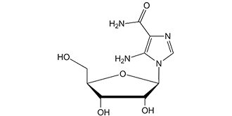
| 规格 | 价格 | 库存 | 数量 |
|---|---|---|---|
| 10 mM * 1 mL in DMSO |
|
||
| 1mg |
|
||
| 5mg |
|
||
| 10mg |
|
||
| 50mg |
|
||
| 100mg |
|
||
| 250mg |
|
||
| 500mg |
|
||
| 1g |
|
||
| 2g |
|
||
| Other Sizes |
|

| 靶点 |
AMPK; Autophagy; Mitophagy; Human Endogenous Metabolite
The primary target of Acadesine (AICAR; NSC-105823) is AMP-activated protein kinase (AMPK), a central regulator of cellular energy homeostasis. - In [1]: It activates rat liver AMPK with a half-maximal effective concentration (EC50) of ~100 μM, and shows no inhibitory activity against protein kinase A (PKA) or protein kinase C (PKC) (IC50 > 1 mM) [1] - In [2]: In human acute myeloid leukemia HL-60 cells, it activates AMPK with an EC50 of ~80 μM, and exhibits no cross-reactivity with Janus kinase 2 (JAK2) or other hematopoietic kinases (IC50 > 500 μM) [2] - In [3]: For human hepatocyte AMPK complexes, the EC50 is ~120 μM; it does not affect the PI3K-Akt signaling pathway (IC50 > 200 μM) [3] |
|---|---|
| 体外研究 (In Vitro) |
Acadesine (500 μM) 经过长达 30-40 分钟的处理后,可增加分离肝细胞提取物中的 ZMP 含量,然后保持相当恒定在约 4 nmol/g。 Acadesine (500 μM) 在 15 分钟内导致大鼠肝细胞中 AMPK 瞬时激活 12 倍,以及脂肪细胞中 AMPK 瞬时激活 2-3 倍,而不影响 ATP、ADP 或 AMP 水平。 Acadesine (500 μM) 会显着抑制大鼠肝细胞中脂肪酸和甾醇的合成。 Acadesine (500 μM) 还会导致 HMG-CoA 还原酶急剧失活。[1] cadesine 的 EC50 为 380 M,以剂量依赖性方式诱导 B-CLL 细胞凋亡。阿卡地辛 (0.5 mM) 将 20 名代表性患者的 B-CLL 细胞活力从 68% 降低至 26%。 Caspase 激活和线粒体细胞色素 c 释放由阿卡地辛 (acadesine) (0.5 mM) 引起。必须摄取 Acadesine (0.5 mM) 并磷酸化,才能引起 B-CLL 细胞凋亡并激活 AMPK。 Acadesine (0.5 mM) 显着降低 B 细胞的活力,但不降低 T 细胞的活力,对 B-CLL 患者的 T 细胞的活力影响极小。 [2] 在 K562、LAMA-84 和 JURL-MK1 细胞中,杜松碱会导致细胞代谢丧失。它还可以有效杀死具有 T315I-BCR-ABL 突变的伊马替尼耐药 K562 细胞和 Ba/F3 细胞。由于 GF109203X 和 Ro-32-0432(经典和新 PKC 的抑制剂)都会抵消 Accadesine 的作用,因此 Accadesine 会导致 K562 细胞中几种 PKC 同工型的运动和激活。第 10 天,阿卡地辛以剂量依赖性方式抑制 K562 集落形成。其生长抑制作用在浓度为 0.25 mM 时就已经很明显,并且在 2.5 mM 时达到最大。 [3] Accadesine 在体外以浓度依赖性方式降低 LPS 刺激的中性粒细胞上 CD18 的表达。 [4] 血液中的 cadesine (1 mM) 可显着抑制由 N-甲酰基-甲硫氨酰-亮氨酰-苯丙氨酸引起的粒细胞 CD11b 上调(平均 61%)。 [5]
1. 调控肝脏糖代谢(来自[1]): - 原代大鼠肝细胞用Acadesine(50 μM、100 μM、200 μM)处理24小时,浓度依赖性抑制胰高血糖素诱导的葡萄糖生成:100 μM时抑制率约40%,200 μM时达65%(葡萄糖氧化酶法检测)。Western blot显示AMPK下游底物乙酰辅酶A羧化酶(ACC)Ser79位点磷酸化水平(p-ACC/ACC比值)较对照升高2.5倍[1] 2. 白血病细胞抗增殖活性(来自[2]): - 人HL-60白血病细胞用Acadesine(25 μM、50 μM、100 μM)处理72小时,细胞活力(MTT法)呈剂量依赖性降低,IC50约为60 μM。流式细胞术显示G2/M期细胞周期阻滞:G2/M期细胞比例从对照的15%升至100 μM处理组的42%。Western blot显示细胞周期抑制剂p21表达上调3.1倍[2] 3. 促进骨骼肌脂肪酸氧化(来自[3]): - 人骨骼肌成肌细胞分化为肌管后,用Acadesine(100 μM、200 μM)处理16小时,脂肪酸氧化率([14C]-棕榈酸掺入法)较对照升高~30%(100 μM)和~55%(200 μM)。实时定量PCR(qPCR)显示200 μM时脂肪酸转运蛋白1(FATP1)mRNA表达上调1.8倍[3] |
| 体内研究 (In Vivo) |
在 K562 细胞异种移植小鼠模型中,cadesine (50 mg/kg) 显着降低肿瘤的发展。 [3] 当施用卡地辛 (10 mg/kg) 时,猪的血流动力学稳定性需要更多的液体。猪死腔通气、恒定潮气量时的峰值吸气压以及 LPS 诱导的肺毛细血管蛋白通透性均受到 cadesine (10 mg/kg) 的抑制。 [4]
1. 白血病异种移植模型的抗肿瘤疗效(来自[2]): - 6~8周龄BALB/c裸鼠右侧胁腹皮下接种HL-60细胞(5×10⁶个细胞/只)。当肿瘤体积达~100 mm³时,小鼠随机分为3组(每组6只):溶剂组(0.9%生理盐水,腹腔注射)、Acadesine 50 mg/kg组(腹腔注射,每日1次)、Acadesine 100 mg/kg组(腹腔注射,每日1次)。治疗21天后,100 mg/kg组肿瘤体积减少~50%(380±45 mm³ vs 溶剂组760±62 mm³),中位生存期延长~30%(35天 vs 溶剂组27天)[2] 2. 高脂饮食(HFD)小鼠代谢紊乱改善(来自[3]): - C57BL/6小鼠高脂饮食(60%脂肪)12周,诱导胰岛素抵抗和肝脂肪变性。随后用Acadesine(150 mg/kg,溶于5% DMSO+95%生理盐水,口服灌胃,每日1次)或溶剂处理14天。Acadesine组空腹血糖(FBG)较溶剂组降低~25%(7.2±0.5 mmol/L vs 9.6±0.8 mmol/L),肝脏甘油三酯含量减少~40%(脂质提取试剂盒检测)[3] |
| 酶活实验 |
在半固体甲基纤维素培养基中,向 K562 细胞系或原代细胞(103 CD34+ 细胞/mL)给予阿卡地辛。细胞系和原代 CD34+ 细胞分别用 MethoCult H4100 或 H4236 培养。培养 10 天后,通过添加 1 mg/mL 3-(4,5-二甲基噻唑-2-基)-2,5-二苯基四唑溴化物 (MTT) 试剂发现菌落,并使用 Image J 对它们进行评分量化软件。
AMP活化蛋白激酶(AMPK)被认为通过关闭生物合成途径来保护细胞免受环境胁迫(如热休克),关键信号是AMP的升高。通过开发一种在完整细胞中激活激酶的特异性试剂,可以促进激酶级联新靶点的鉴定。用5-氨基咪唑-4-甲酰胺核糖核苷(AICAR)孵育大鼠肝细胞会导致单磷酰化衍生物(5-氨基咪唑4-甲酰胺核糖核苷酸;ZMP)在细胞内积累。ZMP模拟AMP对AMPK的激活作用,即直接变构激活和AMPK激酶促进磷酸化。与在完整细胞中激活AMPK的现有方法(如果糖、热休克)不同,AICAR不会干扰ATP、ADP或AMP的细胞含量。用AICAR孵育肝细胞会激活AMPK,因为磷酸化增加,导致AMPK的已知靶标(3-羟基-3-甲基戊二酰辅酶a还原酶)磷酸化和失活,并且几乎完全停止了两种已知的靶标途径,即脂肪酸和甾醇合成。用AICAR孵育分离的脂肪细胞可以拮抗异丙肾上腺素诱导的脂肪分解。这提供了直接证据,表明AMPK对环AMP依赖性蛋白激酶激活激素敏感脂肪酶的抑制作用,以前在无细胞检测中得到证实,也在完整细胞中起作用。AICAR应该是识别由蛋白激酶级联调控的新靶途径和过程的有用工具[1]。 1. 大鼠肝脏AMPK激活实验(来自[1]): - 试剂制备:制备Sf9昆虫细胞表达并经亲和层析纯化的重组大鼠肝脏AMPK;将AMPK特异性底物肽(AMARA肽,序列:AMARAASAAALARRR)溶于反应缓冲液(50 mM Tris-HCl pH7.4、10 mM MgCl₂、1 mM二硫苏糖醇(DTT)),终浓度200 μM;将[γ-³²P]ATP稀释至10 μM,比活度~2000 cpm/pmol[1] - 实验设置:Acadesine用DMSO系列稀释为25 μM、50 μM、100 μM、200 μM、400 μM,加入反应混合物(DMSO终浓度≤1%)。反应混合物含反应缓冲液、AMARA肽和[γ-³²P]ATP。加入重组AMPK(终浓度20 nM)启动反应,30°C孵育30分钟。设置溶剂对照(仅DMSO)和阳性对照(5 mM AMP),每组3个技术重复[1] - 检测与分析:取25 μL反应混合物点样到P81磷酸纤维素滤纸上,用1%磷酸洗涤3次(每次5分钟)去除未结合的[γ-³²P]ATP,丙酮漂洗后风干。液体闪烁计数测放射性,计算相对于溶剂组的AMPK激活倍数,四参数逻辑模型拟合EC50[1] 2. 激酶选择性实验(来自[2]): - 试剂制备:纯化重组人JAK2、PKA、PKCα;为每种激酶优化反应缓冲液(如JAK2缓冲液含50 mM HEPES pH7.5、10 mM MgCl₂、0.1 mM Na₃VO₄)[2] - 实验设置:Acadesine(100 μM、200 μM、500 μM、1000 μM)与各激酶(10 nM)及其特异性底物(如JAK2底物肽:EPQpYEEIPIYEPG)在37°C孵育40分钟,通过ADP-Glo™实验(检测ADP生成)测定激酶活性[2] - 分析:计算相对于溶剂组的抑制率,浓度达500 μM时对所有检测激酶的抑制率均<10%,证实对AMPK的选择性[2] |
| 细胞实验 |
将 HepG2 细胞(5×10⁵ 个细胞)接种到 6 孔培养板中,然后在转染前在无血清培养基中培养 12 小时。 FuGENE6转染试剂用于转染一微克质粒。转染5小时后,除去培养基,然后将添加或不添加AICAR (0.1-1.0 mM)的培养基添加到每个孔中。每 24 小时更换一次刺激介质[1]。
Acadesine/阿卡德新,5-氨基咪唑-4-甲酰胺(AICA)核糖苷,在所有测试样本中诱导B细胞慢性淋巴细胞白血病(B-CLL)细胞凋亡(n=70)。B-CLL细胞的半最大有效浓度(EC(50))为380+/-60微M(n=5)。半胱氨酸天冬氨酸蛋白酶抑制剂Z-VAD.fmk完全阻断了阿卡德辛诱导的细胞凋亡,这涉及半胱氨酸天冬氨酰蛋白酶-3、-8和-9的激活以及细胞色素c的释放。用阿卡德辛孵育B-CLL细胞诱导了腺苷一磷酸活化蛋白激酶(AMPK)的磷酸化,表明它被阿卡德辛激活。核苷转运抑制剂硝基苄硫肌苷(NBTI)、腺苷激酶抑制剂5-碘胸苷和腺苷完全抑制了阿卡塞辛诱导的细胞凋亡和AMPK磷酸化,表明阿卡塞辛掺入细胞及其随后磷酸化为AICA核糖肽(ZMP)是诱导细胞凋亡所必需的。蛋白激酶A和丝裂原活化蛋白激酶的抑制剂并不能保护B-CLL细胞免受阿卡德辛诱导的凋亡。此外,阿卡德新对p53水平或磷酸化没有影响,表明凋亡触发中存在p53非依赖性机制。正常B淋巴细胞与B-CLL细胞对阿卡德新诱导的凋亡一样敏感。然而,在高达4 mM的剂量下,B-CLL患者的T细胞仅受到阿卡德新的轻微影响。用阿卡德新处理的T细胞中没有发生AMPK磷酸化。当B-CLL细胞和T细胞都用0.5 mM阿卡德辛处理时,细胞内ZMP水平高于T细胞,这表明ZMP的积累是激活AMPK和诱导凋亡所必需的。这些结果表明,AMPK参与了B-CLL细胞凋亡控制的新途径,并提高了在B-CLL治疗中使用阿卡德新的可能性[2]。 1. 原代大鼠肝细胞葡萄糖生成实验(来自[1]): - 细胞制备:胶原酶灌注大鼠肝脏分离原代肝细胞,以1×10⁵个细胞/孔接种到24孔板,含10%胎牛血清(FBS)的DMEM培养基中37°C、5% CO₂孵育过夜[1] - 药物处理:更换培养基为含Acadesine(50 μM、100 μM、200 μM)和胰高血糖素(10 nM)的无糖DMEM,孵育24小时,收集培养上清用于葡萄糖检测[1] - 检测:葡萄糖氧化酶试剂盒测上清葡萄糖浓度;RIPA缓冲液提取细胞总蛋白,BCA法定量蛋白用于标准化。胰高血糖素诱导的葡萄糖生成抑制率按[(胰高血糖素组葡萄糖生成量 - Acadesine组葡萄糖生成量)/胰高血糖素组葡萄糖生成量]×100%计算[1] 2. HL-60细胞活力与细胞周期实验(来自[2]): - 活力实验:HL-60细胞以5×10³个细胞/孔接种到96孔板,含10% FBS的RPMI-1640培养基培养24小时。更换培养基为含Acadesine(25 μM、50 μM、100 μM)的培养基,孵育72小时。加入20 μL MTT溶液(5 mg/mL,溶于PBS),继续孵育4小时。吸去上清,加入150 μL DMSO溶解甲瓒结晶,测570 nm吸光度,逻辑回归计算IC50[2] - 细胞周期实验:HL-60细胞以2×10⁵个细胞/孔接种到6孔板,100 μM Acadesine处理48小时,4°C 70%乙醇固定过夜。用碘化丙啶(PI,50 μg/mL)和RNase A(100 μg/mL)37°C染色30分钟,流式细胞术分析,流式软件计算G0/G1、S、G2/M期细胞比例[2] 3. 人骨骼肌肌管脂肪酸氧化实验(来自[3]): - 细胞制备:人骨骼肌成肌细胞在含10% FBS的DMEM中培养至80%汇合度,更换为含2%马血清的DMEM诱导分化为肌管(7天),以5×10⁴个细胞/孔接种到12孔板[3] - 药物处理:Acadesine(100 μM、200 μM)加入培养基,肌管孵育16小时。加入[14C]-棕榈酸(0.5 μCi/孔)继续孵育2小时,孔内插入含CO₂捕获液(2 M NaOH)的小室收集氧化的[14C]-棕榈酸[3] - 检测:将CO₂捕获液转移至闪烁瓶,液体闪烁计数测放射性,按肌管总蛋白含量(BCA法)标准化脂肪酸氧化率[3] |
| 动物实验 |
小鼠:本研究使用14周龄的瘦型(Lepob/+ 或 Lepob/+)和ob/ob(Lepob/Lepob)雄性小鼠。在为期14天的实验处理(AICAR注射后24小时,包括12小时禁食)后,从麻醉小鼠中完整(肌腱对肌腱)切取跖屈肌复合体。迅速称量肌肉重量,随后进行组织学处理或液氮速冻后储存于-80℃。用针头直接穿刺心脏采集血液后,通过切断膈肌并取出整个心脏处死麻醉小鼠。在小鼠背部远端外侧皮下注射AICAR或生理盐水(对照)。AICAR每日口服一次,连续14天,剂量为0.5 mg/g。生理盐水(对照)的注射方式和注射量与AICAR治疗组相同。在死亡前,测量受试者的体重。
大鼠:5周龄雄性ZDF大鼠接受单次皮下注射AICAR(0.5 mg/g体重)或进行单次跑步机运动(60分钟,速度25米/分钟,坡度5%)。对照组(每组n=5)为未经处理的ZDF大鼠。大鼠在皮下注射AICAR后1小时或跑步机运动后立即通过颈椎脱臼处死。为防止肌肉痉挛和缺氧的影响,立即切除红腓肠肌和白腓肠肌,然后立即进行冷冻钳夹,以便后续测量AMPK活性。 1. HL-60异种移植实验(引自[2]): - 动物饲养:BALB/c裸鼠(6-8周龄,雄性)饲养于特定病原体清除(SPF)环境中,光照/黑暗周期为12小时,温度恒定(22±2°C),湿度恒定(50±5%)。小鼠可自由摄取标准啮齿动物饲料和无菌水[2] - 肿瘤接种:将HL-60细胞培养至对数生长期,用胰蛋白酶消化,并重悬于PBS中,浓度为5×10⁷个细胞/mL。每只小鼠右侧腹部皮下注射 0.1 mL 细胞悬液(5×10⁶ 个细胞)[2] - 分组和给药:当肿瘤平均体积生长至约 100 mm³ 时,将小鼠随机分为 3 组(n=6/组):(1)载体组:0.9% 生理盐水,0.2 mL/只,腹腔注射,每日一次;(2)低剂量组:阿卡地辛 50 mg/kg,溶于 0.9% 生理盐水,0.2 mL/只,腹腔注射,每日一次;(3)高剂量组:阿卡地辛 100 mg/kg,溶剂和给药途径/频率与低剂量组相同。治疗持续21天[2] - 监测和取样:每3天测量一次肿瘤体积(计算公式为长×宽²/2)和体重。监测小鼠的生存情况直至达到终点(肿瘤体积>1500 mm³或出现严重并发症)。在终点时,切除肿瘤进行组织学分析(H&E染色)[2] 2. 高脂饮食喂养小鼠代谢实验(引自[3]): - 动物模型:将6周龄雄性C57BL/6小鼠喂食高脂饮食(60%脂肪)12周以诱导代谢紊乱。实验前小鼠禁食12小时以测量基线空腹血糖[3] - 药物制剂和剂量:将阿卡地辛溶解于5% DMSO + 95%生理盐水中,浓度为15 mg/mL。小鼠分为两组(每组 n=6):(1)溶剂组:5% DMSO + 95% 生理盐水,10 mL/kg,灌胃,每日一次;(2)阿卡地辛组:150 mg/kg,10 mL/kg,灌胃,每日一次。治疗持续 14 天 [3] - 疗效评估:每 3 天通过尾静脉采血,使用血糖仪测量空腹血糖 (FBG)。治疗结束后,处死小鼠,收集肝组织,使用脂质提取试剂盒测定甘油三酯含量 [3] |
| 药代性质 (ADME/PK) |
生物半衰期
1周 1. 大鼠药代动力学参数(引自[3]): - 研究设计:雄性Sprague-Dawley (SD) 大鼠(250–300 g)分为2组(每组n=4):口服(150 mg/kg)和静脉注射(50 mg/kg)阿卡地辛[3] - 样本采集:分别于给药后0.083、0.25、0.5、1、2、4、6、8和12小时从颈静脉采集血样。通过离心(3000×g,4°C下10分钟)分离血浆[3] - 主要参数:(1)口服生物利用度:~40%; (2) 半衰期 (t1/2):口服约 2.8 小时,静脉注射约 1.9 小时;(3) 峰浓度 (Cmax):口服约 2.5 μg/mL(1 小时达到);(4) 曲线下面积 (AUC₀-∞):口服约 19.2 μg·h/mL,静脉注射约 24.5 μg·h/mL [3] 2. 代谢谱(引自 [1]):- 在大鼠肝微粒体中,阿卡地辛 (100 μM) 代谢为活性形式 5-氨基咪唑-4-甲酰胺核苷酸 (ZMP),孵育 2 小时后转化率约为 85%。通过 HPLC 分析未检测到其他主要代谢物 [1] |
| 毒性/毒理 (Toxicokinetics/TK) |
蛋白质结合
可忽略不计(约1%) 1. 小鼠急性毒性(引自[3]):- 雌性ICR小鼠单次口服阿卡地辛(150 mg/kg、300 mg/kg、600 mg/kg)。对小鼠进行7天的监测。150 mg/kg和300 mg/kg剂量组未观察到死亡;600 mg/kg剂量组导致20%的死亡率(1/5只小鼠)。300 mg/kg剂量组出现的轻微毒性症状(嗜睡、食欲减少)在48小时内消退。血清丙氨酸转氨酶 (ALT) 和天冬氨酸转氨酶 (AST) 水平保持在正常范围内 [3] 2. 大鼠亚慢性毒性(引自 [2]):- 雄性 SD 大鼠每日一次腹腔注射阿卡地辛 (50 mg/kg,100 mg/kg),持续 28 天。未观察到体重、食物摄入量或器官重量(肝脏、肾脏、心脏)的显著变化。主要器官的组织学检查未见异常病变,血清尿素氮 (BUN) 和肌酐 (Cr) 水平正常 [2] 3. 血浆蛋白结合率(引自 [1]):- 通过超滤法(30 kDa 截留膜)测定阿卡地辛的血浆蛋白结合率。将阿卡地辛(0.1 μM、1 μM、10 μM)加入大鼠血浆中。超滤后,采用高效液相色谱法(HPLC)测定滤液和血浆中阿卡地辛的浓度。所有浓度下的结合率均<10%,表明血浆蛋白结合率极低[1] |
| 参考文献 | |
| 其他信息 |
药效学
研究表明,阿卡地辛可选择性地诱导健康受试者和B细胞慢性淋巴细胞白血病(B-CLL)患者的B细胞发生凋亡,而对T细胞的影响甚微。由于T细胞在抵抗感染中发挥着重要作用,因此预计接受阿卡地辛治疗的患者与接受现有化疗的患者相比,发生严重感染的风险将降低。 1. 作用机制(引自[1]):阿卡地辛是一种核苷类似物,在细胞内转化为ZMP(5-氨基咪唑-4-甲酰胺核苷酸)。ZMP模拟AMP,变构激活AMPK,AMPK进而磷酸化下游底物(例如ACC),从而调节葡萄糖和脂质代谢。它不直接与AMPK结合,而是通过细胞内ZMP的积累发挥作用[1] 2. 在血液学领域的研究应用(引自[2]):阿卡地辛被用作研究AMPK在白血病细胞增殖和存活中作用的工具化合物。它能够诱导HL-60细胞发生G2/M期阻滞,这表明其具有与其他化疗药物联合使用以增强抗肿瘤疗效的潜力,但尚未进入白血病临床试验阶段[2] 3. 在代谢性疾病研究中的价值(引自[3]):由于阿卡地辛能够激活AMPK并改善葡萄糖/脂质代谢,因此被广泛用于2型糖尿病和非酒精性脂肪性肝病(NAFLD)的临床前研究。它可作为 AMPK 激活剂的阳性对照,并有助于验证 AMPK 作为代谢紊乱治疗靶点的有效性 [3] 4. 研发现状(引自 [1][2][3]):阿卡地辛尚未获准用于人体临床。由于其效力相对较低且半衰期较短,它主要用作研究工具,用于研究 AMPK 介导的信号通路和细胞能量代谢,而非候选药物 [1][2][3] |
| 分子式 |
C9H14N4O5
|
|
|---|---|---|
| 分子量 |
258.2313
|
|
| 精确质量 |
258.096
|
|
| 元素分析 |
C, 41.86; H, 5.46; N, 21.70; O, 30.98
|
|
| CAS号 |
2627-69-2
|
|
| 相关CAS号 |
AICAR phosphate;681006-28-0;AICAR-13C2,15N;1609374-70-0
|
|
| PubChem CID |
17513
|
|
| 外观&性状 |
White to light yellow solid powder
|
|
| 密度 |
2.1±0.1 g/cm3
|
|
| 沸点 |
726.3±60.0 °C at 760 mmHg
|
|
| 熔点 |
214-215 °C
|
|
| 闪点 |
393.1±32.9 °C
|
|
| 蒸汽压 |
0.0±2.5 mmHg at 25°C
|
|
| 折射率 |
1.821
|
|
| LogP |
-2.93
|
|
| tPSA |
156.85
|
|
| 氢键供体(HBD)数目 |
5
|
|
| 氢键受体(HBA)数目 |
7
|
|
| 可旋转键数目(RBC) |
3
|
|
| 重原子数目 |
18
|
|
| 分子复杂度/Complexity |
330
|
|
| 定义原子立体中心数目 |
4
|
|
| SMILES |
O1[C@]([H])(C([H])([H])O[H])[C@]([H])([C@]([H])([C@]1([H])N1C([H])=NC(C(N([H])[H])=O)=C1N([H])[H])O[H])O[H]
|
|
| InChi Key |
RTRQQBHATOEIAF-UUOKFMHZSA-N
|
|
| InChi Code |
InChI=1S/C9H14N4O5/c10-7-4(8(11)17)12-2-13(7)9-6(16)5(15)3(1-14)18-9/h2-3,5-6,9,14-16H,1,10H2,(H2,11,17)/t3-,5-,6-,9-/m1/s1
|
|
| 化学名 |
5-amino-1-[(2R,3R,4S,5R)-3,4-dihydroxy-5-(hydroxymethyl)oxolan-2-yl]imidazole-4-carboxamide
|
|
| 别名 |
|
|
| HS Tariff Code |
2934.99.9001
|
|
| 存储方式 |
Powder -20°C 3 years 4°C 2 years In solvent -80°C 6 months -20°C 1 month |
|
| 运输条件 |
Room temperature (This product is stable at ambient temperature for a few days during ordinary shipping and time spent in Customs)
|
| 溶解度 (体外实验) |
DMSO: ~51 mg/mL (~197.5 mM)
Water: <1 mg/mL Ethanol: <1 mg/mL |
|---|---|
| 溶解度 (体内实验) |
配方 1 中的溶解度: ≥ 2.08 mg/mL (8.05 mM) (饱和度未知) in 10% DMSO + 40% PEG300 + 5% Tween80 + 45% Saline (这些助溶剂从左到右依次添加,逐一添加), 澄清溶液。
例如,若需制备1 mL的工作液,可将100 μL 20.8 mg/mL澄清DMSO储备液加入400 μL PEG300中,混匀;然后向上述溶液中加入50 μL Tween-80,混匀;加入450 μL生理盐水定容至1 mL。 *生理盐水的制备:将 0.9 g 氯化钠溶解在 100 mL ddH₂O中,得到澄清溶液。 配方 2 中的溶解度: ≥ 2.08 mg/mL (8.05 mM) (饱和度未知) in 10% DMSO + 90% (20% SBE-β-CD in Saline) (这些助溶剂从左到右依次添加,逐一添加), 澄清溶液。 例如,若需制备1 mL的工作液,可将 100 μL 20.8 mg/mL澄清DMSO储备液加入900 μL 20% SBE-β-CD生理盐水溶液中,混匀。 *20% SBE-β-CD 生理盐水溶液的制备(4°C,1 周):将 2 g SBE-β-CD 溶解于 10 mL 生理盐水中,得到澄清溶液。 View More
配方 3 中的溶解度: ≥ 2.08 mg/mL (8.05 mM) (饱和度未知) in 10% DMSO + 90% Corn Oil (这些助溶剂从左到右依次添加,逐一添加), 澄清溶液。 配方 4 中的溶解度: 2% DMSO+40% PEG 300+2% Tween 80+ddH2O: 6mg/mL 配方 5 中的溶解度: 110 mg/mL (425.98 mM) in PBS (这些助溶剂从左到右依次添加,逐一添加), 澄清溶液; 超声助溶. 1、请先配制澄清的储备液(如:用DMSO配置50 或 100 mg/mL母液(储备液)); 2、取适量母液,按从左到右的顺序依次添加助溶剂,澄清后再加入下一助溶剂。以 下列配方为例说明 (注意此配方只用于说明,并不一定代表此产品 的实际溶解配方): 10% DMSO → 40% PEG300 → 5% Tween-80 → 45% ddH2O (或 saline); 假设最终工作液的体积为 1 mL, 浓度为5 mg/mL: 取 100 μL 50 mg/mL 的澄清 DMSO 储备液加到 400 μL PEG300 中,混合均匀/澄清;向上述体系中加入50 μL Tween-80,混合均匀/澄清;然后继续加入450 μL ddH2O (或 saline)定容至 1 mL; 3、溶剂前显示的百分比是指该溶剂在最终溶液/工作液中的体积所占比例; 4、 如产品在配制过程中出现沉淀/析出,可通过加热(≤50℃)或超声的方式助溶; 5、为保证最佳实验结果,工作液请现配现用! 6、如不确定怎么将母液配置成体内动物实验的工作液,请查看说明书或联系我们; 7、 以上所有助溶剂都可在 Invivochem.cn网站购买。 |
| 制备储备液 | 1 mg | 5 mg | 10 mg | |
| 1 mM | 3.8725 mL | 19.3626 mL | 38.7252 mL | |
| 5 mM | 0.7745 mL | 3.8725 mL | 7.7450 mL | |
| 10 mM | 0.3873 mL | 1.9363 mL | 3.8725 mL |
1、根据实验需要选择合适的溶剂配制储备液 (母液):对于大多数产品,InvivoChem推荐用DMSO配置母液 (比如:5、10、20mM或者10、20、50 mg/mL浓度),个别水溶性高的产品可直接溶于水。产品在DMSO 、水或其他溶剂中的具体溶解度详见上”溶解度 (体外)”部分;
2、如果您找不到您想要的溶解度信息,或者很难将产品溶解在溶液中,请联系我们;
3、建议使用下列计算器进行相关计算(摩尔浓度计算器、稀释计算器、分子量计算器、重组计算器等);
4、母液配好之后,将其分装到常规用量,并储存在-20°C或-80°C,尽量减少反复冻融循环。
计算结果:
工作液浓度: mg/mL;
DMSO母液配制方法: mg 药物溶于 μL DMSO溶液(母液浓度 mg/mL)。如该浓度超过该批次药物DMSO溶解度,请首先与我们联系。
体内配方配制方法:取 μL DMSO母液,加入 μL PEG300,混匀澄清后加入μL Tween 80,混匀澄清后加入 μL ddH2O,混匀澄清。
(1) 请确保溶液澄清之后,再加入下一种溶剂 (助溶剂) 。可利用涡旋、超声或水浴加热等方法助溶;
(2) 一定要按顺序加入溶剂 (助溶剂) 。
| NCT Number | Recruitment | interventions | Conditions | Sponsor/Collaborators | Start Date | Phases |
| NCT00559624 | Completed | Drug: Acadesine | Leukemia, B-Cell, Chronic | Advancell - Advanced In Vitro Cell Technologies, S.A. |
December 2007 | Phase 1 Phase 2 |
|
|
|
|
InvivoChem的所有产品仅用于作科学研究,不面向患者销售
Copyright 2020 InvivoChem LLC | All Rights Reserved 粤ICP备20063088号-1


 COA
COA


 463611831
