| 规格 | 价格 | 库存 | 数量 |
|---|---|---|---|
| 10 mM * 1 mL in DMSO |
|
||
| 1mg |
|
||
| 5mg |
|
||
| 10mg |
|
||
| 25mg |
|
||
| 50mg |
|
||
| 100mg |
|
||
| 250mg |
|
||
| 500mg |
|
||
| 1g |
|
||
| Other Sizes |
|

| 靶点 |
ACAT1 (IC50 = 24 μM); ACAT2 (IC50 = 9.2 μM)
Acyl-coenzyme A:cholesterol acyltransferase (ACAT) [2] |
|---|---|
| 体外研究 (In Vitro) |
当给予阿伐麦布(0、0.25、5、10、20、40和80μM;1、2和3天)时,前列腺癌(PCa)细胞增殖较少[2]。 β-连环蛋白、波形蛋白、N-钙粘蛋白、Snail 和 MMP9 的表达(所有这些都与上皮间质转化 (EMT) 密切相关)被Avasimibe(10 和 20 μM;48 小时)降低 [2] 。在前列腺癌中,avasimibe(10 和 20 μM)通过 E2F-1 信号通路诱导细胞周期停滞。在 PCa 细胞中,avasimibe 导致 G1 期细胞周期停滞 [2]。阿伐麦布(10 和 20 μM)可抑制 PCa 细胞转移 [2]。
1. 对前列腺癌细胞的抗增殖活性:Avasimibe对人前列腺癌细胞系(PC-3、DU145、LNCaP)的增殖具有浓度依赖性抑制作用。以0、5、10、20、40 μM的Avasimibe处理细胞24、48、72小时后,通过CCK-8法检测细胞活力发现,与对照组(0 μM Avasimibe)相比,药物处理组细胞活力显著下降。例如,40 μM Avasimibe处理72小时后,PC-3细胞活力较对照组降低约60% [2] 2. 抑制细胞迁移与侵袭:Transwell迁移和侵袭实验显示,20 μM Avasimibe处理24小时后,PC-3细胞的迁移数量较对照组减少约55%,侵袭数量减少约60%。该效应与肿瘤侵袭相关关键蛋白——基质金属蛋白酶9(MMP-9)的表达下调相关 [2] 3. 调控E2F-1信号通路:Western blot分析表明,10、20、40 μM Avasimibe可剂量依赖性降低PC-3细胞中E2F-1蛋白的表达;同时,E2F-1的下游靶蛋白(细胞周期调控因子Cyclin D1、侵袭相关蛋白MMP-9)也随之下调。此外,20 μM Avasimibe可升高PC-3细胞中凋亡相关蛋白(剪切型caspase-3、剪切型PARP)的表达 [2] 4. 诱导细胞凋亡:Annexin V-FITC/PI双染色结合流式细胞术分析显示,Avasimibe可促进PC-3细胞凋亡。20 μM Avasimibe处理48小时后,PC-3细胞的凋亡率从对照组的约3%升高至约18% [2] |
| 体内研究 (In Vivo) |
在七周内,avasimibe(每隔一天腹腔注射 30 mg/kg)可抑制体内 PCa 细胞的发育和转移。Avasimibe毒性极小,具有良好的生物相容性[2]。
Avasimibe体内抑制前列腺癌细胞生长和转移[2] 通过皮下移植PC-3细胞建立异种移植模型,我们的研究发现,与对照组相比,avasimibe使肿瘤体积减小(图4a, b)。异种移植肿瘤的H&E染色和Ki67免疫荧光染色进一步证实了avasimibe对PCa细胞生长的抑制作用(图4c)。免疫荧光染色也观察到avasimibe组E2F-1表达上调(图4c)。[2] 研究人员通过尾静脉注射表达gfp的PC-3 LV-NC细胞建立肺转移模型,评估表达gfp的PC-3 LV-NC细胞的荧光强度,评价其迁移能力。avasimibe组肺转移瘤的荧光强度弱于对照组(图4d, e)。肺组织H&E染色显示,avasimibe治疗可抑制肺转移瘤的数量(图4f)。[2] 1. 皮下移植瘤模型中的抗肿瘤 efficacy:将PC-3细胞皮下接种于裸鼠背部,待肿瘤平均体积达到约50 mm³时,将裸鼠随机分为两组(每组6只):对照组(给予溶剂0.5%羧甲基纤维素钠,CMC-Na)和Avasimibe处理组(每日口服50 mg/kg Avasimibe)。处理21天后,Avasimibe组的平均肿瘤体积约为280 mm³,显著小于对照组的约650 mm³;Avasimibe组的平均肿瘤重量约为0.22 g,低于对照组的约0.58 g [2] 2. 抑制肺转移:通过尾静脉向裸鼠注射PC-3细胞建立肺转移模型,裸鼠每日口服50 mg/kg Avasimibe或溶剂,持续处理28天。实验结束后,收集肺组织并进行HE染色,结果显示Avasimibe组每只裸鼠肺内转移灶数量约为4个,显著少于对照组的约15个 [2] 3. 调控肿瘤组织中E2F-1及增殖标志物:对皮下移植瘤组织的免疫组化(IHC)染色显示,50 mg/kg Avasimibe可降低E2F-1和增殖标志物Ki-67的阳性表达率。Avasimibe组E2F-1阳性率约为25%(对照组约60%),Ki-67阳性率约为30%(对照组约75%) [2] |
| 细胞实验 |
细胞活力测定[2]
细胞类型: PCa 细胞(PC-3 和 DU 145) 测试浓度: 0、0.25、5、10、 20、40 和 80 µM 孵育时间:1、2 和 3 天 实验结果:剂量依赖性抑制 PC-3 和 DU 145 细胞活力。 蛋白质印迹分析[2] 细胞类型: PCa 细胞(PC-3 和 DU 145) 测试浓度: 10 和20 µM 孵育持续时间: 48 小时 实验结果:降低 EMT 相关蛋白(β-连环蛋白、波形蛋白、N-钙粘蛋白、Snail、MMP9 和 E-钙粘蛋白)。 细胞周期分析[2] 细胞类型: PCa 细胞(PC-3 和 DU 145) 测试浓度: 10 和20 µM 孵育时间: 48 小时 实验结果:诱导 G1 期周期停滞并改变 PCa 细胞中 G1 期相关蛋白水平。 MTT试验[1] 简单地说,将PCa细胞在96孔板中(3000个细胞/孔;200µl培养基)处理1天,并用avasimibe(0、0.25、5、10、20、40和80µM)处理1、2和3天。细胞与20µl MTT (5 mg/ml/孔)在37℃下孵育4小时。丢弃上清后,将MTT甲醛晶体溶解于200µl/孔DMSO中,用微孔板仪测定490 nm处OD值。 克隆生存试验[1] 将PCa细胞置于六孔板上(每孔1500个细胞)。1天后,将正常培养基替换为avasimibe工作液。细胞培养10到15天,直到它们长成菌落。丢弃培养基,用4%多聚甲醛(PFA)固定细胞1 h, 0.1%结晶紫染色半小时。使用Image-Pro Plus计算菌落。 创面愈合试验[1] Avasimibe处理的PCa细胞在六孔板中生长,直到细胞达到95%的合流。然后,用1毫升无菌蓝色微管尖端刮擦细胞单层。将细胞用PBS洗涤2次后,在添加2% FBS和不同浓度avasimibe的培养基中培养12 h。然后在0和12 h用倒置荧光显微镜在预先标记的白光下拍摄细胞。用Photoshop测量划痕边缘之间的水平距离。迁移速率= 1−(12 h划痕距离/0 h初始距离)。 跨井迁移试验[1] 通过Transwell室系统评估细胞迁移。在六孔板上用avasimibe预处理细胞48 h,然后在200µl培养基(无血清)中加入avasimibe处理的PCa细胞(1.2 × 10⁵ PC3或8 × 10⁴ DU 145细胞),在下孔室中放置600µl正常培养基。孵育1天后,用4% PFA固定半小时,0.1%结晶紫染色1小时。在5个随机场倒置相差显微镜下拍照和评价。 流式细胞术用于细胞周期分析[1] 转染1 d后,细胞用avasimibe工作液处理1 d。收集经阿瓦西米处理的PCa细胞,用PBS洗涤三次。共收获1× 10⁶个细胞进行细胞周期染色,然后取1× DNA染色液500µl和通透化液5µl置于黑暗中试管中染色30 min。按照前面的方法对样品的1× 10⁴个细胞进行流式细胞术评估。使用FlowJo软件分析数据。 流式细胞术检测细胞凋亡[1] 细胞凋亡实验步骤如前所述。用100µl结合缓冲液(1×)重悬PCa细胞,用FITC-annexin V(5µl)和碘化丙啶(PI, 5µl)在25°C黑暗条件下染色10 min。然后加入1×结合缓冲液,使其总体积达到500µl,用流式细胞术对样品进行评价。细胞凋亡百分比=晚期细胞凋亡百分比+早期细胞凋亡百分比。 流式细胞术检测活性氧(ROS) [1] 流式细胞术检测细胞内ROS水平。Avasimibe处理的PCa细胞用2 ',7 ' -二氯二氢荧光素二乙酸酯(DCFH-DA,10µM)在25℃下染色20 min,避光,然后用PBS洗涤。流式细胞术检测ROS水平。 1. 细胞增殖实验(CCK-8法):将人前列腺癌细胞(PC-3、DU145、LNCaP)以3×10³个/孔的密度接种于96孔板,用完全培养基培养过夜。向孔中加入不同浓度的Avasimibe(0、5、10、20、40 μM),在37°C、5% CO₂条件下孵育24、48或72小时。孵育结束后,每孔加入10 μL CCK-8试剂,继续孵育2小时,使用酶标仪检测450 nm处的吸光度,按公式(处理组吸光度/对照组吸光度)×100%计算细胞活力 [2] 2. Transwell迁移与侵袭实验:迁移实验中,将PC-3细胞(5×10⁴个/孔)悬浮于含Avasimibe(0或20 μM)的无血清培养基中,加入无Matrigel包被的Transwell上室;下室加入含10%胎牛血清的完全培养基作为趋化因子。37°C、5% CO₂孵育24小时后,用棉签擦去上室表面的细胞,下室细胞用4%多聚甲醛固定15分钟、0.1%结晶紫染色20分钟,在光学显微镜下计数(每孔随机选取5个视野)。侵袭实验中,Transwell上室预先用稀释的Matrigel包被并孵育30分钟形成凝胶,后续步骤与迁移实验一致 [2] 3. Western blot实验:将PC-3细胞接种于6孔板,用不同浓度Avasimibe(0、10、20、40 μM)处理48小时。用冷PBS洗涤细胞2次,加入含蛋白酶和磷酸酶抑制剂的RIPA裂解液,冰上裂解30分钟。裂解液在4°C、12,000 × g条件下离心15分钟,收集上清液(总蛋白),用BCA蛋白定量试剂盒测定蛋白浓度。取30 μg蛋白进行SDS-PAGE电泳,转印至PVDF膜;膜用5%脱脂牛奶封闭1小时,加入一抗(抗E2F-1、Cyclin D1、MMP-9、剪切型caspase-3、剪切型PARP或β-actin)4°C孵育过夜;TBST洗涤3次后,加入HRP标记的二抗室温孵育1小时;用ECL发光试剂盒显影蛋白条带,ImageJ软件定量条带强度(以β-actin为内参) [2] 4. 凋亡实验(Annexin V-FITC/PI染色法):用0或20 μM Avasimibe处理PC-3细胞48小时,用不含EDTA的胰酶消化收集细胞,冷PBS洗涤2次,用1×结合缓冲液重悬细胞至浓度为1×10⁶个/mL。取100 μL细胞悬液,加入5 μL Annexin V-FITC和5 μL PI,室温避光孵育15分钟;加入400 μL 1×结合缓冲液,1小时内用流式细胞仪检测凋亡率 [2] |
| 动物实验 |
动物/疾病模型: SPF雄性裸鼠(BALB/c-nude,4周龄),携带前列腺癌细胞[2]
剂量: 30 mg/kg 给药途径: 腹腔注射,持续7周 实验结果: 与对照组相比,肿瘤体积减小。体内抑制前列腺癌细胞的生长和迁移。 异种移植模型和肺转移模型[2] SPF雄性裸鼠(BALB/c-nude,4周龄)在动物房内适应7天。[2] 将2 × 10⁶个PC-3细胞接种到小鼠侧腹部构建荷瘤小鼠模型(n = 7)。七天后,隔日腹腔注射avasimibe(30 mg/kg,溶于DMSO,并用含1% Tween-80的PBS稀释)和溶剂,持续7周(原液浓度为25 mg/ml)。处死小鼠前,腹腔注射戊巴比妥钠(50 mg/kg)进行麻醉。每隔一天用游标卡尺测量肿瘤体积,持续7周,并按以下公式计算肿瘤体积:肿瘤体积(mm3)= 肿瘤长度×宽度2/2。我们分离了肿瘤组织,然后用4%多聚甲醛固定,并通过苏木精-伊红染色和免疫荧光染色进行验证。[2] 通过将2 × 10⁶个PC-3(LV-NC GFP表达)细胞注射到小鼠(n = 5)尾静脉中构建肺转移模型。如上所述,给予Avasimibe(30 mg/kg)和溶剂,持续7周。使用Fusion FX7光谱成像系统测量肺转移瘤的荧光强度。然后,手术暴露肺组织并收集,用于进一步的苏木精-伊红染色分析。 Avasimibe以明胶胶囊形式口服给药,剂量为mg/kg体重。对照组动物在每项研究中均接受了与高剂量组相同数量的空明胶胶囊。 这项为期2周的重复给药研究是首次在犬类中开展的阿伐西米贝研究(见下文)。剂量选择基于先前使用ACAT抑制剂的经验(Wolfgang等人,1995)。然而,该研究未能确定剂量限制性毒性。因此,开展了一项递增剂量研究以确定最大耐受剂量。在该研究中,两只雄性犬分别在第1-9天接受100 mg/kg的剂量,在第10-16天每天一次接受1000 mg/kg的剂量,在第17-23天每天两次接受1000 mg/kg的剂量。两次给药间隔8小时。在第9天和第16天,分别于给药前、给药后1.5、4、8、12和24小时测定血浆药物浓度;在第23天,分别于给药前、给药后1.5、4、8、9.5、12、16、24和32小时(首次给药)测定血浆药物浓度。在试验前以及第8、15和22天测量血液学和血清化学参数。研究结束时未对动物实施安乐死。参考文献:Toxicol Sci. 2001 Feb;59(2):324-34. 1. 前列腺癌皮下异种移植模型:使用雄性BALB/c裸鼠(4-6周龄)。将PC-3细胞(5×10⁶个细胞溶于0.2 mL PBS中,与Matrigel按1:1比例混合)皮下注射到每只小鼠的右侧背部。每3天使用游标卡尺测量肿瘤体积,并按公式:体积 = (长度 × 宽度²) / 2计算。当肿瘤平均体积达到约50 mm³时,将小鼠随机分为两组(每组n=6):(1)对照组:小鼠每日灌胃一次0.5% CMC-Na(溶剂);(2)Avasimibe治疗组:小鼠每日灌胃一次50 mg/kg Avasimibe(溶于0.5% CMC-Na)。治疗持续21天。实验期间,每3天测量一次小鼠体重,以监测其健康状况。治疗结束后,采用颈椎脱臼法处死小鼠,切除肿瘤,称重,并用4%多聚甲醛固定,用于后续的免疫组化分析[2] 2. 前列腺癌肺转移模型:将1×10⁶个PC-3细胞(悬浮于0.2 mL PBS中)经尾静脉注射到4-6周龄的雄性BALB/c裸鼠体内。细胞注射后1天,将小鼠随机分为两组(每组n=6):对照组(每日灌胃给予0.5% CMC-Na)和Avasimibe治疗组(每日灌胃给予50 mg/kg Avasimibe)。治疗持续28天。处死小鼠后,收集肺组织,用PBS洗涤,用4%多聚甲醛固定48小时,并进行石蜡包埋。将肺组织切成5 μm厚的连续切片,并用HE染色。在光学显微镜下计数肺转移结节的数量(每侧肺取3个切片,每个切片随机选取5个视野)[2] |
| 毒性/毒理 (Toxicokinetics/TK) |
Avasimibe 是一种新型酰基辅酶 A:胆固醇酰基转移酶 (ACAT) 抑制剂,目前正在开发作为抗动脉粥样硬化药物。该化合物的临床前安全性和毒代动力学已在比格犬中通过递增剂量研究和持续 2 周、13 周和 52 周的重复给药研究进行了评估。递增剂量研究评估了口服(胶囊)剂量,最高剂量为 1000 mg/kg,每日两次;而 2 周、13 周和 52 周的研究分别评估了每日一次剂量,最高剂量分别为 300 mg/kg、1000 mg/kg 和 1000 mg/kg。阿伐西米贝被发现是肝脏CYP3A的底物和诱导剂,导致血浆药物浓度在第1天后显著下降。当剂量超过100 mg/kg时,血浆药物浓度明显趋于稳定。显著的毒理学反应仅限于较高剂量(≥300 mg/kg),包括呕吐、粪便性状改变、流涎、体重减轻、肝毒性的显微镜和临床病理学证据以及红细胞形态改变。1000 mg/kg剂量组出现肝毒性导致的死亡。毒性与本研究中使用的高剂量阿伐西米贝所表现出的过度药效学效应(例如,血清胆固醇显著降低)密切相关,而非与全身暴露量(Cmax或AUC)相关。仅在为期 52 周的研究中观察到肾上腺效应,表现为剂量 ≥ 300 mg/kg 时出现轻微至中度的皮质细胞质空泡化和纤维化,肾上腺重量无变化。总之,avasimibe 是一种 ACAT 抑制剂,对犬的肾上腺影响极小,其剂量限制性毒性表现为易于监测且可逆的肝功能变化。Toxicol Sci. 2001 Feb;59(2):324-34. 1. 裸鼠体内毒性评估:在皮下异种移植模型 21 天的治疗期和肺转移模型 28 天的治疗期内,与对照组相比,Avasimibe (50 mg/kg) 治疗组未观察到明显的体重减轻。实验结束时,采集小鼠血清样本,检测丙氨酸氨基转移酶(ALT)、天冬氨酸氨基转移酶(AST)(肝功能指标)和肌酐(Cr)(肾功能指标)的水平。结果显示,Avasimibe组与对照组的ALT、AST和Cr水平无显著差异,表明50 mg/kg剂量的Avasimibe对裸鼠无明显的急性肝肾毒性[2]。
|
| 参考文献 |
[1]. Taichi Ohshiro,et al. Pyripyropene A, an acyl-coenzyme A:cholesterol acyltransferase 2-selective inhibitor, attenuates hypercholesterolemia and atherosclerosis in murine models of hyperlipidemia. Arterioscler Thromb Vasc Biol. 2011 May;31(5):1108-15.
[2]. Kangping Xiong, et al. The cholesterol esterification inhibitor avasimibe suppresses tumour proliferation and metastasis via the E2F-1 signalling pathway in prostate cancer.Cancer Cell Int. 2021 Aug 30;21(1):461. |
| 其他信息 |
阿瓦西米贝是一种单萜类化合物。
阿瓦西米贝是一种口服生物利用度高的酰基辅酶A:胆固醇酰基转移酶(ACAT)抑制剂,可防止胆固醇沉积在动脉壁上。由于难以评估阿瓦西米贝预防斑块形成的效果,以及其能够增加细胞色素P450 3A4的活性,从而加速体内其他药物的清除,因此相关研究已终止。 药物适应症 已研究用于治疗/治疗外周血管疾病。 背景:迫切需要新的有效药物来治疗前列腺癌(PCa)。阿瓦西米贝最近被认为是一种有前景的抗癌药物。本研究的主要目的是探讨阿瓦西米贝在前列腺癌中的作用及其潜在机制。 方法:本研究采用MTT法和克隆形成存活率实验来检测阿瓦西米贝治疗后细胞的增殖情况。通过划痕实验和Transwell迁移实验检测了avasimibe对细胞迁移的影响。采用流式细胞术检测了细胞周期分布和凋亡情况。采用免疫荧光染色和Western blot分析检测了细胞周期相关蛋白和上皮-间质转化(EMT)相关蛋白的表达。体内实验中,采用异种移植瘤模型和肺转移瘤模型评估了avasimibe的抗肿瘤作用。结果:研究发现avasimibe能够抑制肿瘤生长并诱导G1期阻滞。此外,avasimibe治疗后,细胞周期相关蛋白CDK2/4/6、Cyclin D1和Cyclin A1+A2的表达显著升高,而p21的表达降低。阿伐西米贝治疗后,前列腺癌细胞的迁移能力减弱,随后上皮间质转化(EMT)相关蛋白N-钙黏蛋白、β-连环蛋白、波形蛋白、Snail和MMP9的表达下调,而E-钙黏蛋白的表达上调。此外,阿伐西米贝治疗后E2F-1的表达升高。敲低E2F-1的表达后,阿伐西米贝引起的细胞增殖和迁移抑制作用显著恢复。异种移植模型的结果显示,阿伐西米贝在体内抑制肿瘤生长。免疫荧光染色显示,与对照组相比,阿伐西米贝组肿瘤组织中Ki67水平降低,而E2F-1水平升高。肺转移模型也证实了阿伐西米贝对前列腺癌转移的抑制作用。与对照组相比,阿伐西米贝组的肺转移灶数量显著减少。结论:我们的研究结果表明,avasimibe可通过E2F-1信号通路抑制肿瘤增殖和转移。这些发现证明了avasimibe作为一种新型有效前列腺癌治疗药物的潜力。[2] Avasimibe是一种新型口服生物利用度高的ACAT抑制剂,目前正在进行临床开发(III期临床试验)。在对大鼠、犬和人体的研究中,该药物安全性良好。体外研究表明,avasimibe不仅通过增强游离胆固醇外流来减少泡沫细胞的形成,而且还通过抑制修饰的低密度脂蛋白(LDL)的摄取来减少泡沫细胞的形成。这些细胞中胆固醇酯含量的浓度依赖性降低并未伴随细胞内游离胆固醇的增加,这与avasimibe良好的安全性相符。在肝脏中,avasimibe显著降低了载脂蛋白B(apo B)和含apo B的脂蛋白向血浆中的分泌。阿瓦西米贝可诱导培养的大鼠肝细胞中胆固醇7α-羟化酶的表达并增加胆汁酸的合成,且其给药并未导致大鼠胆汁结石指数升高。该化合物的降血脂功效在喂食胆固醇和未喂食胆固醇的动物中均得到证实。在这些模型中,血浆胆固醇水平降低,主要归因于非高密度脂蛋白胆固醇(非HDL-C)的减少。临床数据有限,但在一项纳入130名合并高脂血症和低α-脂蛋白血症的男性和女性的研究中,每日服用50-500毫克阿瓦西米贝可显著降低血浆总甘油三酯和极低密度脂蛋白胆固醇(VLDL-C)水平。尽管总胆固醇、低密度脂蛋白胆固醇(LDL-C)和高密度脂蛋白胆固醇(HDL-C)水平未发生变化,但必须强调的是,动物实验数据表明,阿瓦西米贝除了具有降胆固醇作用外,还可能具有直接的抗动脉粥样硬化活性。阿伐西米贝治疗还可以提高斑块稳定性,因为它能减少动脉壁脂质的积累,抑制巨噬细胞浸润到中膜,并降低基质金属蛋白酶的表达和活性。此外,阿伐西米贝和他汀类药物已被证实具有协同作用,联合治疗不仅可以抑制动脉粥样硬化病变的进展,还可以诱导病变消退,且与血浆胆固醇水平的变化无关。参考文献:Cardiovasc Drug Rev. 2003 Spring;21(1):33-50. 1. 阿伐西米贝的既往临床应用背景:阿伐西米贝最初是作为一种ACAT抑制剂开发的,用于治疗高胆固醇血症,因为它能抑制胆固醇酯化并降低血浆胆固醇水平。然而,由于III期临床试验疗效有限,其用于治疗高胆固醇血症的临床开发后来被终止[2] 2.阿伐西米贝的新型抗肿瘤机制:该研究发现阿伐西米贝具有一种新的药理作用——抑制前列腺癌的进展。其机制涉及靶向E2F-1信号通路:阿伐西米贝下调E2F-1的表达,进而降低细胞周期蛋白D1(阻断细胞周期从G1期向S期的进程)和MMP-9(抑制细胞迁移和侵袭)的表达,同时促进凋亡蛋白(裂解型caspase-3、裂解型PARP)的表达,从而诱导癌细胞凋亡[2] 3. 潜在的临床意义:该研究结果表明,阿伐西米贝可能成为前列腺癌的潜在治疗药物,尤其适用于E2F-1高表达或转移潜能增加的晚期前列腺癌患者[2] |
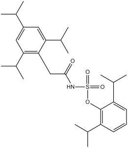
| 分子式 |
C29H43NO4S
|
|
|---|---|---|
| 分子量 |
501.72
|
|
| 精确质量 |
501.291
|
|
| 元素分析 |
C, 69.42; H, 8.64; N, 2.79; O, 12.76; S, 6.39
|
|
| CAS号 |
166518-60-1
|
|
| 相关CAS号 |
166518-61-2 (sodium); 166518-60-1 (free form);
|
|
| PubChem CID |
166558
|
|
| 外观&性状 |
Typically exists as white to off-white solids at room temperature
|
|
| 密度 |
1.1±0.1 g/cm3
|
|
| 熔点 |
178-180° (Lee); mp 169.5-170.4° (Dozeman)
|
|
| 折射率 |
1.529
|
|
| LogP |
9.34
|
|
| tPSA |
80.85
|
|
| 氢键供体(HBD)数目 |
1
|
|
| 氢键受体(HBA)数目 |
4
|
|
| 可旋转键数目(RBC) |
10
|
|
| 重原子数目 |
35
|
|
| 分子复杂度/Complexity |
734
|
|
| 定义原子立体中心数目 |
0
|
|
| SMILES |
S(N([H])C(C([H])([H])C1C(=C([H])C(C([H])(C([H])([H])[H])C([H])([H])[H])=C([H])C=1C([H])(C([H])([H])[H])C([H])([H])[H])C([H])(C([H])([H])[H])C([H])([H])[H])=O)(=O)(=O)OC1C(=C([H])C([H])=C([H])C=1C([H])(C([H])([H])[H])C([H])([H])[H])C([H])(C([H])([H])[H])C([H])([H])[H]
|
|
| InChi Key |
PTQXTEKSNBVPQJ-UHFFFAOYSA-N
|
|
| InChi Code |
InChI=1S/C29H43NO4S/c1-17(2)22-14-25(20(7)8)27(26(15-22)21(9)10)16-28(31)30-35(32,33)34-29-23(18(3)4)12-11-13-24(29)19(5)6/h11-15,17-21H,16H2,1-10H3,(H,30,31)
|
|
| 化学名 |
((2,4,6-Tris(1-methylethyl)phenyl)acetyl)sulfamic acid 2,6-bis(1-methylethyl)phenyl ester
|
|
| 别名 |
|
|
| HS Tariff Code |
2934.99.9001
|
|
| 存储方式 |
Powder -20°C 3 years 4°C 2 years In solvent -80°C 6 months -20°C 1 month |
|
| 运输条件 |
Room temperature (This product is stable at ambient temperature for a few days during ordinary shipping and time spent in Customs)
|
| 溶解度 (体外实验) |
|
|||
|---|---|---|---|---|
| 溶解度 (体内实验) |
配方 1 中的溶解度: 7.5 mg/mL (14.95 mM) in 10% DMSO + 40% PEG300 + 5% Tween80 + 45% Saline (这些助溶剂从左到右依次添加,逐一添加), 悬浮液;超声助溶。
例如,若需制备1 mL的工作液,可将100 μL 75.0 mg/mL澄清DMSO储备液加入到400 μL PEG300中,混匀;然后向上述溶液中加入50 μL Tween-80,混匀;加入450 μL生理盐水定容至1 mL。 *生理盐水的制备:将 0.9 g 氯化钠溶解在 100 mL ddH₂O中,得到澄清溶液。 配方 2 中的溶解度: 7.5 mg/mL (14.95 mM) in 10% DMSO + 90% (20% SBE-β-CD in Saline) (这些助溶剂从左到右依次添加,逐一添加), 悬浊液; 超声助溶。 例如,若需制备1 mL的工作液,可将 100 μL 75.0mg/mL澄清的DMSO储备液加入到900μL 20%SBE-β-CD生理盐水中,混匀。 *20% SBE-β-CD 生理盐水溶液的制备(4°C,1 周):将 2 g SBE-β-CD 溶解于 10 mL 生理盐水中,得到澄清溶液。 View More
配方 3 中的溶解度: 7.5 mg/mL (14.95 mM) in 10% DMSO + 90% Corn Oil (这些助溶剂从左到右依次添加,逐一添加), 澄清溶液; 超声助溶. 配方 4 中的溶解度: 2% DMSO+corn oil: 5mg/mL 1、请先配制澄清的储备液(如:用DMSO配置50 或 100 mg/mL母液(储备液)); 2、取适量母液,按从左到右的顺序依次添加助溶剂,澄清后再加入下一助溶剂。以 下列配方为例说明 (注意此配方只用于说明,并不一定代表此产品 的实际溶解配方): 10% DMSO → 40% PEG300 → 5% Tween-80 → 45% ddH2O (或 saline); 假设最终工作液的体积为 1 mL, 浓度为5 mg/mL: 取 100 μL 50 mg/mL 的澄清 DMSO 储备液加到 400 μL PEG300 中,混合均匀/澄清;向上述体系中加入50 μL Tween-80,混合均匀/澄清;然后继续加入450 μL ddH2O (或 saline)定容至 1 mL; 3、溶剂前显示的百分比是指该溶剂在最终溶液/工作液中的体积所占比例; 4、 如产品在配制过程中出现沉淀/析出,可通过加热(≤50℃)或超声的方式助溶; 5、为保证最佳实验结果,工作液请现配现用! 6、如不确定怎么将母液配置成体内动物实验的工作液,请查看说明书或联系我们; 7、 以上所有助溶剂都可在 Invivochem.cn网站购买。 |
| 制备储备液 | 1 mg | 5 mg | 10 mg | |
| 1 mM | 1.9931 mL | 9.9657 mL | 19.9314 mL | |
| 5 mM | 0.3986 mL | 1.9931 mL | 3.9863 mL | |
| 10 mM | 0.1993 mL | 0.9966 mL | 1.9931 mL |
1、根据实验需要选择合适的溶剂配制储备液 (母液):对于大多数产品,InvivoChem推荐用DMSO配置母液 (比如:5、10、20mM或者10、20、50 mg/mL浓度),个别水溶性高的产品可直接溶于水。产品在DMSO 、水或其他溶剂中的具体溶解度详见上”溶解度 (体外)”部分;
2、如果您找不到您想要的溶解度信息,或者很难将产品溶解在溶液中,请联系我们;
3、建议使用下列计算器进行相关计算(摩尔浓度计算器、稀释计算器、分子量计算器、重组计算器等);
4、母液配好之后,将其分装到常规用量,并储存在-20°C或-80°C,尽量减少反复冻融循环。
计算结果:
工作液浓度: mg/mL;
DMSO母液配制方法: mg 药物溶于 μL DMSO溶液(母液浓度 mg/mL)。如该浓度超过该批次药物DMSO溶解度,请首先与我们联系。
体内配方配制方法:取 μL DMSO母液,加入 μL PEG300,混匀澄清后加入μL Tween 80,混匀澄清后加入 μL ddH2O,混匀澄清。
(1) 请确保溶液澄清之后,再加入下一种溶剂 (助溶剂) 。可利用涡旋、超声或水浴加热等方法助溶;
(2) 一定要按顺序加入溶剂 (助溶剂) 。
|
|---|
|
|
 氟康唑
氟康唑
 伊曲康唑
伊曲康唑
 PF-4981517
 阿利札林
阿利札林
InvivoChem的所有产品仅用于作科学研究,不面向患者销售
Copyright 2020 InvivoChem LLC | All Rights Reserved 粤ICP备20063088号-1


 COA
COA


 463611831
