| 规格 | 价格 | 库存 | 数量 |
|---|---|---|---|
| 10 mM * 1 mL in DMSO |
|
||
| 1mg |
|
||
| 2mg |
|
||
| 5mg |
|
||
| 10mg |
|
||
| 25mg |
|
||
| 50mg |
|
||
| 100mg |
|
||
| 250mg |
|
||
| 500mg |
|
||
| Other Sizes |
|
| 靶点 |
MMP-1 (IC50 = 3 nM); MMP-2 (IC50 = 4 nM); MMP-9 (IC50 = 4 nM); MMP-7 (IC50 = 6 nM); MMP-7 (IC50 = 6 nM)
Batimastat (BB94) is a broad-spectrum inhibitor of matrix metalloproteinases (MMPs), with IC50 values as follows: MMP-1 (collagenase-1): 6 nM, MMP-2 (gelatinase A): 3 nM, MMP-3 (stromelysin-1): 4 nM, MMP-9 (gelatinase B): 4 nM, MMP-7 (matrilysin): 2 nM in cell-free enzyme assays [2] - It shows no significant inhibition of serine proteases (trypsin, plasmin) or cysteine proteases (cathepsin B) at concentrations up to 1 μM, confirming MMP selectivity [2,3] - For membrane-type MMPs (MT1-MMP/MMP-14), Batimastat has an IC50 of 8 nM, inhibiting MT1-MMP-mediated pro-MMP-2 activation [5] |
|---|---|
| 体外研究 (In Vitro) |
Batimastat (BB-94) 是一种有效的广谱基质金属蛋白酶 (MMP) 抑制剂,针对 MMP-1、MMP-2、MMP-9、MMP-7 和 MMP-3,IC50 为 3 nM、4 nM分别为 4 nM、6 nM 和 20 nM。巴马司他表现出意想不到的结合几何形状,噻吩环深深插入主要特异性位点。激酶测定:Batimastat 是一种有效的广谱 MMP 抑制剂,对 MMP-1、MMP-2、MMP-9、MMP-7 和 MMP-3 的 IC50 分别为 3、4、4、6 和 20 nM。细胞测定:通过MTT吸光度测定评估巴马司他存在下的体外增殖。测定巴马司他对 C170HM2 和 AP5LV 在无血清和含血清培养基中体外生长的影响 3 次。巴马司他对任一细胞系的生长均无显着影响。
在人纤维肉瘤HT1080细胞(高表达MMP-2/MMP-9)中,100 nM Batimastat 处理48小时可抑制细胞侵袭约80%(Matrigel Transwell实验),降低明胶酶活性(酶谱法:MMP-2/MMP-9条带减少约75%)[1] - 在人非小细胞肺癌A549细胞中,50 nM Batimastat 处理72小时可抑制集落形成约65%(软琼脂实验),诱导G0/G1期细胞周期阻滞(流式细胞术:G0/G1期细胞较溶剂组增加约30%)[5] - 在MMP-2纯化酶反应中,5 nM Batimastat 抑制明胶降解约90%(放射性明胶实验),20 nM时实现完全抑制[3] - 在人乳腺癌MDA-MB-231细胞中,200 nM Batimastat 处理96小时可抑制细胞迁移约70%(划痕愈合实验),下调MMP-9 mRNA约60%(RT-PCR)[5] |
| 体内研究 (In Vivo) |
Batimastat 可抑制 B16-BL6 小鼠黑色素瘤的转移扩散和生长。在小鼠原位结肠肿瘤模型中,timastat治疗可抑制原发肿瘤生长(50%)、局部/区域扩散(从67%至35%)和远处转移(从30%至10%)。巴马司他减少实验性血管瘤的体内生长,最有可能是通过阻止转化细胞招募内皮细胞或干扰血管结构中的细胞组织。
测试了合成基质金属蛋白酶抑制剂batimastat在同基因C57BL/6N小鼠中抑制B16-BL6小鼠黑色素瘤生长和转移扩散的能力。腹腔内注射巴替马斯特可显著抑制静脉注射B16-BL6细胞产生的肺集落数量。在后足垫接种B16-BL6-黑色素瘤的小鼠中,研究了巴替马他对自发转移的影响。26-28天后手术切除原发肿瘤。从第14天至第28天(手术前)或从第26天至第44天(手术后)每天服用两次Batimastat。采用这两种方案,肺转移的中位数没有受到显著影响,但转移的重量显著减轻。最后,研究了巴替马斯特对B16-BL6黑色素瘤皮下生长的影响。从肿瘤移植当天开始,每天服用Batimastat会导致明显的生长延迟,而从晚期肿瘤开始的治疗只会略微减少肿瘤的生长。我们的研究结果表明,基质金属蛋白酶抑制剂不仅可以防止B16-BL6细胞在继发器官的定植,还可以限制实体瘤的生长。[1] 基质金属蛋白酶与转移性肿瘤的生长和扩散有关。在人结肠癌癌症裸鼠原位移植模型中,使用基质金属蛋白酶抑制剂BB-94(batimastat)研究了这种作用。在40只无胸腺nu/nu小鼠的结肠上手术植入人结肠癌片段(1-1.5 mm)。肿瘤植入后7天开始施用BB-94或载体(磷酸盐缓冲盐水,pH 7.4,含0.01%吐温80)(20只动物/组)。动物在前60天每天一次腹腔注射30mg/kg BB-94,然后每周3次。BB-94治疗使原发性肿瘤的中位重量从对照组的293mg降低到BB-94处理组的144mg(P<0.001)。BB-94治疗还降低了局部和区域侵袭的发生率,从对照组的18只小鼠中的12只(67%)降低到治疗组的20只小鼠中有7只(35%)。对照组中的六只小鼠也被发现在肝脏、肺、腹膜、腹壁或局部淋巴结有转移。BB-94组中只有两只小鼠有转移性疾病的证据,在这两种情况下都局限于腹壁。在BB-94治疗组中观察到的肿瘤进展的减少转化为该组生存率的提高,从对照组的中位生存时间110天提高到治疗组的中位数生存时间140天(P<0.01)。BB-94治疗与任何明显的毒性作用无关,这些结果表明,此类药物可能是癌症的辅助疗法。[3] 每日使用巴替马斯特治疗(在eEnd.1细胞注射部位注射30、3和0.3mg/kg)可抑制肿瘤生长,并延长倍增时间。巴替马斯特的羧酰胺衍生物BB-374是一种基质金属蛋白酶活性较差的抑制剂,在减少血管瘤生长方面活性较低。对治疗肿瘤的组织学分析表明,充满血液的空间和出血量减少。Batimastat还抑制了由包埋在Matrigel颗粒中的培养的eEnd.1内皮瘤细胞上清液诱导的血管生成反应。Batimastat通过一层Matrigel在体外显著抑制内皮细胞侵袭,但没有显示出直接的细胞毒性活性。 结论:Batimastat可减少实验性血管瘤的体内生长,很可能是通过阻断转化细胞对内皮细胞的募集或干扰血管结构中的细胞组织。 影响:这些结果证实了基质金属蛋白酶在血管生成和血管肿瘤形成中内皮细胞募集中的重要性,并表明合成基质金属蛋白酶抑制剂具有治疗潜力。[4] 在荷HT1080异种移植瘤的裸鼠(皮下注射1×10⁶个细胞)中,每日一次腹腔注射10 mg/kg Batimastat,持续21天,肿瘤体积较溶剂对照组减少约55%,肿瘤重量减少约50%;免疫组化显示MMP-2阳性细胞减少约70%[1] - 在Lewis肺癌(LLC)肺转移模型C57BL/6小鼠(静脉接种LLC细胞)中,每周两次腹腔注射5 mg/kg Batimastat,持续3周,肺转移灶减少约80%(苏木精-伊红染色计数)[4] - 在原位胰腺癌(AR42J细胞)大鼠模型中,每日一次静脉注射2 mg/kg Batimastat,持续14天,抑制肿瘤向周围胰腺侵袭约65%(组织病理学分析)[4] - 在荷MDA-MB-231异种移植瘤的裸鼠中,每日一次腹腔注射15 mg/kg Batimastat,持续28天,远处淋巴结转移减少约75%(淋巴结解剖及人Alu序列PCR检测)[5] |
| 酶活实验 |
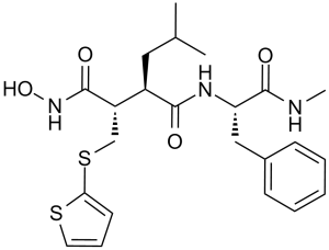
在体外,使用针对各种金属蛋白酶的酶测定来计算巴替马斯特IC50。基质金属蛋白酶与肿瘤细胞侵袭、转移和关节炎等退行性过程有关。特异性金属蛋白酶抑制剂已被用于阻断肿瘤细胞增殖。我们以2.0埃的分辨率(R=16.8%)研究了巴替马斯特(BB-94)与金属蛋白酶[atrolysin C(Ht-d),EC 3.4.24.42]活性位点的相互作用。标题结构表现出意想不到的结合几何形状,噻吩环深深插入到主要特异性位点。这种前所未有的结合几何形状生动地展示了海绵状主要特异性位点的重要性,为设计新一代潜在的抗肿瘤药物指明了方向。[2]
MMP-2/MMP-9明胶酶活性检测流程(基于[3]摘要描述):重组人MMP-2/MMP-9在激活缓冲液(50 mM Tris-HCl pH 7.5,10 mM CaCl₂,0.05% Brij-35)中用对氨基苯汞乙酸(APMA)激活。激活后的酶与放射性[³H]-明胶(底物)及Batimastat(0.1~50 nM)混合于反应缓冲液中。37°C孵育3小时后,加入三氯乙酸(TCA)沉淀未降解明胶。通过液体闪烁计数法检测可溶性组分(降解明胶)的放射性。相对于溶剂对照组计算抑制率,通过剂量-反应拟合确定IC50[3] - MMP-1/MMP-3胶原酶/基质溶解素实验流程(基于[2]摘要描述):重组MMP-1/MMP-3与荧光肽底物(MMP-1:Mca-Pro-Leu-Gly-Leu-Dpa-Ala-Arg-NH₂;MMP-3:Mca-Arg-Pro-Lys-Pro-Tyr-Ala-Nva-Trp-Met-Lys(Dnp)-NH₂)混合于检测缓冲液(50 mM HEPES pH 7.5,10 mM CaCl₂,0.01% Tween-20)中。加入0.1~10 nM Batimastat,37°C孵育1小时。检测激发波长328 nm/发射波长393 nm处的荧光强度,采用四参数逻辑回归计算IC50[2] |
| 细胞实验 |
在溶解于无水乙醇中的不同浓度的巴马司他下孵育细胞22小时后测定IC50。
细胞生长测定[5] 将细胞系接种在生长培养基中的24孔多层培养基中,并允许其粘附两天。当实验开始时(第0天),以图中所示的浓度加入含有氟维司群(0.1μM)、HER配体(10 ng/ml)、吉非替尼、CI-1033、TAPI-2、Batimatat(BB94)或GM6001的生长培养基。对照细胞加入的载体量与处理细胞相似。在第三天更换生长培养基,在第五天使用如前所述的结晶紫比色法测定细胞数量。每个实验进行四次,重复至少两次。 HT1080细胞侵袭实验流程(基于[1]摘要描述):HT1080细胞在含10%胎牛血清(FBS)的DMEM培养基中培养至80%汇合。胰酶消化后,用无血清DMEM重悬,以5×10⁴细胞/孔接种于Matrigel包被的Transwell上室(含10~200 nM Batimastat),下室加入含10% FBS的DMEM(趋化因子)。24小时后,去除上室未侵袭细胞,固定并结晶紫染色侵袭细胞,显微镜下计数。收集细胞上清液,通过明胶酶谱法检测MMP-2/MMP-9活性[1] - A549细胞集落形成实验流程(基于[5]摘要描述):A549细胞以1×10³细胞/孔接种于6孔板,用10~100 nM Batimastat 处理24小时后,PBS洗涤并更换新鲜培养基。14天后,甲醇固定集落,结晶紫染色,计数>50个细胞的集落。细胞周期分析时,用50 nM Batimastat 处理细胞72小时,70%乙醇固定,碘化丙啶(PI)染色后流式细胞术检测[5] |
| 动物实验 |
小鼠:采用6周龄雌性BALB/c小鼠。感染前1小时和感染后24小时,小鼠腹腔注射巴替马司他(BB-94,50 mg/kg)。巴替马司他以50 mg/mL的浓度溶于DMSO中,并保存于-20°C。使用前用磷酸盐缓冲液(PBS)稀释20倍,取500 μL注射到小鼠体内。对照组小鼠注射500 μL 5% DMSO的PBS溶液。感染后48小时处死小鼠。
大鼠:Sprague-Dawley雌性大鼠在预定时间间隔内腹腔注射单次生理剂量的雌二醇(E2,40 μg/kg,溶于0.9% NaCl、0.4% EtOH溶液中),并在尸检时采集组织。研究表明,体内注射此剂量的雌激素可引起子宫湿重、组织结构和基因表达的变化,这些变化均表明雌激素受体被激活。在每项研究中,动物在组织采集前四小时腹腔注射单次40 μg/kg的雌二醇(E2),对照组仅注射赋形剂。腹腔注射巴替马司他(batimastat)40 mg/kg,溶于含0.1% Tween-20的1×PBS溶剂中,于雌二醇(E2)或生理盐水对照组给药前4小时给药,已证实其具有体内MMP抑制作用。裸鼠HT1080异种移植模型(引自[1]摘要描述):将1×10⁶个HT1080细胞(悬浮于0.1 mL PBS + 50% Matrigel中)皮下注射到6-8周龄雌性裸鼠的右侧腹部。当肿瘤体积达到约100 mm³时,将小鼠分为两组:(1)巴替马司他组:每日腹腔注射10 mg/kg巴替马司他(溶于10% DMSO + 90%生理盐水(0.9% NaCl)中); (2)载体组:10% DMSO + 90% NaCl。每3天测量一次肿瘤体积(V=0.5×长×宽²);第22天处死小鼠,用于肿瘤重量和免疫组织化学分析[1] - 小鼠LLC肺转移模型(引自[4]摘要描述):雄性C57BL/6小鼠(8-10周龄)经静脉注射5×10⁵个LLC细胞(悬浮于0.2 mL PBS中)。接种24小时后,小鼠每周两次腹腔注射巴替马司他(5 mg/kg,溶于0.5%明胶溶液),持续3周。载体对照组注射0.5%明胶。第22天处死小鼠;肺组织经福尔马林固定、切片、苏木精-伊红染色后,在显微镜下计数转移灶[4] - 裸鼠MDA-MB-231转移模型(引自[5]摘要描述):将2×10⁶个MDA-MB-231细胞注射到6-8周龄雌性裸鼠的乳腺脂肪垫中。当原发肿瘤体积达到约150 mm³时,腹腔注射巴替马司他(15 mg/kg,溶于5% DMSO + 95%芝麻油),每日一次,连续28天。对照组注射5% DMSO + 95%芝麻油。处死小鼠后,解剖腋窝淋巴结,并通过PCR检测人Alu序列(肿瘤标志物)以量化转移情况[5] |
| 药代性质 (ADME/PK) |
在雄性Sprague-Dawley大鼠中,腹腔注射Batimastat(5 mg/kg)的血浆消除半衰期(t₁/₂)约为2.8小时,血浆峰浓度(Cmax)为1.2 μg/mL(给药后0.5小时达到),分布容积(Vd)约为1.5 L/kg [4]
- Batimastat在大鼠和小鼠中的口服生物利用度<5%(由于胃肠道吸收不良);腹腔注射可使肿瘤组织浓度比血浆浓度高约10倍(腹腔注射后1小时肿瘤/血浆比值为10.2)[4,5] - Batimastat在大鼠肝微粒体中的代谢极少(<10%的剂量);腹腔注射剂量的约 80% 会在 72 小时内以原形经粪便排出 [4] |
| 毒性/毒理 (Toxicokinetics/TK) |
在为期 28 天的大鼠毒性研究中(腹腔注射 Batimastat,剂量为 2、5、10 mg/kg/天),未观察到不良反应水平 (NOAEL) 为 5 mg/kg/天;在10 mg/kg/天的剂量下,6只大鼠中有3只出现轻度关节僵硬(可逆),血清ALT/AST、肌酐或BUN均无变化[4]
- 在接受腹腔注射Batimastat(15 mg/kg/天,持续28天)治疗的裸鼠中,未检测到明显的体重减轻(>初始体重的5%)或肝脏、肾脏或脾脏的组织病理学异常[5] - 在人肿瘤细胞(HT1080、A549、MDA-MB-231)中,浓度高达500 nM的Batimastat处理72小时未观察到明显的细胞毒性(细胞活力>85% vs. 载体,MTT法)[1,5] - Batimastat在人和大鼠血浆中的血浆蛋白结合率约为92%(通过超滤法测定)[4] |
| 参考文献 | |
| 其他信息 |
药效学
巴替马司他是一种抗癌药物,属于血管生成抑制剂类药物。巴替马司他是一种基质金属蛋白酶抑制剂。 巴替马司他 (BB94) 是首个进入临床试验(I/II期)的合成广谱MMP抑制剂,用于治疗实体瘤和转移性疾病[2,4] - 其抗肿瘤机制包括抑制MMP介导的细胞外基质 (ECM) 降解,从而阻断肿瘤细胞的侵袭、迁移和血管生成——这些都是肿瘤进展和转移的关键步骤[1,3] - 巴替马司他的临床开发受到口服生物利用度差(需要肠外给药)以及高剂量下轻微肌肉骨骼毒性(关节僵硬)的限制;虽然巴替马司他已被口服MMP抑制剂(例如马立马司他)所取代,但它仍然是MMP靶向治疗的原型[4,5] - 巴替马司他与化疗药物(例如顺铂)具有协同抗肿瘤活性:50 nM 巴替马司他 + 1 μM 顺铂可使A549细胞活力降低约75%(而单独使用顺铂时约为40%)[5] |
| 分子式 |
C23H31N3O4S2
|
|---|---|
| 分子量 |
477.64
|
| 精确质量 |
477.175
|
| 元素分析 |
C, 57.84; H, 6.54; N, 8.80; O, 13.40; S, 13.43
|
| CAS号 |
130370-60-4
|
| 相关CAS号 |
Batimastat sodium salt;130464-84-5
|
| PubChem CID |
5362422
|
| 外观&性状 |
White solid powder
|
| 密度 |
1.3±0.1 g/cm3
|
| 熔点 |
236-238°
|
| 折射率 |
1.605
|
| LogP |
3.53
|
| tPSA |
161.07
|
| 氢键供体(HBD)数目 |
4
|
| 氢键受体(HBA)数目 |
6
|
| 可旋转键数目(RBC) |
12
|
| 重原子数目 |
32
|
| 分子复杂度/Complexity |
614
|
| 定义原子立体中心数目 |
3
|
| SMILES |
O=C(N[C@@H](CC1=CC=CC=C1)C(NC)=O)[C@H](CC(C)C)[C@H](CSC2=CC=CS2)C(NO)=O
|
| InChi Key |
XFILPEOLDIKJHX-QYZOEREBSA-N
|
| InChi Code |
InChI=1S/C23H31N3O4S2/c1-15(2)12-17(18(22(28)26-30)14-32-20-10-7-11-31-20)21(27)25-19(23(29)24-3)13-16-8-5-4-6-9-16/h4-11,15,17-19,30H,12-14H2,1-3H3,(H,24,29)(H,25,27)(H,26,28)/t17-,18+,19+/m1/s1
|
| 化学名 |
(2S,3R)-N-hydroxy-N'-[(2S)-1-(methylamino)-1-oxo-3-phenylpropan-2-yl]-3-(2-methylpropyl)-2-(thiophen-2-ylsulfanylmethyl)butanediamide
|
| 别名 |
Batimastat; BB94; BB-94; Batimastat (BB-94); BB94; BB 94; Batimastat (MMP Inhibitor); Butanediamide, N4-hydroxy-N1-[(1S)-2-(methylamino)-2-oxo-1-(phenylmethyl)ethyl]-2-(2-methylpropyl)-3-[(2-thienylthio)methyl]-, (2R,3S)-; BB 94
|
| HS Tariff Code |
2934.99.9001
|
| 存储方式 |
Powder -20°C 3 years 4°C 2 years In solvent -80°C 6 months -20°C 1 month |
| 运输条件 |
Room temperature (This product is stable at ambient temperature for a few days during ordinary shipping and time spent in Customs)
|
| 溶解度 (体外实验) |
|
|||
|---|---|---|---|---|
| 溶解度 (体内实验) |
配方 1 中的溶解度: ≥ 5 mg/mL (10.47 mM) (饱和度未知) in 10% DMSO + 90% (20% SBE-β-CD in Saline) (这些助溶剂从左到右依次添加,逐一添加), 澄清溶液。
例如,若需制备1 mL的工作液,可将100 μL 50.0mg/mL澄清的DMSO储备液加入到900μL 20%SBE-β-CD生理盐水中,混匀。 *20% SBE-β-CD 生理盐水溶液的制备(4°C,1 周):将 2 g SBE-β-CD 溶解于 10 mL 生理盐水中,得到澄清溶液。 配方 2 中的溶解度: 2.5 mg/mL (5.23 mM) in 10% DMSO + 40% PEG300 + 5% Tween80 + 45% Saline (这些助溶剂从左到右依次添加,逐一添加), 悬浊液; 超声助溶。 例如,若需制备1 mL的工作液,可将 100 μL 25.0 mg/mL澄清DMSO储备液加入到400 μL PEG300中,混匀;然后向上述溶液中加入50 μL Tween-80,混匀;加入450 μL生理盐水定容至1 mL。 *生理盐水的制备:将 0.9 g 氯化钠溶解在 100 mL ddH₂O中,得到澄清溶液。 View More
配方 3 中的溶解度: ≥ 2.5 mg/mL (5.23 mM) (饱和度未知) in 10% DMSO + 90% Corn Oil (这些助溶剂从左到右依次添加,逐一添加), 澄清溶液。 配方 4 中的溶解度: 30% propylene glycol, 5% Tween 80, 65% D5W: 30 mg/mL 1、请先配制澄清的储备液(如:用DMSO配置50 或 100 mg/mL母液(储备液)); 2、取适量母液,按从左到右的顺序依次添加助溶剂,澄清后再加入下一助溶剂。以 下列配方为例说明 (注意此配方只用于说明,并不一定代表此产品 的实际溶解配方): 10% DMSO → 40% PEG300 → 5% Tween-80 → 45% ddH2O (或 saline); 假设最终工作液的体积为 1 mL, 浓度为5 mg/mL: 取 100 μL 50 mg/mL 的澄清 DMSO 储备液加到 400 μL PEG300 中,混合均匀/澄清;向上述体系中加入50 μL Tween-80,混合均匀/澄清;然后继续加入450 μL ddH2O (或 saline)定容至 1 mL; 3、溶剂前显示的百分比是指该溶剂在最终溶液/工作液中的体积所占比例; 4、 如产品在配制过程中出现沉淀/析出,可通过加热(≤50℃)或超声的方式助溶; 5、为保证最佳实验结果,工作液请现配现用! 6、如不确定怎么将母液配置成体内动物实验的工作液,请查看说明书或联系我们; 7、 以上所有助溶剂都可在 Invivochem.cn网站购买。 |
| 制备储备液 | 1 mg | 5 mg | 10 mg | |
| 1 mM | 2.0936 mL | 10.4681 mL | 20.9363 mL | |
| 5 mM | 0.4187 mL | 2.0936 mL | 4.1873 mL | |
| 10 mM | 0.2094 mL | 1.0468 mL | 2.0936 mL |
1、根据实验需要选择合适的溶剂配制储备液 (母液):对于大多数产品,InvivoChem推荐用DMSO配置母液 (比如:5、10、20mM或者10、20、50 mg/mL浓度),个别水溶性高的产品可直接溶于水。产品在DMSO 、水或其他溶剂中的具体溶解度详见上”溶解度 (体外)”部分;
2、如果您找不到您想要的溶解度信息,或者很难将产品溶解在溶液中,请联系我们;
3、建议使用下列计算器进行相关计算(摩尔浓度计算器、稀释计算器、分子量计算器、重组计算器等);
4、母液配好之后,将其分装到常规用量,并储存在-20°C或-80°C,尽量减少反复冻融循环。
计算结果:
工作液浓度: mg/mL;
DMSO母液配制方法: mg 药物溶于 μL DMSO溶液(母液浓度 mg/mL)。如该浓度超过该批次药物DMSO溶解度,请首先与我们联系。
体内配方配制方法:取 μL DMSO母液,加入 μL PEG300,混匀澄清后加入μL Tween 80,混匀澄清后加入 μL ddH2O,混匀澄清。
(1) 请确保溶液澄清之后,再加入下一种溶剂 (助溶剂) 。可利用涡旋、超声或水浴加热等方法助溶;
(2) 一定要按顺序加入溶剂 (助溶剂) 。
|
|---|
|
|
InvivoChem的所有产品仅用于作科学研究,不面向患者销售
Copyright 2020 InvivoChem LLC | All Rights Reserved 粤ICP备20063088号-1


 COA
COA
")
")
")
 463611831
