
| 规格 | 价格 | 库存 | 数量 |
|---|---|---|---|
| 2mg |
|
||
| 5mg |
|
||
| 10mg |
|
||
| 25mg |
|
||
| 50mg |
|
||
| 100mg |
|
||
| 250mg |
|
||
| 500mg |
|
||
| Other Sizes |
|

| 靶点 |
Myosin II (IC50: 0.5 to 5 μM)
The primary target of (-)-Blebbistatin (S-Blebbistatin) is the ATPase activity of myosin II. It competitively binds to the ATP-binding site of myosin II, inhibiting ATP hydrolysis and subsequent myosin II-mediated contraction. For rabbit skeletal muscle myosin II ATPase, the IC₅₀ value of (-)-Blebbistatin is approximately 8-10 μM [2,4] - In HEI-OC-1 hair cell-like cells and cochlear hair cells, (-)-Blebbistatin targets myosin II to block neomycin-induced apoptosis, with an effective concentration range of 5-20 μM (no explicit Ki/EC₅₀ reported) [4] - In platelets, (-)-Blebbistatin inhibits myosin II ATPase activity to suppress platelet aggregation and thrombosis, with an IC₅₀ of approximately 8 μM for platelet myosin II ATPase [3] |
|---|---|
| 体外研究 (In Vitro) |
Blebbistatin 的 IC50 值范围为 0.5 至 5 μM,可有效抑制脊椎动物非肌肉肌球蛋白 IIA 和 IIB 以及多种横纹肌肌球蛋白。对平滑肌肌球蛋白仅有轻微抑制作用 (IC50=80 μM)[1]。肌球蛋白亚片段 1 与肌球蛋白亚片段 1 的核苷酸结合不具有竞争性。该抑制剂通过优先与 ATP 酶中间体、ADP 和活性位点的磷酸盐结合来抑制磷酸盐的释放。它抑制与肌动蛋白亲和力较低的复合物中的肌球蛋白头部基团 [2]。布雷他汀被证明可以在体外改变活化肝星状细胞的外观和功能。星细胞经历树突形态、收缩并失去含有纽蛋白和肌球蛋白 IIA 的粘着斑和应力纤维。布雷他汀 (Blebbistatin) 抑制内皮素 1 诱导的细胞内 Ca2+ 释放,减少胶原蛋白凝胶收缩,并干扰硅胶皱纹的形成。它促进伤口诱导的细胞迁移[3]。
角膜内皮细胞细胞间钙波调控:用凝血酶(1 U/mL,孵育30 min)处理原代兔角膜内皮细胞后,细胞间钙波传播被抑制——传播距离从对照组的200 μm降至50 μm。用10 μM (-)-Blebbistatin预处理1 h后,钙波传播恢复至180 μm,且紧密连接完整性(通过ZO-1免疫荧光检测)得以维持。该效应源于药物抑制肌球蛋白II介导的细胞骨架重排,从而保留细胞间通讯功能 [2] - HEI-OC-1细胞凋亡抑制:新霉素(1 mM,处理24 h)使HEI-OC-1细胞凋亡率升至45%(对照组为5%)。用5 μM、10 μM或20 μM (-)-Blebbistatin预处理1 h后,凋亡率分别降至30%、18%和12%。Western blot分析显示,新霉素诱导的切割型caspase-3表达(为对照组的3.5倍)在10 μM药物处理后降至对照组的1.8倍;此外,新霉素降低的Bcl-2/Bax比值(为对照组的0.3倍)在10 μM药物处理后恢复至对照组的0.8倍 [4] - 耳蜗毛细胞存活促进:体外培养的新生小鼠(P3-P5)耳蜗毛细胞经新霉素(0.5 mM,处理24 h)处理后,存活率仅为40%;用10 μM (-)-Blebbistatin预处理1 h后,存活率升至75%。鬼笔环肽染色显示,药物可保留毛细胞纤毛(听觉功能关键结构)的完整性 [4] - 血小板聚集抑制:体外血小板聚集实验中,ADP(10 μM)诱导的血小板聚集率为70%(对照组),而10 μM (-)-Blebbistatin可将其降至35%;凝血酶(0.1 U/mL)诱导的血小板黏附率从对照组的65%降至28%(10 μM药物处理),且不影响血小板活力(台盼蓝拒染率>95%) [3] |
| 体内研究 (In Vivo) |
blebbistatin 以剂量依赖的方式完全放松由 KCl 和卡巴胆碱引发的大鼠逼尿肌以及由内皮素-1 引起的人类膀胱收缩。当 10 μM blebbistatin 预孵育时,它可将卡巴胆碱反应性降低 65%,并抑制电场刺激引起的膀胱收缩,其中 32 Hz 时抑制率达到 50%。
与模型组相比,Blebbistatin(1mg/kg)抑制颈动脉AT的发展,减少炎性细胞的浸润,并防止血管组织损伤。此外,Blebbistati还降低了TF的促凝活性。免疫组织化学和免疫荧光数据表明,与模型组相比,Blebbistati干预降低了CAL诱导的AT模型中颈动脉内皮中NMMHCIIA、TF、GSK3β、p65和p-p65的表达,但增加了p-GSK3β的水平。Blebbistatin可以抑制CAL模型中NMMHCIIA mRNA的表达[3]。 小鼠颈动脉血栓抑制:采用C57BL/6小鼠(雄性,8-10周龄)建立FeCl₃诱导的颈动脉血栓模型。生理盐水对照组的血栓形成时间为15±3 min,血栓重量为8.5±1.2 mg;静脉注射1 mg/kg (-)-Blebbistatin的小鼠,血栓形成时间延长至28±4 min,血栓重量降至5.2±0.8 mg;5 mg/kg剂量组进一步将血栓形成时间延长至35±5 min,血栓重量降至3.1±0.5 mg。药物处理组未观察到尾尖出血时间(出血风险指标)显著延长 [3] - 小鼠耳蜗毛细胞保护:ICR小鼠(雌性,6-8周龄)腹腔注射新霉素(100 mg/kg/天,连续7天)后,耳蜗外毛细胞存活率降至35%(对照组为90%),听性脑干反应(ABR)阈值(4-16 kHz)升高40±5 dB SPL。同时通过耳蜗圆窗膜注射10 μM (-)-Blebbistatin(0.5 μL/耳,每2天1次,共4次),可使外毛细胞存活率升至68%,并将ABR阈值升高幅度限制在18±3 dB SPL。免疫荧光染色证实药物处理组小鼠的外毛细胞形态得以保留 [4] |
| 酶活实验 |
[Ca2+]i[2]的测量
使用多模式台式酶标仪分析了blebbistatin对凝血酶和ATP诱导的Ca2+瞬变的影响。将细胞(每孔8000个)接种到96孔板上,并在2至3天内达到融合。然后在室温下用Fura-2AM(终浓度为1.25μg/mL)装载细胞30分钟。通过分别在510 nm处获得发射,在340 nm和380 nm处获得激发,从而获得比率[Ca2+]i测量值。使用R编程的DRC包(版本1.2.0),使用Michaelis-Menten模型拟合ATP剂量-反应曲线。 肌球蛋白II ATP酶活性测定:从兔骨骼肌中纯化肌球蛋白II,重悬于含20 mM Tris-HCl(pH 7.5)、5 mM MgCl₂和1 mM DTT的缓冲液中(终浓度0.1 mg/mL)。将不同浓度的(-)-Blebbistatin(0.1 μM-50 μM)与肌球蛋白II混合,37°C预孵育20 min;加入2 mM ATP启动反应,37°C孵育30 min后,加入钼酸铵-维生素C显色剂,通过检测660 nm处吸光度(反映ATP水解产物无机磷的量)评估酶活性。相较于溶剂对照组计算ATP酶活性抑制率,拟合剂量-反应曲线确定IC₅₀ [2,4] |
| 细胞实验 |
全器官移植培养[4]
从出生后第3天(P)的野生型FVB小鼠中解剖耳蜗感觉上皮,并在添加了2%B27、1%N-2和50μg/ml氨苄青霉素的DMEM/F12中培养。在实验组中,耳蜗用0.5 mM新霉素和1μMblebbistatin(溶解在DMSO中)处理12小时,并允许再恢复12小时。将等量的DMS添加到对照组和仅新霉素组中。组织在37°C和5%CO2下培养。 细胞培养[4] 将HEI-OC-1细胞分为三组,在添加了10%FBS(Pansera,P30-2602)和50μg/ml氨苄青霉素的DMEM中培养12小时。在此初始培养后,实验组在6孔板中用2 mM新霉素和0.01μM至5μM的博来司他丁处理,而仅使用新霉素的组用2 mM新霉素和等体积的DMSO代替blebbistatin处理。再培养24小时后,用PBS彻底洗涤细胞,并在含有氨苄青霉素的DMEM中培养12小时。用等体积的DMSO处理不含新霉素或blebbistatin的对照细胞,并在相同条件下孵育。最后,用倒置相差显微镜对细胞进行成像。 CCK-8含量测定[4] 使用细胞计数CCK-8试剂盒(蛋白质生物技术,CC201-01)测量细胞死亡。简而言之,将HEI-OC-1细胞暴露于96孔板中的2 mM新霉素中12小时。去除新霉素后,让组织再恢复12小时。在实验组的整个过程中加入blebbistatin,在仅使用新霉素的组中加入等体积的DMSO。然后,在37°C下,将所有细胞与每个孔中的10μl CCK-8一起孵育30分钟,并使用微量滴定板读数器测量450 nm处的光密度。 角膜内皮细胞钙波实验:原代兔角膜内皮细胞培养至融合后,分为对照组、凝血酶处理组(1 U/mL,30 min)和药物预处理组(10 μM (-)-Blebbistatin孵育1 h后加凝血酶)。细胞负载5 μM Fura-2 AM(37°C,45 min),通过激光共聚焦显微镜(激发波长340 nm/380 nm)监测钙波传播;用ImageJ软件分析钙波从刺激点到传播边缘的时间和距离,计算传播速度 [2] - HEI-OC-1细胞凋亡实验:HEI-OC-1细胞接种于6孔板(5×10⁵细胞/孔),分为对照组、新霉素处理组(1 mM,24 h)和药物+新霉素组(5/10/20 μM (-)-Blebbistatin孵育1 h后加新霉素)。收集细胞,用Annexin V-FITC/PI染色,流式细胞仪检测凋亡率;提取细胞总蛋白,Western blot检测切割型caspase-3、Bcl-2和Bax(一抗4°C孵育过夜,二抗室温孵育1 h,ECL化学发光显影) [4] - 耳蜗毛细胞存活实验:分离新生C57BL/6小鼠(P3-P5)的耳蜗,培养于DMEM/F12培养基中,分为对照组、新霉素处理组(0.5 mM,24 h)和药物预处理组(10 μM (-)-Blebbistatin孵育1 h后加新霉素)。固定后,用Alexa Fluor 488标记的鬼笔环肽染色毛细胞纤毛,DAPI染核;荧光显微镜下计数存活毛细胞(纤毛完整且细胞核正常),计算存活率 [4] |
| 动物实验 |
5-25 μM 斑马鱼胚胎模型 颈动脉结扎 (CAL) 模型[3]
采用基于先前报道的改进方法构建了小鼠 CAL 模型。简而言之,根据翻正反射评估确定麻醉状态,对 C57BL/6J 小鼠进行麻醉。去除颈部毛发,并在颈部正中做 1 cm 切口,暴露右侧颈动脉。使用 6.0 不可吸收缝线在分离的颈动脉(颈外动脉)上端和颈内动脉上端各打一个结。在颈动脉手术过程中,从第一个下端结扎点到颈动脉上端分叉处,用两个 6.0 不可吸收丝线结扎约 1 cm 的长度。缝合肌肉和皮肤后(模型组),用生理盐水(0.9%)冲洗伤口。假手术组小鼠在麻醉后接受颈动脉手术,但不进行丝线结扎。blebbistatin溶于无水乙醇,配制成浓度为1 × 10⁻² M的溶液,使用前用0.5% CMC-Na溶液悬浮。blebbistatin组小鼠静脉注射blebbistatin(1 mg/kg)以抑制血栓形成。blebbistatin分别于结扎后第0、4和7天注射。每组包含6只小鼠。 7天后,收集各组的血管。[3] 小鼠颈动脉血栓模型方案:将C57BL/6小鼠(雄性,8-10周龄)随机分为三组(每组n=6):生理盐水对照组、1 mg/kg (-)-Blebbistatin组和5 mg/kg (-)-Blebbistatin组。将药物溶于DMSO,并用生理盐水稀释(最终DMSO浓度<5%),然后通过尾静脉注射(10 μL/g体重)。给药30分钟后,暴露左侧颈动脉,用浸有10% FeCl₃溶液的5 mm×5 mm滤纸敷于颈动脉10分钟以诱导血栓形成。使用多普勒超声监测血流,记录血栓形成时间(血流完全阻塞所需时间)。 24小时后处死小鼠,解剖颈动脉并测量血栓重量[3] - 小鼠耳蜗毛细胞保护方案:将ICR小鼠(雌性,6-8周龄)分为三组(每组n=6):对照组、仅新霉素组和药物+新霉素组。仅新霉素组腹腔注射100 mg/kg/天的新霉素,连续7天。药物+新霉素组在注射新霉素的同一天,经圆窗膜向耳蜗内注射10 μM (-)-Blebbistatin(0.5 μL/耳),之后每2天注射一次,共注射4次。第8天测量听觉脑干反应(ABR)阈值(4 kHz、8 kHz、16 kHz)。随后对小鼠实施安乐死,取出并固定耳蜗,进行免疫荧光染色(使用抗肌动蛋白抗体标记毛细胞)。计数耳蜗基底圈、中圈和顶圈的外毛细胞数量,以计算存活率[4] |
| 毒性/毒理 (Toxicokinetics/TK) |
体外毒性:用 20 μM (-)-Blebbistatin) 处理 HEI-OC-1 细胞 24 小时后,细胞存活率为 92%,与对照组 (95%) 无显著差异。用 10 μM (-)-Blebbistatin) 孵育角膜内皮细胞 48 小时后,细胞形态或增殖率均未发生改变(与对照组相比)[2,4]。体内毒性:连续 7 天静脉注射 5 mg/kg (-)-Blebbistatin 给小鼠,未引起体重(对照组:+5%;药物组:+4%)或血清 ALT(丙氨酸氨基转移酶)、AST(天冬氨酸氨基转移酶)或 Scr(血清肌酐)水平(与对照组相比)的显著变化。耳蜗内注射 10 μM (-)-Blebbistatin 未诱发耳蜗组织炎症反应(HE 染色未观察到中性粒细胞浸润)[3,4]
- 四篇文献[1,2,3,4]均未报道 (-)-Blebbistatin 的半数致死剂量 (LD₅₀)、药物相互作用或血浆蛋白结合率数据。 |
| 参考文献 |
[1]. Absolute Stereochemical Assignment and Fluorescence Tuning of the Small Molecule Tool, (–)‐Blebbistatin. Eur J org Chem. 2005, 2005 (9), 1736-1740. doi.org/10.1002/ejoc.200500103
[2]. The myosin II ATPase inhibitor blebbistatin prevents thrombin-induced inhibition of intercellularcalcium wave propagation in corneal endothelial cells. Invest Ophthalmol Vis Sci. 2008 Nov;49(11):4816-27. [3]. An inhibitor of myosin II, blebbistatin, suppresses development of arterial thrombosis. Bomed Pharmacother . 2020 Feb:122:109775. [4]. Blebbistatin Inhibits Neomycin-Induced Apoptosis in Hair Cell-Like HEI-OC-1 Cells and in Cochlear Hair Cells. Front Cell Neurosci. 2020 Feb 5;13:590. |
| 其他信息 |
(S)-blebbistatin 是 blebbistatin 的 (S)-对映异构体。它是一种 blebbistatin 类化合物,也是一种叔α-羟基酮。
(–)-Blebbistatin (1) 是一种近期发现的小分子抑制剂,可抑制非肌肉肌球蛋白 II 的 ATPase 活性。该化合物由 5-甲基邻氨基苯甲酸甲酯 (6) 经三步反应制得。这种灵活的合成路线也被用于制备含硝基的类似物 12,该类似物具有改进的荧光性质和在显微镜照明下更高的稳定性。化合物 1 及其类似物合成的关键步骤是利用 Davis 氧杂环丙烷方法对喹诺酮中间体 3 进行不对称羟基化。通过对含重原子(溴)的类似物 11 进行 X 射线晶体结构分析,确定了 (–)-blebbistatin (1) 的绝对立体化学构型为 S,随后对 11 进行还原,并证实其与 1 相同。[1] 目的:凝血酶通过依赖于肌球蛋白轻链 (MLC) 磷酸化的机制抑制牛角膜内皮细胞 (BCEC) 中的细胞间 Ca(2+) 波传播。本研究使用选择性肌球蛋白 II ATPase 抑制剂 blebbistatin,探讨凝血酶的作用是否由肌动蛋白-肌球蛋白收缩力增强介导。方法:将 BCEC 暴露于凝血酶 (2 U/mL) 中 5 分钟。采用免疫细胞化学法检测 MLC 磷酸化。使用 Fluo-4AM 共聚焦显微镜观察 Ca(2+) 波。采用荧光漂白后恢复 (FRAP) 技术研究了通过间隙连接进行的细胞间通讯 (IC)。ATP 释放通过荧光素-荧光素酶法测定。荧光素黄 (LY) 摄取用于研究半通道活性,Fura-2 用于检测凝血酶和 ATP 介导的 Ca²⁺ 反应。结果:用 blebbistatin(5 μM,20 分钟)或其硝基衍生物预处理可阻止凝血酶诱导的 Ca²⁺ 波抑制。光灭活的 blebbistatin 及其非活性对映体均不能阻止凝血酶的作用。blebbistatin 还阻止了凝血酶诱导的 LY 摄取、ATP 释放和 FRAP 抑制,表明其阻止了凝血酶对旁分泌和间隙连接 IC 的影响。在没有凝血酶的情况下,blebbistatin 对旁分泌或间隙连接 IC 没有显著影响。该药物对肌球蛋白轻链(MLC)磷酸化或凝血酶或ATP诱导的细胞内钙离子浓度([Ca(2+)](i))瞬变均无影响。结论:Blebbistatin可阻止凝血酶对细胞间钙离子波传播的抑制作用。研究结果表明,肌球蛋白II介导的肌动蛋白-肌球蛋白收缩在凝血酶诱导的间隙连接钙离子浓度(IC)和半通道介导的旁分泌钙离子浓度(IC)抑制中起着核心作用。[2]动脉血栓形成(AT)可导致多种缺血相关疾病,给全球带来严重的医疗负担。作为肌球蛋白II抑制剂,blebbistatin在血栓形成过程中发挥着重要作用。我们研究了blebbistatin对颈动脉结扎(CAL)诱导的颈动脉血栓形成的影响及其潜在机制。我们通过CAL建立了小鼠颈动脉血栓形成模型。小鼠被分为三组:CAL模型组、blebbistatin治疗组和假手术组。7天后,从各组小鼠中采集血管。采用显色法检测组织因子(TF)的促凝活性,并通过组织病理学评分评估血栓严重程度。采用免疫组织化学和免疫荧光染色检测非肌肉肌球蛋白重链II A(NMMHCIIA)、TF、糖原合成酶激酶3β(GSK3β)和核因子-κB(NF-κB)的表达。采用定量聚合酶链式反应(qPCR)检测mRNA表达。与模型组相比,blebbistatin(1 mg/kg)抑制了颈动脉AT的形成,减少了炎症细胞浸润,并预防了血管组织损伤。此外,blebbistatin还降低了TF的促凝活性。免疫组织化学和免疫荧光数据表明,与模型组相比,blebbistatin干预降低了CAL诱导的AT模型中颈动脉内皮细胞中NMMHCIIA、TF、GSK3β、p65和p-p65的表达,但增加了p-GSK3β的水平。blebbistatin能够抑制CAL模型中NMMHCIIA mRNA的表达。总的来说,我们的数据表明,blebbistatin能够(至少部分地)通过GSK3β/NF-κB信号通路抑制动脉内皮细胞中TF的表达和AT的发生发展。[3] 衰老、噪声和耳毒性药物引起的毛细胞(HC)丢失是感音神经性听力损失的主要原因。氨基糖苷类抗生素在临床上常用,但由于氧自由基的积累以及随后诱导毛细胞凋亡,这些药物常常具有耳毒性副作用。 blebbistatin是一种肌球蛋白II抑制剂,可调节微管组装和肌球蛋白-肌动蛋白相互作用,目前的研究主要集中于其调节心脏或膀胱收缩力的能力。blebbistatin通过调节细胞骨架结构和减少活性氧(ROS)的积累,可以抑制多种细胞类型的凋亡。然而,目前尚无关于blebbistatin对毛细胞(HC)凋亡影响的报道。本研究发现,blebbistatin显著抑制了新霉素诱导的毛细胞样HEI-OC-1细胞的凋亡。此外,我们还发现,blebbistatin处理显著提高了线粒体膜电位(MMP),降低了ROS的积累,并抑制了新霉素处理后毛细胞样HEI-OC-1细胞和外植体培养的耳蜗毛细胞中促凋亡基因的表达。同时,blebbistatin 可以保护毛细胞与耳蜗螺旋神经节神经元之间的突触连接。本研究表明,blebbistatin 可以维持线粒体功能并降低活性氧(ROS)水平,从而在暴露于新霉素后维持毛细胞的存活率和内耳的神经功能,提示 blebbistatin 在预防耳毒性药物引起的毛细胞丢失方面具有潜在的临床应用价值。[4] 立体化学和荧光性质:文献[1]证实,(-)-Blebbistatin的绝对构型为S构型,比旋光度为[α]D²⁵ = -123° (c=0.1, 甲醇)。该化合物在365 nm紫外光激发下发出蓝色荧光(发射波长:450 nm),其荧光强度随溶剂极性的增加而增强,可用于细胞内药物定位成像[1] - 作用机制:(-)-Blebbistatin与肌球蛋白II的ATP结合位点结合,竞争性抑制ATP水解。这阻断了肌球蛋白II头部与肌动蛋白的相互作用,从而抑制肌球蛋白II介导的细胞收缩、黏附和细胞骨架重组。在血栓形成模型中,它抑制血小板肌球蛋白II,从而减少血小板聚集和血栓收缩;在毛细胞中,它通过抑制肌球蛋白II介导的凋亡信号通路(例如,减少Bax向线粒体的转位)来阻断新霉素诱导的细胞凋亡[2,3,4]。 - 工具化合物特性:文献[1]指出,(-)-Blebbistatin是研究肌球蛋白II功能的关键小分子工具。与外消旋混合物相比,S构型的(-)-Blebbistatin对肌球蛋白II的抑制活性高100倍,且具有更高的特异性,使其适用于在生物学研究中选择性靶向肌球蛋白II[1]。 |
| 分子式 |
C18H16N2O2
|
|---|---|
| 分子量 |
292.33
|
| 精确质量 |
292.121
|
| 元素分析 |
C, 73.95; H, 5.52; N, 9.58; O, 10.95
|
| CAS号 |
856925-71-8
|
| 相关CAS号 |
Blebbistatin;674289-55-5 (racemic); 856925-71-8 (S-isomer); 1177356-70-5 (R-isomer)
|
| PubChem CID |
5287792
|
| 外观&性状 |
Light yellow to yellow solid powder
|
| 密度 |
1.3±0.1 g/cm3
|
| 沸点 |
486.7±55.0 °C at 760 mmHg
|
| 熔点 |
210-212ºC
|
| 闪点 |
248.1±31.5 °C
|
| 蒸汽压 |
0.0±1.3 mmHg at 25°C
|
| 折射率 |
1.681
|
| LogP |
0.93
|
| tPSA |
52.9
|
| 氢键供体(HBD)数目 |
1
|
| 氢键受体(HBA)数目 |
3
|
| 可旋转键数目(RBC) |
1
|
| 重原子数目 |
22
|
| 分子复杂度/Complexity |
497
|
| 定义原子立体中心数目 |
1
|
| SMILES |
CC1=CC2=C(C=C1)N=C3[C@](C2=O)(CCN3C4=CC=CC=C4)O
|
| InChi Key |
LZAXPYOBKSJSEX-GOSISDBHSA-N
|
| InChi Code |
InChI=1S/C18H16N2O2/c1-12-7-8-15-14(11-12)16(21)18(22)9-10-20(17(18)19-15)13-5-3-2-4-6-13/h2-8,11,22H,9-10H2,1H3/t18-/m1/s1
|
| 化学名 |
1,2,3,3a-tetrahydro-3aS-hydroxy-6-methyl-1-phenyl-4H-Pyrrolo[2,3-b]quinolin-4-one
|
| 别名 |
(S)-Blebbistatin; (-)-Blebbistatin; 856925-71-8; (S)-(-)-Blebbistatin; (S)-blebbistatin; Blebbistatin, (-)-; (-)Blebbistatin; Blebbistatin (S)-form [MI]; CHEBI:75388; Blebbistatin.
|
| HS Tariff Code |
2934.99.9001
|
| 存储方式 |
Powder -20°C 3 years 4°C 2 years In solvent -80°C 6 months -20°C 1 month 注意: (1). 请将本产品存放在密封且受保护的环境中(例如氮气保护),避免吸湿/受潮。 (2). 该产品在溶液状态不稳定,请现配现用。 |
| 运输条件 |
Room temperature (This product is stable at ambient temperature for a few days during ordinary shipping and time spent in Customs)
|
| 溶解度 (体外实验) |
|
|||
|---|---|---|---|---|
| 溶解度 (体内实验) |
配方 1 中的溶解度: ≥ 1 mg/mL (3.42 mM) (饱和度未知) in 10% DMSO + 40% PEG300 + 5% Tween80 + 45% Saline (这些助溶剂从左到右依次添加,逐一添加), 澄清溶液。
例如,若需制备1 mL的工作液,可将100 μL 10.0 mg/mL澄清DMSO储备液加入400 μL PEG300中,混匀;然后向上述溶液中加入50 μL Tween-80,混匀;加入450 μL生理盐水定容至1 mL。 *生理盐水的制备:将 0.9 g 氯化钠溶解在 100 mL ddH₂O中,得到澄清溶液。 配方 2 中的溶解度: 1 mg/mL (3.42 mM) in 10% DMSO + 90% (20% SBE-β-CD in Saline) (这些助溶剂从左到右依次添加,逐一添加), 悬浊液; 超声助溶。 例如,若需制备1 mL的工作液,可将 100 μL 10.0 mg/mL澄清DMSO储备液加入900 μL 20% SBE-β-CD生理盐水溶液中,混匀。 *20% SBE-β-CD 生理盐水溶液的制备(4°C,1 周):将 2 g SBE-β-CD 溶解于 10 mL 生理盐水中,得到澄清溶液。 View More
配方 3 中的溶解度: ≥ 1 mg/mL (3.42 mM) (饱和度未知) in 10% DMSO + 90% Corn Oil (这些助溶剂从左到右依次添加,逐一添加), 澄清溶液。 1、请先配制澄清的储备液(如:用DMSO配置50 或 100 mg/mL母液(储备液)); 2、取适量母液,按从左到右的顺序依次添加助溶剂,澄清后再加入下一助溶剂。以 下列配方为例说明 (注意此配方只用于说明,并不一定代表此产品 的实际溶解配方): 10% DMSO → 40% PEG300 → 5% Tween-80 → 45% ddH2O (或 saline); 假设最终工作液的体积为 1 mL, 浓度为5 mg/mL: 取 100 μL 50 mg/mL 的澄清 DMSO 储备液加到 400 μL PEG300 中,混合均匀/澄清;向上述体系中加入50 μL Tween-80,混合均匀/澄清;然后继续加入450 μL ddH2O (或 saline)定容至 1 mL; 3、溶剂前显示的百分比是指该溶剂在最终溶液/工作液中的体积所占比例; 4、 如产品在配制过程中出现沉淀/析出,可通过加热(≤50℃)或超声的方式助溶; 5、为保证最佳实验结果,工作液请现配现用! 6、如不确定怎么将母液配置成体内动物实验的工作液,请查看说明书或联系我们; 7、 以上所有助溶剂都可在 Invivochem.cn网站购买。 |
| 制备储备液 | 1 mg | 5 mg | 10 mg | |
| 1 mM | 3.4208 mL | 17.1040 mL | 34.2079 mL | |
| 5 mM | 0.6842 mL | 3.4208 mL | 6.8416 mL | |
| 10 mM | 0.3421 mL | 1.7104 mL | 3.4208 mL |
1、根据实验需要选择合适的溶剂配制储备液 (母液):对于大多数产品,InvivoChem推荐用DMSO配置母液 (比如:5、10、20mM或者10、20、50 mg/mL浓度),个别水溶性高的产品可直接溶于水。产品在DMSO 、水或其他溶剂中的具体溶解度详见上”溶解度 (体外)”部分;
2、如果您找不到您想要的溶解度信息,或者很难将产品溶解在溶液中,请联系我们;
3、建议使用下列计算器进行相关计算(摩尔浓度计算器、稀释计算器、分子量计算器、重组计算器等);
4、母液配好之后,将其分装到常规用量,并储存在-20°C或-80°C,尽量减少反复冻融循环。
计算结果:
工作液浓度: mg/mL;
DMSO母液配制方法: mg 药物溶于 μL DMSO溶液(母液浓度 mg/mL)。如该浓度超过该批次药物DMSO溶解度,请首先与我们联系。
体内配方配制方法:取 μL DMSO母液,加入 μL PEG300,混匀澄清后加入μL Tween 80,混匀澄清后加入 μL ddH2O,混匀澄清。
(1) 请确保溶液澄清之后,再加入下一种溶剂 (助溶剂) 。可利用涡旋、超声或水浴加热等方法助溶;
(2) 一定要按顺序加入溶剂 (助溶剂) 。
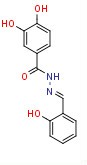 KM 91104
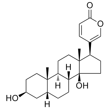 Bufalin
 土霉素
土霉素
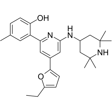 BRITE-338733
InvivoChem的所有产品仅用于作科学研究,不面向患者销售
Copyright 2020 InvivoChem LLC | All Rights Reserved 粤ICP备20063088号-1


 COA
COA
 463611831
