| 规格 | 价格 | 库存 | 数量 |
|---|---|---|---|
| 10 mM * 1 mL in DMSO |
|
||
| 1mg |
|
||
| 5mg |
|
||
| 10mg |
|
||
| 25mg |
|
||
| 50mg |
|
||
| 100mg |
|
||
| 250mg |
|
||
| 500mg |
|
||
| 1g |
|
||
| Other Sizes |
|
| 靶点 |
Microtubule; tubulin polymerization; β-tubulin (Kd = 0.4 μM)
Combretastatin A4 (CA-4; CRC 87-09) specifically targets β-tubulin, binding to the colchicine-binding site to inhibit microtubule polymerization, with IC50 values of 0.8 nM (human nasopharyngeal carcinoma CNE-1 cells), 1.2 nM (CNE-2 cells), and 2.5 nM for inhibiting tubulin polymerization [3][4] It shows no significant binding to other cytoskeletal proteins or kinases at therapeutic concentrations [3][4] |
|---|---|
| 体外研究 (In Vitro) |
使用Combretastatin A4/考布他汀 A4 磷酸盐 (≥ 50 μM) 时,前向散射大大减少,膜联蛋白 V 结合细胞的比例显着增加。考布他汀 A4 磷酸盐不会显着增加溶血量。 Combretastatin A4 磷酸盐浓度为数百 μM 时可显着增强 Fluo3 荧光。当细胞外 Ca2+ 被去除时,Combretastatin A4 磷酸盐 (100 μM) 对膜联蛋白-V 结合的影响大大降低,但并未完全消除。考布他汀 A4 磷酸盐 (≥ 50 μM) 不会显着增加 ROS 和神经酰胺,但会显着降低 GSH 丰度和 ATP 水平 [2]。共封装阿霉素-考布他汀-A4磷酸盐(1:10)的聚合物胶囊对人鼻咽上皮癌(KB)细胞具有强协同细胞毒性[3]。这些重要分子的表达和 3-D 细胞中 VM 的数量不受考布他汀 A4 磷酸盐预处理的影响 [4]。
将人红细胞暴露于Combretastatin A4/CA4P(≥50µM)48小时后,膜联蛋白-V结合细胞的百分比显著增加,前向散射显著降低Combretastatin A4/CA4P没有明显增加溶血。100µM CA4P显著增加了Fluo3荧光。通过去除细胞外Ca2+,CA4P(100µM)对膜联蛋白-V结合的影响显著减弱,但并未完全消除。CA4P(≥50µM)显著降低了GSH丰度和ATP水平,但没有显著增加ROS或神经酰胺。 结论:Combretastatin A4CA4P引发红细胞膜的细胞收缩和磷脂紊乱,这种作用至少部分是由于细胞外Ca2+的进入和能量消耗。[2] 使用三维培养的体外模型来测试Combretastatin A4/CA4P对Walker 256细胞管形成的影响。进行Western blot分析以评估缺氧诱导因子(HIF)-1α和VM相关标志物的表达。在体外缺氧条件下48小时,W256细胞形成与VM标志物表达增加相关的VM网络。CA4P预处理不影响三维培养中VM的量以及这些关键分子的表达[4]。 在人类鼻咽癌细胞系(CNE-1、CNE-2)中,游离 Combretastatin A4 抑制细胞增殖,IC50 值为 0.8 nM(CNE-1)和 1.2 nM(CNE-2);共载 Combretastatin A4 与阿霉素的聚合物囊泡增强抗增殖活性,使 IC50 降至 0.3 nM(CNE-1)和 0.4 nM(CNE-2)[3] - 1 nM Combretastatin A4 处理 24 小时后,75% 的 CNE-1 细胞发生 G2/M 期阻滞,与阿霉素(0.5 μM)联用时阻滞率提升至 88% [3] - 在 W256 乳腺癌细胞中,Combretastatin A4(0.5-5 nM)剂量依赖性诱导血管拟态(VM)形成,2 nM 浓度下 VM 密度增加 2.3 倍;该效应与 VE- 钙粘蛋白和 MMP-2 表达分别上调 1.8 倍和 2.1 倍相关 [4] - Combretastatin A4(1-10 μM)诱导人类红细胞自杀性死亡,5 μM 浓度处理 48 小时后,磷脂酰丝氨酸暴露率从 3% 升至 42%,细胞内钙浓度升高 2.5 倍 [2] - 2 nM Combretastatin A4 诱导 CNE-2 细胞凋亡,48 小时后膜联蛋白 V 阳性细胞比例从 4% 升至 55%;聚合物囊泡制剂进一步将凋亡率提升至 72% [3] - Western blot 分析显示,1-2 nM Combretastatin A4 下调鼻咽癌细胞中 α/β- 微管蛋白聚合,激活半胱天冬酶 -3/PARP 切割,使 Bax/Bcl-2 比值上调 3.2 倍 [3] |
| 体内研究 (In Vivo) |
治疗30分钟后,给予120 mg/10 mL/kg Combretastatin A4磷酸二钠的大鼠具有更高的DBP和MBP。用康布他汀 A4 磷酸二钠 120 mg/10 mL/kg 治疗的大鼠显示出康布他汀 A4 及其磷酸盐的以下毒代动力学特征:康布他汀 A4 的 Cmax、T1/2 和 AUC0-inf 值为 156± 13 μM、5.87±1.69 h、89.4±10.1 h·μM[1]。 W256 肿瘤在考布他汀 A4 磷酸盐治疗后显示出明显的瘤内缺氧,这与 VM 发展的增加有关。 cercopetastatin A4 磷酸盐仅延迟肿瘤生长两天,但肿瘤生长很快恢复。第 8 天时,VM 密度与肿瘤重量和体积呈正相关。通过 HIF-1α/EphA2/PI3K/基质金属蛋白酶 (MMP) 信号通路,磷酸西西他汀 A4 刺激 W256 肿瘤中缺氧和 VM 形成,从而损害肿瘤更新[4]。
在本研究中,我们设计了可生物降解的多聚体,用于联合递送抗血管生成药物Combretastatin A4磷酸盐(CA4P)和阿霉素(DOX),以破坏肿瘤新生血管系统并抑制癌症细胞增殖,目的是实现协同抗肿瘤效果。以甲氧基聚乙二醇-b-聚乳酸(mPEG-PLA)嵌段共聚物为药物载体,通过溶剂蒸发法制备了共包封DOX和CA4P的聚合物体(Ps-DOX-CA4P)。所得Ps-DOX-CA4P具有囊泡形状,大小均匀,约为50nm,DOX与CA4P的共包封率可控。更重要的是,Ps-DOX-CA4P(1:10)对人鼻咽表皮癌(KB)细胞显示出强烈的协同细胞毒性(组合指数CI=0.31)。此外,Ps-DOX-CA4P在裸鼠KB组织异种移植物中显著积累。与这些观察结果一致,Ps-DOX-CA4P(1:10)由于体内肿瘤血管系统的快速破坏和持续的肿瘤细胞增殖抑制而具有显著的抗肿瘤效力。总体研究结果表明,在多聚体中联合递送抗血管生成药物和化学治疗剂是癌症治疗的一种潜在的有前景的策略。[3] 在体内,W256肿瘤在Combretastatin A4/CA4P治疗后表现出明显的瘤内缺氧,并伴有VM形成增加。CA4P在2天内仅表现出肿瘤生长的延迟,但随后肿瘤迅速再生。VM密度与第8天的肿瘤体积和肿瘤重量呈正相关。CA4P引起缺氧,通过HIF-1α/EphA2/PI3K/基质金属蛋白酶(MMP)信号通路诱导W256肿瘤中VM的形成,从而导致受损肿瘤的再生[4]。 在 BALB/c 裸鼠 CNE-1 异种移植模型中,静脉注射 Combretastatin A4- 阿霉素共载聚合物囊泡(等效 10 mg/kg Combretastatin A4,隔日一次,连续 21 天)的肿瘤生长抑制率(TGI)达 86%,显著高于游离 Combretastatin A4(52% TGI)或游离阿霉素(48% TGI)[3] - 在 Wistar 大鼠心肌损伤模型中,腹腔注射 Combretastatin A4 二钠磷酸盐(20 mg/kg,单次给药)诱导心肌损伤,表现为血清肌酸激酶同工酶(CK-MB)和乳酸脱氢酶(LDH)水平升高(较对照组分别增加 2.8 倍和 3.1 倍)、心肌细胞凋亡(35% TUNEL 阳性细胞)及间质水肿 [1] - 在裸鼠 W256 乳腺癌异种移植模型中,Combretastatin A4(15 mg/kg,静脉给药,每周一次,连续 4 周)使肿瘤 VM 密度增加 2.1 倍,但与 VM 抑制剂联用时肿瘤体积减少 68% [4] - Combretastatin A4 聚合物囊泡处理组小鼠的肿瘤组织中,微血管密度降低 65%,半胱天冬酶 -3 激活增加 4.5 倍,Ki-67 增殖指数降至 22%(溶媒组为 70%)[3] |
| 酶活实验 |
体外微管蛋白聚合测定[5,6]
根据Wang等人描述的方法,将猪脑微管蛋白(纯度>97%)与普通微管蛋白缓冲液(80 mM PIPES、2.0 mM MgCl2、0.5 mM EGTA和1 mM GTP)混合,在4°C下达到3 mg/mL的最终浓度。在96孔板中混合微管蛋白溶液和测试化合物后,立即在37°C的SYNERGY 4微孔板读取器中孵育微管蛋白聚合测定,并在340nm下每30秒监测65分钟。以紫杉醇作为微管蛋白聚合的阳性对照,秋水仙碱和ABI-274作为微管蛋白解聚的阳性对照进行重复实验。 用于亲和性测定的SPR[5,6] 在配备有葡聚糖SPR传感器芯片(Reichert Polycarboxylate Hydrogel chip P/N 13206067)的Reicher4SPR系统中使用SPR技术分析与微管蛋白的结合亲和力。然后,将50μg/mL微管蛋白固定在传感器芯片表面,以获得12μRIU。芯片上的四个流动池中的一个作为阴性对照。在传感器芯片表面上注射不同浓度的4v或秋水仙碱进行缔合分析,然后进行离解分析。实验数据在25°C下使用运行缓冲液PBST(8 mM Na2HPO4、136 mM NaCl、2 mM KH2PO4、2.6 mM KCl和0.05%(v/v)Tween 20,pH 7.4)获得。平衡离解常数(KD)是用TraceDrawer软件通过稳态拟合模式计算的。 微管聚合抑制实验:纯化微管蛋白(10 μM)与系列浓度的 Combretastatin A4(0.1 nM 至 30 nM)在聚合缓冲液中 37°C 孵育。60 分钟内通过检测 340 nm 吸光度监测微管聚合,从聚合抑制的剂量 - 反应曲线计算 IC50 值 [3][4] - β- 微管蛋白结合实验:荧光标记的秋水仙碱(秋水仙碱结合位点配体)与重组 β- 微管蛋白(5 μM)及系列浓度的 Combretastatin A4(0.5 nM 至 20 nM)25°C 孵育 40 分钟。荧光偏振法检测竞争性结合,Combretastatin A4 与 β- 微管蛋白的解离常数(Kd)为 1.1 nM [3] |
| 细胞实验 |
细胞阻抗评估[1]
参照和修改早期研究中的方法,使用xCELLigence心脏分析仪对hiPS-CM的细胞阻抗进行了分析。简而言之,iCell hiPS CM是从Cellular Dynamics International购买的。根据制造商的方案,使用iCell hiPS CM专用的平板培养基和维持培养基,在96孔xCELLigence Cardio E-plate中以20000个细胞/孔和37°C在5%CO2中解冻和培养hiPS CM。在潜伏期,根据制造商的说明,使用xCELLigence心脏分析仪连续监测阻抗值。以12.9ms的间隔连续采样阻抗,并在每个测量点以20秒的扫描持续时间进行监测。孵育14天后,将测试化合物(100 nM、1μM和10μM CA4DP;100 nM,1μM,和10μMCombretastatin A4/CA4;CA4DP为0.1%H2O,CA4[载体]为0.1%DMSO)(n=3孔)加入培养物中。然后,使用专用软件计算阻抗细胞指数(CI)和搏动率16、18、20。CI和打浆率的数据由添加试验化合物前的值进行归一化。给药后36小时的CI用于检测细胞毒性作用。给药后15分钟、3小时和12小时的搏动率用于检测收缩性的变化。 背景/目的:Combretastatin A4/康布他汀A4磷酸二钠(CA4P)用于治疗恶性肿瘤。该物质之前已被证明会引发自杀性细胞死亡或凋亡。与有核细胞的凋亡类似,红细胞也可能进入自杀性死亡或红细胞凋亡,其特征是细胞收缩和细胞膜紊乱,磷脂酰丝氨酸易位到红细胞表面。红细胞下垂的刺激因素包括细胞质Ca2+活性([Ca2+]i)、神经酰胺、氧化应激和ATP耗竭的增加。本研究探讨了CA4P是否会诱导红细胞下垂,如果是,则深入了解相关机制。 方法:采用流式细胞术从膜联蛋白-V结合、前向散射的细胞体积、Fluo3荧光的[Ca2+]i、DCF荧光的活性氧(ROS)丰度、CMF荧光的谷胱甘肽(GSH)丰度和荧光抗体的神经酰胺丰度估算细胞表面的磷脂酰丝氨酸暴露量。此外,利用基于萤光素酶的测定法定量细胞质ATP水平,并根据上清液中的血红蛋白浓度估算溶血[2]。 抗增殖实验:CNE-1/CNE-2 细胞接种于 96 孔板(3×10³ 个细胞 / 孔),用系列浓度的游离 Combretastatin A4、游离阿霉素或共载聚合物囊泡(等效 0.01 nM 至 20 nM Combretastatin A4)处理 72 小时。MTT 法评估细胞活力,计算 IC50 值及协同指数 [3] - 细胞周期分析:CNE-1 细胞用 Combretastatin A4(0.5-2 nM)或聚合物囊泡(等效 0.2-1 nM)处理 24 小时,70% 乙醇固定,碘化丙啶染色,流式细胞术定量 G2/M 期比例 [3] - 凋亡实验:CNE-2/W256 细胞用 Combretastatin A4(1-2 nM)或聚合物囊泡(等效 0.5-1 nM)处理 48 小时,用膜联蛋白 V-FITC/碘化丙啶染色,流式细胞术分析。Western blot 检测半胱天冬酶 -3/PARP 切割 [3][4] - 血管拟态实验:W256 细胞接种于基质胶包被的培养板,用 Combretastatin A4(0.5-5 nM)处理 24 小时。相差显微镜观察 VM 结构,计数管状结构量化 VM 密度 [4] - 红细胞自杀性死亡实验:人类红细胞悬浮于缓冲液中,用 Combretastatin A4(1-10 μM)处理 48 小时。膜联蛋白 V-FITC 染色检测磷脂酰丝氨酸暴露,Fluo-3 AM 荧光检测细胞内钙 [2] |
| 动物实验 |
溶于DMSO;100 mg/kg;腹腔注射至荷NT2和MDA-MB-231肿瘤的FVB/N或裸鼠NMRI雌性小鼠。组织病理学变化评估[1]
共14只大鼠被分为四组,如表1所示。6周龄时,通过尾静脉推注给予CA4DP/康布瑞他汀A4(30或60 mg/10 mL/kg,间隔24小时,共4次;或120 mg/10 mL/kg,间隔72小时,共2次)或生理盐水(间隔72小时,共2次)。末次给药后第二天,用异氟烷麻醉大鼠,并进行尸检。此外,一只大鼠在接受四次 60 mg/10 mL/kg CA4DP 给药后,因 CA4DP 中毒在尸检前意外死亡。死因被认为是 CA4DP 的心脏毒性,因为在该大鼠中观察到了严重的心肌坏死。放血后,取出大鼠心脏并立即固定于 10% 中性磷酸盐缓冲福尔马林溶液中。按照先前报道7所述,将固定的心脏沿心室进行两层横切。将固定的心脏包埋于石蜡中,并切片至 4-6 μm 厚。标本用苏木精-伊红 (HE) 染色。使用光学显微镜观察这些标本。 心电图数据评估[1] 使用了两只大鼠(1 号和 2 号动物)。在5周龄时,于戊巴比妥钠麻醉下,将一个用于传输心电图数据的微型遥测装置(重量3.9克,体积1.9毫升)植入大鼠背部皮下。该遥测装置自带的成对电极分别置于大鼠背部和腹部皮下,用于记录心尖-心底(A-B)导联心电图。术后一周,将每只大鼠置于信号接收板上的笼子中,记录其心电信号。心电图数据以1毫秒的间隔连续采样,所有心电图波形成分的数据分析均使用连接模数转换器的个人电脑上的心电图处理器分析系统进行;心电图数据存储在外部硬盘上。在心电图记录期间,通过尾静脉以推注方式向两只大鼠分别给予 CA4DP/康布瑞他汀 A4 50 mg/10 mL/kg,每隔 24 小时给药一次,共给药 3 次。心电图记录持续至第三次给药后 12 小时。连续记录 4 秒的心电图波形并取平均值,分析心电图波形成分(RR 间期、QRS 波时限、PR 间期和 QT 间期)。血压评估 [1] 共使用 9 只大鼠。6 周龄大鼠用异氟烷麻醉,并仰卧固定。暴露股动脉,插入充满肝素化生理盐水的聚乙烯导管。导管通过压力传感器连接至压力传感器放大设备,记录动脉血压。血压以1毫秒的间隔连续采样,所有数据分析均使用连接模数转换器的个人电脑上的心电图处理器分析系统进行。在血压记录期间,通过尾静脉单次推注给予CA4DP/康布瑞他汀A4(120毫克/10毫升/公斤)或生理盐水(10毫升/公斤)(CA4DP组n=5,生理盐水组n=4)。记录血压直至给药后30分钟。取连续4秒的血压波形平均值,并分析血压成分(收缩压[SBP]、舒张压[DBP]和平均动脉压[MBP])和心率(HR)。 毒代动力学分析[1] 大鼠单次静脉注射120 mg/10 mL/kg的CA4DP/康布瑞他汀A4(n = 3)。分别于给药后10分钟、1小时、3小时、6小时和24小时,经颈静脉采血,并收集于肝素抗凝管中。采血后立即离心分离血浆。离心后,取等体积血浆与等体积的1%甲酸混合,并于-20℃保存。解冻后的血浆样品经固相萃取纯化,采用液相色谱-串联质谱法(LC-MS/MS)测定血浆中康布瑞他汀A4磷酸盐(CA4DP的游离碱;CA4P)和康布瑞他汀A4(CA4DP的代谢物;CA4)的浓度。采用 Phoenix WinNonlin 6.3 进行非房室分析,获得毒代动力学参数[最大浓度 (Cmax)、末端半衰期 (T1/2) 和浓度-时间曲线下面积(从零到无穷大)]。 CNE-1 鼻咽癌异种移植模型:将 5×10⁶ 个 CNE-1 细胞皮下植入雌性 BALB/c 裸鼠(6-8 周龄)。当肿瘤体积达到 100-150 mm3 时,将小鼠随机分组(每组 n=8),并分别进行以下治疗:(1)静脉注射载体(PBS + 0.1% DMSO),(2)静脉注射游离的康布瑞他汀 A4(10 mg/kg),每两天一次,持续 21 天,(3)静脉注射游离的阿霉素(5 mg/kg),每两天一次,持续 21 天,(4)静脉注射康布瑞他汀 A4-阿霉素共载聚合物囊泡(相当于 10 mg/kg 康布瑞他汀 A4 + 相当于 5 mg/kg 阿霉素),每两天一次,持续 21 天。每3天测量一次肿瘤体积和重量[3] - 心肌损伤模型:将雄性Wistar大鼠(200-250 g)随机分组(n=6/组),腹腔注射康布瑞他汀A4磷酸二钠(20 mg/kg)或溶剂对照。注射后24小时处死大鼠,收集血清和心肌组织进行生化和组织病理学分析[1] - W256乳腺癌异种移植模型:将5×10⁶个W256细胞皮下植入雌性裸鼠(6-8周龄)。当肿瘤体积达到100-150 mm³时,将小鼠随机分组(n=8/组),每周静脉注射康布瑞他汀A4(15 mg/kg),持续4周。收集肿瘤组织用于血管生成模拟(VM)检测和免疫组织化学染色[4] - 通过将康布瑞他汀A4和阿霉素包封在可生物降解的聚合物中,制备了康布瑞他汀A4聚合物囊泡,粒径控制在120-180 nm[3] |
| 药代性质 (ADME/PK) |
代谢/代谢物
康布瑞他汀 A4 已知的代谢物包括 (2S,3S,4S,5R)-3,4,5-三羟基-6-[2-甲氧基-5-[(Z)-2-(3,4,5-三甲氧基苯基)乙烯基]苯氧基]氧杂环己烷-2-羧酸。 |
| 毒性/毒理 (Toxicokinetics/TK) |
康布瑞他汀A4(20 mg/kg,腹腔注射)可诱导大鼠心肌毒性,表现为血清CK-MB和LDH水平升高、心肌细胞凋亡和间质炎症[1]
- 康布瑞他汀A4(1-10 μM)可诱导人红细胞凋亡,在4.2 μM浓度下,50%的细胞磷脂酰丝氨酸暴露[2] - 游离的康布瑞他汀A4(10 mg/kg,静脉注射)可导致裸鼠轻度体重下降(6%),而聚合物囊泡制剂可将体重下降减少至<3%[3] - 用康布瑞他汀A4(10-15 mg/kg,静脉注射)治疗的小鼠未观察到明显的肝脏或肾脏组织病理学异常[3][4] |
| 参考文献 |
|
| 其他信息 |
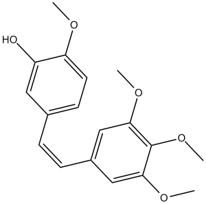
康布瑞他汀A4是一种芪类化合物。
已有报道称康布瑞他汀A4存在于非洲柳树(Combretum caffrum)中,并有相关数据。 康布瑞他汀A4是一种微管聚合抑制剂,提取自南非柳树,可导致有丝分裂停滞,并选择性地靶向、减少或破坏现有血管,从而减少肿瘤的血液供应。 另见:磷布瑞他汀(注释已移至)。 康布瑞他汀A4是一种微管聚合抑制剂,提取自南非柳树,可导致有丝分裂停滞,并选择性地靶向、减少或破坏现有血管,从而减少肿瘤的血液供应。 评估了康布瑞他汀A4磷酸二钠(CA4DP)诱导的心肌损伤的组织病理学和心电图特征,并探讨了心肌损伤与血管变化之间的关系以及CA4DP对心肌细胞的直接毒性作用。我们通过给大鼠注射CA4DP诱导心肌损伤,并通过组织病理学检查和心电图评估心肌损伤情况。我们评估了CA4DP处理组大鼠的血压,以及CA4DP对人诱导多能干细胞来源的心肌细胞(hiPS-CMs)基于细胞阻抗的收缩力的影响。结果显示,心肌出现多灶性坏死,主要累及室间隔和左心室心尖部心内膜下区域,同时伴有毛细血管损伤、ST段形态改变和QT间期延长。心肌损伤的组织病理学特征表明,CA4DP导致心肌血流减少。CA4DP可升高舒张压,并对hiPS-CMs产生直接影响。这些结果表明,CA4DP可诱导小动脉和毛细血管功能障碍,并对心肌细胞具有直接毒性。因此,人们认为CA4DP通过导致心肌微循环崩溃而引起毛细血管和心肌损伤。此外,CA4DP对心肌细胞的直接毒性作用以协同的方式诱导心肌损伤。[1] 本研究旨在探讨单次给药后,磷酸康布瑞他汀A4 (CA4P) 对体外和体内血管生成拟态 (VM) 通道形成的影响及其支持 VM 的潜在机制。采用三维培养的体外模型检测CA4P对Walker 256细胞管状结构形成的影响。通过Western blot分析评估缺氧诱导因子 (HIF)-1α和 VM 相关标志物的表达。本研究建立了W256荷瘤大鼠模型,通过双重染色和缺氧标记物匹莫尼达唑(pimonidazole)检测,探讨了CA4P对血管生成拟态(VM)形成和肿瘤缺氧的影响。通过肿瘤生长曲线评估了CA4P的抗肿瘤疗效。体外缺氧48小时后,W256细胞形成VM网络,并伴有VM标记物表达增加。CA4P预处理不影响三维培养中VM的数量以及这些关键分子的表达。体内实验表明,CA4P治疗后W256肿瘤出现明显的瘤内缺氧,并伴有VM形成增加。CA4P仅在2天内延缓了肿瘤生长,之后肿瘤迅速复发。在第8天,VM密度与肿瘤体积和肿瘤重量呈正相关。CA4P引起缺氧,并通过HIF-1α/EphA2/PI3K/基质金属蛋白酶(MMP)信号通路诱导W256肿瘤中VM的形成,从而导致受损肿瘤的再生。[4] 我们近期报道了微管蛋白与秋水仙碱结合位点抑制剂(CBSI)ABI-231(含有2-芳基-4-苯甲酰基咪唑,简称ABI)复合物的晶体结构。基于此及其他晶体结构,我们在此报道了一系列新型ABI-231吡啶类似物的构效关系研究,其中化合物4v对多种癌细胞系表现出最强的活性(平均IC50约为1.8 nM)。我们解析了另一种强效CBSI ABI-274和4v与微管蛋白复合物的晶体结构,并证实了它们直接结合于秋水仙碱结合位点。4v抑制微管蛋白聚合,显著抑制A375黑色素瘤肿瘤生长,诱导肿瘤坏死,破坏肿瘤血管生成,并在体内诱导肿瘤细胞凋亡。综上所述,这些研究表明4v代表了一种很有前景的新一代微管蛋白抑制剂。[5] 基于我们之前报道的ABI-I和ABI-II类似物,我们设计并合成了新型ABI-III化合物。ABI-III化合物对多种黑色素瘤和前列腺癌细胞系具有高度活性,其中活性最佳的化合物平均IC50值为3.8 nM。它们不是Pgp的底物,因此可能有效克服Pgp介导的多药耐药性。 ABI-III类似物通过抑制微管蛋白聚合来维持其作用机制。[6] 康布瑞他汀A4是一种从非洲风车子树皮中分离得到的天然产物,是一种强效的微管蛋白聚合抑制剂。[3][4] 其作用机制涉及与β-微管蛋白的秋水仙碱结合位点结合,破坏微管动力学,诱导癌细胞发生G2/M期细胞周期阻滞和caspase依赖性凋亡。[3][4] 康布瑞他汀A4可通过上调VE-钙黏蛋白和MMP-2诱导乳腺癌细胞的血管生成拟态,这可能会限制其单药治疗的疗效,但可通过与血管生成拟态抑制剂联合使用来逆转。[4] 与阿霉素共包封于聚合物囊泡中可增强康布瑞他汀A4的抗肿瘤疗效并降低全身用药风险。毒性,提高治疗指数[3] 康布瑞他汀A4具有潜在的心脏毒性和血液毒性(红细胞自杀性死亡),在临床应用中需要注意[1][2] |
| 分子式 |
C18H20O5
|
|
|---|---|---|
| 分子量 |
316.35
|
|
| 精确质量 |
316.131
|
|
| 元素分析 |
C, 68.34; H, 6.37; O, 25.29
|
|
| CAS号 |
117048-59-6
|
|
| 相关CAS号 |
168555-66-6; 222030-63-9; 117048-59-6
|
|
| PubChem CID |
5351344
|
|
| 外观&性状 |
White to light yellow solid powder
|
|
| 密度 |
1.2±0.1 g/cm3
|
|
| 沸点 |
490.3±45.0 °C at 760 mmHg
|
|
| 熔点 |
84.5-85.5ºC
|
|
| 闪点 |
250.3±28.7 °C
|
|
| 蒸汽压 |
0.0±1.3 mmHg at 25°C
|
|
| 折射率 |
1.607
|
|
| LogP |
3.57
|
|
| tPSA |
57.15
|
|
| 氢键供体(HBD)数目 |
1
|
|
| 氢键受体(HBA)数目 |
5
|
|
| 可旋转键数目(RBC) |
6
|
|
| 重原子数目 |
23
|
|
| 分子复杂度/Complexity |
358
|
|
| 定义原子立体中心数目 |
0
|
|
| SMILES |
COC1=C(C=C(C=C1)/C=C\C2=CC(=C(C(=C2)OC)OC)OC)O
|
|
| InChi Key |
HVXBOLULGPECHP-WAYWQWQTSA-N
|
|
| InChi Code |
InChI=1S/C18H20O5/c1-20-15-8-7-12(9-14(15)19)5-6-13-10-16(21-2)18(23-4)17(11-13)22-3/h5-11,19H,1-4H3/b6-5-
|
|
| 化学名 |
2-methoxy-5-[(Z)-2-(3,4,5-trimethoxyphenyl)ethenyl]phenol
|
|
| 别名 |
|
|
| HS Tariff Code |
2934.99.9001
|
|
| 存储方式 |
Powder -20°C 3 years 4°C 2 years In solvent -80°C 6 months -20°C 1 month |
|
| 运输条件 |
Room temperature (This product is stable at ambient temperature for a few days during ordinary shipping and time spent in Customs)
|
| 溶解度 (体外实验) |
|
|||
|---|---|---|---|---|
| 溶解度 (体内实验) |
配方 1 中的溶解度: ≥ 3 mg/mL (9.48 mM) (饱和度未知) in 10% DMSO + 40% PEG300 + 5% Tween80 + 45% Saline (这些助溶剂从左到右依次添加,逐一添加), 澄清溶液。
例如,若需制备1 mL的工作液,可将100 μL 30.0 mg/mL 澄清的 DMSO 储备液加入到400 μL PEG300中,混匀;再向上述溶液中加入50 μL Tween-80,混匀;然后加入450 μL 生理盐水定容至1 mL。 *生理盐水的制备:将 0.9 g 氯化钠溶解在 100 mL ddH₂O中,得到澄清溶液。 配方 2 中的溶解度: ≥ 3 mg/mL (9.48 mM) (饱和度未知) in 10% DMSO + 90% Corn Oil (这些助溶剂从左到右依次添加,逐一添加), 澄清溶液。 例如,若需制备1 mL的工作液,可将 100 μL 30.0 mg/mL 澄清 DMSO 储备液添加到 900 μL 玉米油中并混合均匀。 View More
配方 3 中的溶解度: 5% DMSO +50% PEG 300 +ddH2O: 30mg/mL 1、请先配制澄清的储备液(如:用DMSO配置50 或 100 mg/mL母液(储备液)); 2、取适量母液,按从左到右的顺序依次添加助溶剂,澄清后再加入下一助溶剂。以 下列配方为例说明 (注意此配方只用于说明,并不一定代表此产品 的实际溶解配方): 10% DMSO → 40% PEG300 → 5% Tween-80 → 45% ddH2O (或 saline); 假设最终工作液的体积为 1 mL, 浓度为5 mg/mL: 取 100 μL 50 mg/mL 的澄清 DMSO 储备液加到 400 μL PEG300 中,混合均匀/澄清;向上述体系中加入50 μL Tween-80,混合均匀/澄清;然后继续加入450 μL ddH2O (或 saline)定容至 1 mL; 3、溶剂前显示的百分比是指该溶剂在最终溶液/工作液中的体积所占比例; 4、 如产品在配制过程中出现沉淀/析出,可通过加热(≤50℃)或超声的方式助溶; 5、为保证最佳实验结果,工作液请现配现用! 6、如不确定怎么将母液配置成体内动物实验的工作液,请查看说明书或联系我们; 7、 以上所有助溶剂都可在 Invivochem.cn网站购买。 |
| 制备储备液 | 1 mg | 5 mg | 10 mg | |
| 1 mM | 3.1611 mL | 15.8053 mL | 31.6106 mL | |
| 5 mM | 0.6322 mL | 3.1611 mL | 6.3221 mL | |
| 10 mM | 0.3161 mL | 1.5805 mL | 3.1611 mL |
1、根据实验需要选择合适的溶剂配制储备液 (母液):对于大多数产品,InvivoChem推荐用DMSO配置母液 (比如:5、10、20mM或者10、20、50 mg/mL浓度),个别水溶性高的产品可直接溶于水。产品在DMSO 、水或其他溶剂中的具体溶解度详见上”溶解度 (体外)”部分;
2、如果您找不到您想要的溶解度信息,或者很难将产品溶解在溶液中,请联系我们;
3、建议使用下列计算器进行相关计算(摩尔浓度计算器、稀释计算器、分子量计算器、重组计算器等);
4、母液配好之后,将其分装到常规用量,并储存在-20°C或-80°C,尽量减少反复冻融循环。
计算结果:
工作液浓度: mg/mL;
DMSO母液配制方法: mg 药物溶于 μL DMSO溶液(母液浓度 mg/mL)。如该浓度超过该批次药物DMSO溶解度,请首先与我们联系。
体内配方配制方法:取 μL DMSO母液,加入 μL PEG300,混匀澄清后加入μL Tween 80,混匀澄清后加入 μL ddH2O,混匀澄清。
(1) 请确保溶液澄清之后,再加入下一种溶剂 (助溶剂) 。可利用涡旋、超声或水浴加热等方法助溶;
(2) 一定要按顺序加入溶剂 (助溶剂) 。
|
|---|
 |
InvivoChem的所有产品仅用于作科学研究,不面向患者销售
Copyright 2020 InvivoChem LLC | All Rights Reserved 粤ICP备20063088号-1


 COA
COA

 463611831
