| 规格 | 价格 | 库存 | 数量 |
|---|---|---|---|
| 10 mM * 1 mL in DMSO |
|
||
| 1mg |
|
||
| 5mg |
|
||
| 10mg |
|
||
| 25mg |
|
||
| 50mg |
|
||
| 100mg |
|
||
| 250mg |
|
||
| Other Sizes |
|

| 靶点 |
HIF hydroxylase
|
|---|---|
| 体外研究 (In Vitro) |
提高成绩的物质和方法已成为竞技体育中的一个严重问题。缺氧诱导因子(HIF)稳定剂可以增强生物体的分子氧运输能力,并可能被滥用为运动中的性能增强剂。本文描述了使用马肝微粒体在QExactive高分辨率质谱仪上测定的流行缺氧诱导因子脯氨酰羟化酶(HIF-PH)抑制剂,即daprodustat、Desidustat和vadadustat的代谢转化。在这项研究中,共检测到10种达布达司他代谢物(均为I期)、10种Desidustat (I期和II期各5种)和15种vadadustat代谢物(I期6种,II期9种)。当前研究的重要发现如下:(1)所有三种HIF-PH抑制剂候选药物都容易氧化,从而产生相应的羟基化代谢物;(2) 在Desidustat 中,还观察到肟键的水解和解离;(3) 观察到母体药物的葡糖醛酸结合物(达普司他除外)以及单羟基类似物;(4) 仅观察到vadadustat的磺酸结合代谢物[1]。
|
| 体内研究 (In Vivo) |
在体内,desindusat(口服;10-100 mg/kg)表现出良好的有效性[1]。
慢性肾病(CKD)与激活的炎症反应有关。Desidustat是一种脯氨酰羟化酶(PHD)抑制剂,可用于治疗与CKD相关的贫血,但其对CKD炎症和纤维化变化的影响尚未得到评估。在这项研究中,我们研究了地塞米松对急性和慢性肾损伤临床前模型中炎症和纤维化变化的影响。通过缺血再灌注在雄性Sprague-Dawley大鼠中诱导急性肾损伤,其中评估了去沙尘暴(15mg/kg,PO)的影响。在另一项实验中,雄性C57小鼠用腺嘌呤处理14天以诱导CKD。这些小鼠接受去沙司他(15mg/kg,口服,隔日一次)治疗14天,并继续服用腺嘌呤。去沙司他可预防血清肌酐、尿素、IL-1β、IL-6和肾损伤分子-1(KIM-1)的升高,并提高急性肾损伤大鼠的促红细胞生成素水平。用腺嘌呤治疗的小鼠会出现CKD和贫血,去粉尘治疗可改善血清肌酐、尿素,还可改善血红蛋白,降低肝脏和血清hepcidin。脱尘处理可显著降低IL-1β、IL-6、髓过氧化物酶(MPO)和氧化应激。通过组织学分析和羟脯氨酸含量观察到,去沙司他治疗也减少了肾纤维化。脱尘治疗减少了肾纤维化和炎症,同时减少了肾损伤临床前模型中的贫血,这可能转化为对CKD患者的保护作用[1] 自身免疫性溶血性贫血(AIHA)是一组由针对红细胞的自身抗体介导的异质性疾病,导致溶血和贫血。根据触发因素,AIHA会迅速或随着时间的推移而发展。Desidustat是一种脯氨酰羟化酶抑制剂,临床上用于治疗慢性肾病(CKD)引起的贫血。在这项研究中,我们研究了desidustat在AIHA临床前模型中的作用。我们使用大鼠红细胞诱导小鼠AIHA。然后用去沙司他(15mg/kg,口服,每天一次)治疗这些小鼠八周。去尘治疗增加了血红蛋白、红细胞和红细胞压积,降低了白细胞和淋巴细胞。这种治疗抑制了血清LDH、红细胞氧化应激、抗体滴度和红细胞表面抗体沉积,延长了红细胞寿命。去铁可降低血清和脾脏铁含量,同时减轻脾脏重量和氧化应激。通过治疗去铁,骨髓铁含量增加,骨髓中CD71(早期红系祖细胞的细胞表面标志物)和TER-119(晚期红系祖动物的细胞表面标记物)的表达升高。这种治疗还抑制了膜结合抗体在晚期红系细胞中的沉积。治疗显示脾脏总细胞、CD71和TER-119阳性细胞减少。因此,去粉尘治疗增加了红细胞生成,骨髓红系细胞的早期成熟具有更长的红细胞寿命,这是由于抗体介导的红细胞及其祖细胞的裂解减少,导致氧化应激减少。因此,desidustat可以成为治疗AIHA的良好治疗选择[2]。 |
| 细胞实验 |
在体外,检查了Caco2细胞通透性、血浆蛋白结合、代谢、细胞色素P450(CYP)抑制和CYP诱导[4]。
|
| 动物实验 |
动物/疾病模型: C57 小鼠[1]
剂量: 10、30、50、100 mg/kg;20 mg/kg 给药途径: 灌胃(po);口服,每日一次,连续7天。 实验结果: EPO 和 Hb 水平显著升高。 SD 大鼠急性肾损伤[1] 雄性 SD 大鼠(7-8 周龄,体重 210-240 g)用氯胺酮(50 mg/kg,腹腔注射)和赛拉嗪(10 mg/kg,腹腔注射)麻醉,开腹后暴露双侧肾蒂。用止血钳夹闭双侧肾蒂 25 分钟。双侧肾脏缺血结束后,移除血管夹以恢复肾脏血流灌注。皮肤和肌肉用3.0号手术缝线缝合。缺血再灌注损伤(I/R)组(n = 9)分别给予载体或Desidustat(15 mg/kg,口服,n = 9),于肾脏缺血开始前30分钟和开始后2小时给药。假手术(正常)对照组动物仅在开腹后暴露肾蒂。再灌注24小时后,所有动物在异氟烷麻醉下经眼眶后静脉丛取血。使用括号中提及的试剂盒分析血清样本中的肌酐、尿素、IL-1β、IL-6、促红细胞生成素和肾损伤分子KIM-1水平。 C57小鼠慢性肾病[1] 通过补充腺嘌呤诱导C57小鼠(7-8周龄,体重25-30 g)发生慢性肾病。根据体重将动物随机分为两组:腺嘌呤组(n = 8)和Desidustat组(15 mg/kg,n = 8)。所有组均以50 mg/kg的剂量经口服(PO)给予腺嘌呤,持续14天。接下来的14天,在继续补充腺嘌呤的同时,隔天给予Desidustat治疗。另设一组饲喂标准饲料的对照组。第15天,将动物置于代谢笼中收集尿液。次日,在异氟烷麻醉下,经眼眶后静脉丛取血,分离血清。使用Labcare Diagnostics试剂盒测定白蛋白、肌酐和尿素水平。肾小球滤过率估算值(eGFR)的计算方法参照文献(Pestel、Krzykalla和Weckesser,2007)。IL-1β和IL-6水平的测定采用括号内所列的试剂盒。血清和肝脏中的铁调素采用ELISA试剂盒测定。处死后立即收集肾脏,并在-70℃下保存直至测定,以硫代巴比妥酸反应物(TBARS)表示脂质过氧化产物。TBARS采用分光光度法测定(Buege和Aust,1978)。脂质过氧化以丙二醛 (MDA) 当量表示,消光系数为 1.56 × 10⁵ L/mol/cm,结果以 nmol MDA/g 组织表示。肾脏中超氧化物歧化酶 (SOD) 的活性通过分光光度法测定,即在 420 nm 波长下抑制焦棓酚的自氧化 10 分钟(Marklund & Marklund, 1974)。一个活性单位定义为抑制焦棓酚氧化 50% 所需的酶量。髓过氧化物酶 (MPO) 在血清和肾脏中进行测定(Kim et al., 2012)。此外,还对肾脏进行处理,使用 Quickzyme 羟脯氨酸测定试剂盒测定羟脯氨酸含量。 自身免疫性溶血性贫血的诱导[2] 通过重复注射大鼠红细胞 (RBC) 在小鼠中诱导自身免疫性溶血性贫血 (AIHA)。在异氟烷麻醉下,通过眼眶后穿刺采集 SD 大鼠的血液,血液样本置于含有乙二胺四乙酸二钠 (EDTA) 作为抗凝剂的试管中。去除血浆后,用磷酸盐缓冲液 (PBS,pH 7.4) 洗涤细胞三次,并将细胞浓度调整至 1 × 10⁹ 个细胞/ml(PBS 配制)。每周一次,通过腹腔注射途径向小鼠注射约 2 × 10⁸ 个大鼠红细胞。在开始每周注射大鼠红细胞六周后,从小鼠尾静脉采集血液进行全血细胞计数,并根据红细胞计数和血红蛋白含量进行随机分组。分组包括溶剂对照组(10 mL/kg)和Desidustat治疗组(15 mg/10 mL/kg,每日一次,口服),治疗持续八周。在此治疗期间,继续注射大鼠红细胞八周。治疗结束后,采集全血(用于血清和全血细胞计数)、骨髓、肝脏和脾脏。使用Cobas 6000仪器和试剂盒测定血清样本中的乳酸脱氢酶活性。 已开展体内药代动力学研究,包括小鼠、大鼠、犬和猴的口服生物利用度、剂量线性、组织分布和排泄情况。[4] |
| 药代性质 (ADME/PK) |
在Caco-2细胞中,地西司他(desidustat)在低pH值下表观渗透性高,在中性pH值下表观渗透性低。地西司他的口服生物利用度(%F)为43%~100%,各物种达峰时间(Tmax)中位数约为0.25~1.3小时。地西司他的平均血浆清除率(CL)较低,为1.3~4.1 mL/min/kg(约占肝血流量的1.8%~7.4%),平均稳态分布容积(Vss)为0.2~0.4 L/kg(约占体液总量的30%~61%)。在15~100 mg/kg的剂量范围内,地西司他的暴露量呈剂量依赖性增加。他能迅速分布于各种组织,其中肝脏(1.8)和肾脏(1.7)的组织/血液比值最高。 Desidustat 具有较高的血浆蛋白结合率,并且在人肝微粒体、肝细胞和重组 CYP 酶中代谢稳定。它对主要药物代谢 CYP 酶没有显著抑制作用(IC50 > 300 µM),也没有在 HepG2 细胞中诱导 CYP1A2 和 CYP3A4/5 的潜力(浓度高达 100 µM)。当与铁补充剂或磷酸盐结合剂联合使用时,其发生临床药物相互作用的可能性极低。在大鼠体内,Desidustat 主要以原形经尿液(占口服剂量的 25%)和胆汁(占口服剂量的 25%)排泄。静脉注射和口服给药后,Desidustat 的平均消除半衰期在不同物种间分别为 1.0 至 5.3 小时和 1.3 至 5.7 小时。[4] 综上所述,Desidustat 口服吸收良好。该药物的暴露量呈剂量依赖性增加,不会在组织中蓄积,并通过双重途径消除。它代谢稳定,引起临床药物相互作用(DDI)的可能性极小,并表现出治疗贫血的可区分的药代动力学特性。[4]
|
| 参考文献 | |
| 其他信息 |
Desidustat 是一种 N-酰基氨基酸。
Desidustat 目前正在进行临床试验 NCT04012957(Desidustat 治疗慢性肾脏病贫血)。 Desidustat 是一种口服生物利用度高的缺氧诱导因子脯氨酰羟化酶 (HIF-PH) 抑制剂 (HIF-PHI),具有潜在的抗贫血和抗炎活性。Desidustat 给药后,可与 HIF-PH 结合并抑制其活性。HIF-PH 是一种在正常氧条件下负责降解 HIF 家族转录因子的酶。这可以防止 HIF 分解并促进 HIF 活性。HIF 活性的增加会导致内源性促红细胞生成素的产生增加,从而增强红细胞生成。它还可以降低肽类激素铁调素的表达,提高铁的利用率,并提高血红蛋白 (Hb) 水平。 HIF 响应低氧水平调节基因表达,包括红细胞生成和铁代谢所需的基因。此外,HIF 1α (HIF1A) 可能通过 HIF 依赖性地调控肺泡上皮细胞的葡萄糖代谢,在急性肺损伤 (ALI) 期间减轻炎症。 作用机制 小分子缺氧诱导因子 desidustat 可抑制脯氨酰水解酶并刺激红细胞生成。目前正在研究其对炎症性贫血和 COVID-19 的疗效。 |
| 分子式 |
C16H16N2O6
|
|---|---|
| 分子量 |
332.308044433594
|
| 精确质量 |
332.1
|
| 元素分析 |
C, 57.83; H, 4.85; N, 8.43; O, 28.89
|
| CAS号 |
1616690-16-4
|
| 相关CAS号 |
1616690-16-4;
|
| PubChem CID |
75593290
|
| 外观&性状 |
White to off-white solid powder
|
| 密度 |
1.5±0.1 g/cm3
|
| 折射率 |
1.676
|
| LogP |
0.54
|
| tPSA |
116
|
| 氢键供体(HBD)数目 |
3
|
| 氢键受体(HBA)数目 |
6
|
| 可旋转键数目(RBC) |
6
|
| 重原子数目 |
24
|
| 分子复杂度/Complexity |
583
|
| 定义原子立体中心数目 |
0
|
| SMILES |
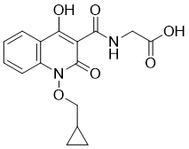
O=C(O)CNC(C1=C(O)C2=C(N(OCC3CC3)C1=O)C=CC=C2)=O
|
| InChi Key |
IKRKQQLJYBAPQT-UHFFFAOYSA-N
|
| InChi Code |
InChI=1S/C16H16N2O6/c19-12(20)7-17-15(22)13-14(21)10-3-1-2-4-11(10)18(16(13)23)24-8-9-5-6-9/h1-4,9,21H,5-8H2,(H,17,22)(H,19,20)
|
| 化学名 |
2-[[1-(cyclopropylmethoxy)-4-hydroxy-2-oxoquinoline-3-carbonyl]amino]acetic acid
|
| 别名 |
Desidustat; 1616690-16-4; ZYAN1; Zyan-1; Desidustat [INN]; Oxemia; ZYAN1 compound; Y962PQA4KS;
|
| HS Tariff Code |
2934.99.9001
|
| 存储方式 |
Powder -20°C 3 years 4°C 2 years In solvent -80°C 6 months -20°C 1 month |
| 运输条件 |
Room temperature (This product is stable at ambient temperature for a few days during ordinary shipping and time spent in Customs)
|
| 溶解度 (体外实验) |
DMSO : ~10 mg/mL (~30.09 mM)
|
|---|---|
| 溶解度 (体内实验) |
配方 1 中的溶解度: ≥ 1 mg/mL (3.01 mM) (饱和度未知) in 10% DMSO + 40% PEG300 + 5% Tween80 + 45% Saline (这些助溶剂从左到右依次添加,逐一添加), 澄清溶液。
例如,若需制备1 mL的工作液,可将100 μL 10.0 mg/mL澄清DMSO储备液加入400 μL PEG300中,混匀;然后向上述溶液中加入50 μL Tween-80,混匀;加入450 μL生理盐水定容至1 mL。 *生理盐水的制备:将 0.9 g 氯化钠溶解在 100 mL ddH₂O中,得到澄清溶液。 配方 2 中的溶解度: ≥ 1 mg/mL (3.01 mM) (饱和度未知) in 10% DMSO + 90% Corn Oil (这些助溶剂从左到右依次添加,逐一添加), 澄清溶液。 例如,若需制备1 mL的工作液,可将 100 μL 10.0 mg/mL 澄清 DMSO 储备液加入到 900 μL 玉米油中并混合均匀。 请根据您的实验动物和给药方式选择适当的溶解配方/方案: 1、请先配制澄清的储备液(如:用DMSO配置50 或 100 mg/mL母液(储备液)); 2、取适量母液,按从左到右的顺序依次添加助溶剂,澄清后再加入下一助溶剂。以 下列配方为例说明 (注意此配方只用于说明,并不一定代表此产品 的实际溶解配方): 10% DMSO → 40% PEG300 → 5% Tween-80 → 45% ddH2O (或 saline); 假设最终工作液的体积为 1 mL, 浓度为5 mg/mL: 取 100 μL 50 mg/mL 的澄清 DMSO 储备液加到 400 μL PEG300 中,混合均匀/澄清;向上述体系中加入50 μL Tween-80,混合均匀/澄清;然后继续加入450 μL ddH2O (或 saline)定容至 1 mL; 3、溶剂前显示的百分比是指该溶剂在最终溶液/工作液中的体积所占比例; 4、 如产品在配制过程中出现沉淀/析出,可通过加热(≤50℃)或超声的方式助溶; 5、为保证最佳实验结果,工作液请现配现用! 6、如不确定怎么将母液配置成体内动物实验的工作液,请查看说明书或联系我们; 7、 以上所有助溶剂都可在 Invivochem.cn网站购买。 |
| 制备储备液 | 1 mg | 5 mg | 10 mg | |
| 1 mM | 3.0092 mL | 15.0462 mL | 30.0924 mL | |
| 5 mM | 0.6018 mL | 3.0092 mL | 6.0185 mL | |
| 10 mM | 0.3009 mL | 1.5046 mL | 3.0092 mL |
1、根据实验需要选择合适的溶剂配制储备液 (母液):对于大多数产品,InvivoChem推荐用DMSO配置母液 (比如:5、10、20mM或者10、20、50 mg/mL浓度),个别水溶性高的产品可直接溶于水。产品在DMSO 、水或其他溶剂中的具体溶解度详见上”溶解度 (体外)”部分;
2、如果您找不到您想要的溶解度信息,或者很难将产品溶解在溶液中,请联系我们;
3、建议使用下列计算器进行相关计算(摩尔浓度计算器、稀释计算器、分子量计算器、重组计算器等);
4、母液配好之后,将其分装到常规用量,并储存在-20°C或-80°C,尽量减少反复冻融循环。
计算结果:
工作液浓度: mg/mL;
DMSO母液配制方法: mg 药物溶于 μL DMSO溶液(母液浓度 mg/mL)。如该浓度超过该批次药物DMSO溶解度,请首先与我们联系。
体内配方配制方法:取 μL DMSO母液,加入 μL PEG300,混匀澄清后加入μL Tween 80,混匀澄清后加入 μL ddH2O,混匀澄清。
(1) 请确保溶液澄清之后,再加入下一种溶剂 (助溶剂) 。可利用涡旋、超声或水浴加热等方法助溶;
(2) 一定要按顺序加入溶剂 (助溶剂) 。
InvivoChem的所有产品仅用于作科学研究,不面向患者销售
Copyright 2020 InvivoChem LLC | All Rights Reserved 粤ICP备20063088号-1


 COA
COA
 463611831
