| 规格 | 价格 | 库存 | 数量 |
|---|---|---|---|
| 50mg |
|
||
| 100mg |
|
||
| 250mg |
|
||
| 500mg |
|
||
| Other Sizes |
|

| 靶点 |
Natural triterpenoid saponinl; HMGB1; anti-tumor; anti-diabetic
|
|---|---|
| 体外研究 (In Vitro) |
甘草酸二钾(0-400 μM,4 天)可抑制 IL-4 和 IL-13 产生的细胞中特应性皮炎相关基因(NELL2、CA2、AQP3 和 HAS3 基因水平)的 mRNA 水平 [1]。甘草酸二钾(0-400 μM,4 天)可部分恢复 IL-4/IL-13 诱导的 AD 样皮肤等效小鼠的特应性皮炎样表型(海绵状细胞间隙)[1]。 0-100 μM,24 小时)可减少舒尼替尼诱导的 CCC-HEH-2 细胞自噬和细胞死亡 [5]。
特应性皮炎(AD)是一种慢性炎症性皮肤病,目前尚未完全了解。皮肤屏障功能缺陷和Th2免疫反应失调被认为是AD发病的关键。在本研究中,我们使用角质形成细胞和使用Th2细胞因子IL-4和IL-13的ad样皮肤等效模型。采用角化细胞和AD样皮肤模型研究甘草酸二钾/dipotassium glycyrrhizinate (KG)对AD治疗的影响。KG在受Th2细胞因子刺激的角质形成细胞中降低ad相关基因表达。在ad样皮肤等效模型中,KG减轻ad样表型和基因表达模式,并抑制ad相关细胞因子的释放。这些发现表明KG在AD治疗中具有潜在的有效性,AD样皮肤等效模型可能有助于了解AD的发病机制 甘草酸(GA)/dipotassium glycyrrhizinate (KG)对转染TGR5[2]细胞中TGR5的直接影响 [2] 在我们之前的报道中,我们使用CHO-K1细胞转染外源性TGR5受体基因。然后应用Western blot分析证实实验成功。这些CHO-K1细胞中表达的TGR5具有功能性。[2] 然后,应用这些细胞来估计GA对TGR5受体的直接影响。在表达TGR5的细胞中,GA处理显著增强了以细胞内2-NBDG含量为指标的葡萄糖摄取(图3A)。Triamterene剂量依赖性地阻断了这些细胞中ga诱导的葡萄糖摄取(图3B)。此外,GA在TGR5-CHO-K1细胞中也以剂量依赖性的方式增加了环AMP (cAMP)水平(图3C),并以同样的方式降低了三萜烯(图3D)。然而,在2-NBDG摄取增加(图3A)或cAMP水平升高(图3B)方面,GA并未影响缺乏TGR5表达的CHO-K1细胞。 甘草酸(GA)/dipotassium glycyrrhizinate (KG)通过TGR5促进肠细胞[2]中GLP-1的分泌 [2] 具有TGR5受体的培养肠道NCI-H716细胞常被用于研究GLP-1的释放。在与GA孵育的NCI-H716细胞中也观察到GLP-1分泌明显升高(图4A)或钙内流(图4B)。然而,在沉默NCI-H716细胞中表达的TGR5基因后,GA的这两种作用都被删除了。这与我们之前的报道完全相似,丁烯酸通过激活TGR5促进GLP-1的释放。 |
| 体内研究 (In Vivo) |
甘草酸二钾(150 mg/kg,腹腔注射)可提高心血管 GLP-1 水平并减轻链脲佐菌素(65 mg/kg,腹腔注射)引起的 1 型糖尿病症状 [2]。 (50 mg/kg,腹腔注射,每天一次,持续 4 周)可预防大鼠碘化钾引起的自身免疫炎症模型中前列腺炎的发生[3]。腹腔注射50–200 mg/kg甘草酸二钾。
甘草酸升高链脲佐菌素诱导的 1 型糖尿病大鼠(STZ 治疗的大鼠)血浆 GLP-1 水平,而氨苯蝶啶足以抑制武田 G 蛋白偶联受体 5 (TGR5) 被阻断 [1]。在小鼠中,甘草酸(50 mg/kg,腹腔注射)可显着降低 TgAb、HMGB1、TNF-α、IL-6 和 IL-1β 水平 [3]。
甘草酸对1型糖尿病大鼠血糖变化的影响 [2] 在STZ诱导的糖尿病大鼠中,注射甘草酸(GA)以剂量依赖的方式减轻了高血糖(图1A)。此外,与正常大鼠(141.4±10.2 pmol/l,n=8)相比,STZ诱导的糖尿病大鼠的血浆胰岛素水平(5.35±2.11 pmol/l,n=8)显著降低。然而,最高剂量的GA未能改变这些糖尿病大鼠的血浆胰岛素水平(5.66±1.39 pmol/l,n=8)。这意味着内源性胰岛素在该动物模型中不参与GA的作用。西格列汀以有效抑制二肽基肽酶-4(DPP-4)的剂量也减轻了这些糖尿病大鼠的高血糖。西格列汀显著增强了糖尿病大鼠注射GA后高血糖的降低(图1A)。此外,无论西格列汀预处理如何,氨苯蝶啶都能剂量依赖性地抑制这些糖尿病大鼠中GA诱导的变化(图1B)。 甘草酸(GA)对糖尿病大鼠血浆GLP-1水平变化的影响 [2] 在1型糖尿病大鼠中,甘草酸(GA)诱导血浆GLP-1水平呈剂量依赖性升高(图2A)。通过以有效抑制GLP-1失活酶DPP-4的剂量用西格列汀预处理,糖尿病大鼠中甘草酸(GA)的这种作用显著增强(图2A)。此外,与血糖的变化类似,在这些糖尿病大鼠中,无论是否用西格列汀预处理,氨苯蝶啶也以剂量依赖性方式逆转了GA诱导的血浆GLP-1水平的升高(图2B)。 NaI组在8周和16周时HMGB1的mRNA表达明显高于对照组。NaI组甲状腺球蛋白抗体、HMGB1、肿瘤坏死因子α、IL-6和IL-1β的血清水平显著升高,但甘草酸(GL)注射液显著降低了这些水平。NaI+甘草酸(GL)组的甲状腺炎患病率和淋巴细胞浸润率显著降低甘草酸(GL)给药也显著降低了甲状腺中TLR2、MyD88、HMGB1和核转录因子κB的蛋白表达,减轻了甲状腺炎的严重程度。[3] 结论:HMGB1可能通过引起炎症浸润在自身免疫性甲状腺炎中起关键作用,从而增加自身免疫性甲状腺疾病的严重程度甘草酸(GL)有效地减轻了碘诱导的NOD中的甲状腺炎。H-2h4小鼠通过与抑制TLR2-HMGB1信号传导相关的分子机制[3]。 特发性肺纤维化是一种进行性和致死性肺间质性疾病,目前缺乏有效的治疗方法。甘草酸/Glycyrrhizic acid (GA)是一种从传统中草药甘草中提取的天然化合物,最近有报道称其在动物模型中对肺损伤和肝纤维化有改善作用,但GA是否对肺纤维化有治疗作用尚不清楚。在这项研究中,我们研究了GA对博来霉素(BLM)诱导的肺纤维化大鼠模型肺纤维化的潜在治疗作用。结果表明,GA治疗显著改善了blm诱导的肺纤维化,减轻了blm诱导的炎症、氧化应激、上皮-间质转化和肺部转化生长因子- β信号通路的激活。此外,我们在体外证明了GA处理抑制3T6成纤维细胞的增殖,诱导细胞周期阻滞并促进细胞凋亡,这意味着GA介导的纤维增殖抑制可能有助于抗blm诱导的肺纤维化。总之,我们的研究表明GA在治疗肺纤维化方面具有治疗潜力[6].。 |
| 酶活实验 |
抗菌试验[4]
通过生长抑制效率和最小抑菌浓度(MIC)来评估GL凝胶的抗菌活性。金黄色葡萄球菌(S.aureus)和大肠杆菌(E.coli)分别被用作代表性的革兰氏阳性菌和革兰氏阴性菌。简而言之,将单个菌落接种到5 mL Luria Bertani(LB)肉汤培养基中,在37℃下以200 rpm的速度振荡培养过夜。过夜预培养后,将细菌悬浮液在LB培养基中稀释至106cfu mL-1,然后补充终浓度为0至2 mM的PBS或GL凝胶。然后将培养物在37℃下轻轻摇晃过夜。使用Synergy H1酶标仪在600nm波长下测量培养物的光密度。当没有观察到可见的细菌生长时,MIC被确定为抗菌材料的最低浓度。1遵循相同的程序实时监测用不同浓度的PBS或GL凝胶处理的细菌的生长动力学,除了在培养过程中的不同时间点记录每种培养物的OD600。特别是,将106cfu mL-1的金黄色葡萄球菌和大肠杆菌分别与终浓度为MIC的PBS或GL凝胶一起孵育。孵育2小时后,用PBS将细菌悬浮液稀释100倍,将100μL稀释的细菌铺在琼脂培养板上,在37℃下孵育过夜。 活/死荧光染色[4] 在MIC下用PBS或GL凝胶处理2小时后,通过离心收获金黄色葡萄球菌和大肠杆菌,并用0.9%NaCl洗涤3次。然后在黑暗中用SYTO 9和来自活/死染色试剂盒(L7012)的碘化丙啶(PI)对细菌进行染色30分钟。然后用LSM 700共聚焦激光扫描显微镜成像系统对细菌进行可视化。 细菌的形态学特征[4] SEM用于研究GL凝胶处理后细菌的形态。将106cfu mL-1的金黄色葡萄球菌和大肠杆菌与终浓度为MIC的PBS或GL凝胶一起孵育2小时,通过离心收集并用2.5%(w/w)戊二醛固定2小时。用PBS洗涤3次后,将细菌滴在硅片上并干燥过夜。硅片表面的细菌依次用分级乙醇水溶液(30%、50%、70%、80%、90%和100%)脱水10分钟。干燥过夜后,用金涂覆样品,并使用Phenom World Pro X扫描电子显微镜成像。 |
| 细胞实验 |
细胞活力测定[4]
对L929成纤维细胞进行细胞活力测定,以确定GL凝胶的体外细胞毒性。简而言之,L929细胞在添加了10%胎牛血清(FBS)的Dulbecco's Modified Eagle's Medium(DMEM)中在37℃、5%CO2的气氛中生长◦C.收获细胞,以每孔1×10⁴个细胞的接种密度接种在96孔板中。孵育过夜后,将DMEM培养基替换为含有一定浓度的制备好的GL凝胶的新鲜培养基。孵育24小时后,用含有10%CellTiter BlueTM试剂的100μL培养基代替培养基,在37℃下孵育2小时◦C.使用Synergy H1酶标仪在550/590nm(Ex/Em)下测量荧光强度。通过与阴性对照(用PBS处理的L929细胞)进行比较来计算相对细胞存活率。 细胞成像[4] L929细胞接种在24孔板中,接种密度为每孔2×10⁴个细胞。过夜培养后,用含有2mM GL凝胶的DMEM处理细胞,并在37℃下共培养◦用PBS洗涤处理过的细胞,并在室温下用4%(w/v)多聚甲醛固定15分钟。然后,用FITCphalloidin对细胞染色1小时,然后对细胞核酸进行DAPI染色15分钟。洗涤后,用LSM 700共聚焦激光扫描显微镜成像系统对细胞进行成像。 溶血试验[4] 通过以2000rpm离心5分钟从大鼠血液中分离红细胞(RBC)。沉淀的RBC在使用前用PBS洗涤3次。将红细胞悬浮在体积浓度为2%的PBS中,并补充终浓度为1-2mM的GL凝胶。用PBS和水处理的红细胞分别设置为阴性对照和阳性对照。在37℃下孵育2小时后,将处理过的红细胞以2000 rpm离心5分钟。使用酶标仪测量上清液在545 nm处的吸光度。溶血率使用以下方程式计算。 用于转染TGR5的CHO-K1细胞[2] 如前所述,使用成年中国仓鼠的卵巢从CHO细胞系制备CHO-K1细胞。在目前的实验中,根据我们之前的报告,使用人TGR5 cDNA的表达载体转染CHO-K1细胞。第二天,Western blot证实转染成功。然后,表达TGR5的细胞用于甘草酸(GA)处理。 细胞葡萄糖摄取的测量[2] 2-(N-(7-硝基苯-2-氧杂-1,3-二唑-4-基)氨基)-2-脱氧葡萄糖(2-NBDG)用于使用这种荧光葡萄糖类似物测定葡萄糖摄取,如我们之前的报告所述。使用荧光分光光度计测定每个细胞样品中的荧光强度。用BCA试剂盒测定蛋白质。然后,在接受甘草酸(GA)处理的细胞中定量2-NBDG的摄取。在预处理30分钟后,还将氨苯蝶啶的有效性与赋形剂治疗组进行了比较。 细胞中cAMP的测定[2] 按照我们之前的方法,用甘草酸(GA)处理细胞72小时。然后用ELISA试剂盒测定细胞内cAMP水平,类似于我们之前的法。每次测量都进行了两次。 细胞外GLP-1的测定[2] 我们使用NCI-H716细胞(每孔5×10⁵个细胞)在37°C下用指定浓度的甘草酸(GA)处理1小时。然后,根据我们之前的方法,使用ELISA试剂盒分析细胞外GLP-1水平。对指定样品进行每次重复测量。 细胞培养与处理[6] 小鼠成纤维细胞系3T6购自中国科学院细胞库。细胞在添加10% FBS的DMEM中培养,在湿度为95%空气和5% CO2的37℃环境中培养。 甘草酸/Glycyrrhizic acid (GA)在DMSO中溶解成180mm浓原液,用PBS稀释成9mm工作液。分别用5、10、25、50、100、200 μM GA处理3T6细胞24 h,采用乳酸脱氢酶活性测定试剂盒测定条件培养基中乳酸脱氢酶(LDH)活性,分析细胞毒性。在后续实验中,分别选择低剂量(25 μM)、中剂量(50 μM)和高剂量(100 μM)的GA处理细胞。 增殖试验[6] 采用3-(4,5-二甲基噻唑-2-基)-2,5-二苯基溴化四氮唑(MTT)法测定细胞增殖情况。3T6细胞以每孔3000个细胞的密度接种于96孔微孔板中,37°C培养24 h。每孔中加入Glycyrrhizic acid (GA)/甘草酸(GA)至指定的终浓度(0、25、50或100 μM)。GA处理24或48 h后,将MTT加入培养基中,终浓度为0.2 mg/ml, 37℃孵育4 h。然后,抽吸培养基,在每孔200 μl DMSO中使甲醛晶体完全溶解。用ELX-800型微孔板仪记录490 nm处的光密度。每个分析点在5个重复中进行。 流式细胞术分析细胞周期和凋亡[6] 甘草酸/Glycyrrhizic acid (GA)处理24 h后,流式细胞术分析细胞周期和凋亡情况。为了分析细胞周期,收集细胞,在70%乙醇中4°C固定2 h,与碘化丙啶(PI)溶液在37°C下黑暗孵育30 min,然后在FACSCalibur流式细胞仪上分析。按照说明书使用Annexin V-FITC/PI细胞凋亡检测试剂盒检测细胞凋亡。染色后用流式细胞术分析细胞凋亡情况。 抓伤试验[6] 细胞迁移是通过成熟的体外划伤实验来评估的(Liang等,2007)。用5 μM丝裂霉素c孵育3T6细胞2 h,抑制细胞增殖。用200 μl移液管尖端水平穿过细胞单层表面均匀划痕。将分离的细胞用无血清培养基洗净,在37℃5% CO2培养箱中,用浓度Glycyrrhizic acid (GA)的无血清培养基培养24 h。在抓伤后0、6、12、24 h倒置显微镜下拍摄细胞,计算创面愈合率为(原始间隙距离-指定时间点的间隙距离)/原始间隙距离× 100%。 Transwell Assay [6] 3T6细胞用5 μM丝裂霉素- c预处理2 h,在浓度甘草酸的培养基中重悬。将200 μl悬液中的2 × 10⁴个细胞置于预涂有Matrigel的Transwell腔中。将Transwell室置于24孔板中,每孔含800 μl培养基,培养基中添加20% FBS。37℃,5% CO2气氛下培养24 h。然后擦拭Transwell膜上表面的细胞和基质,用多聚甲醛固定膜下表面的细胞,并用结晶紫染色。在200 ×倒置显微镜下观察细胞,在每个膜上的5个视野中计数入侵细胞的数量。 |
| 动物实验 |
动物/疾病模型:碘诱导的自身免疫性甲状腺炎小鼠模型[3]
剂量:50 mg/kg 给药途径:腹腔注射(ip),每日一次,持续4周:血清TgAb、HMGB1、TNF-α、IL-6、IL-1β水平降低。降低甲状腺炎的发生率和淋巴细胞浸润,并减轻甲状腺炎的严重程度。 糖尿病大鼠血浆葡萄糖和GLP-1水平的测定[2] 糖尿病大鼠在给予甘草酸(GA)前两周,每日口服5 mg/kg西格列汀以抑制DPP-4或给予溶剂。实验前,大鼠禁食过夜,但不限制饮水。然后,将所需剂量的甘草酸(GA)通过腹腔注射给予大鼠,部分大鼠预先用西格列汀处理30分钟。在戊巴比妥麻醉(30 mg/kg,腹腔注射)下,于注射甘草酸(GA)1小时后,从大鼠股静脉采集血样。血浆葡萄糖水平采用全自动生化分析仪测定,方法如前所述。血浆GLP-1水平采用商业化ELISA试剂盒测定,方法如我们之前的报告所述。 将80只4周龄的雄性NOD.H-2h4小鼠随机分为对照组和碘补充剂(NaI)组,对照组饲喂普通水,NaI组饲喂0.005%碘化钠溶液。另取24只雄性NOD.H-2h4小鼠随机分为三组(每组8只):对照组、碘化钠(NaI)组和碘补充后给予GL治疗组(NaI + GL)。NOD.H-2h4小鼠饲喂0.005%碘化钠溶液8周以诱导自身免疫性甲状腺炎。碘补充后,小鼠腹腔注射GL,持续4周。采用组织病理学方法评估甲状腺淋巴细胞浸润的严重程度。采用酶联免疫吸附试验(ELISA)检测血清中HMGB1、肿瘤坏死因子α、白细胞介素(IL)-6、IL-1β和甲状腺球蛋白抗体滴度。采用免疫组织化学染色和实时聚合酶链反应(RT-PCR)检测HMGB1的表达。采用蛋白质印迹法检测TLR2、HMGB1、MyD88和核转录因子κB的表达。[3] 大鼠博来霉素诱导肺纤维化的建立及甘草酸(GA)治疗[6] 本研究使用了40只8周龄、体重约250克的雄性Sprague-Dawley大鼠。将大鼠随机分为五组,每组8只:对照组、博来霉素组、博来霉素+GA50组、博来霉素+GA100组和博来霉素+GA200组。采用先前描述的方法(Thrall等,1979)诱导博来霉素肺纤维化。所有大鼠均以3.5 ml/kg体重的10%氯化钠溶液麻醉。麻醉后,沿颈部正中线切开皮肤,钝性分离暴露气管。将1 ml注射器的针头插入气管,将溶于100 μl无菌生理盐水中的博来霉素以5 mg/kg体重的剂量注入大鼠肺部,对照组大鼠则注射等体积的生理盐水。注射后立即翻转大鼠,以确保博来霉素在肺部均匀分布,然后缝合颈部皮肤切口。之后,BLM+GA50组、BLM+GA100组和BLM+GA200组的大鼠分别腹腔注射50、100和200 mg/kg体重的戊二醛(GA),连续28天,其余动物注射生理盐水。在 BLM 诱导后 28 天处死大鼠,通过气管内滴注和引流 1.5 ml 生理盐水三次收集支气管肺泡灌洗液 (BALF),然后切除肺脏进行进一步分析。 |
| 药代性质 (ADME/PK) |
吸收、分布和排泄
甘草酸主要经首过水解生成甘草次酸后被吸收。因此,口服100 mg甘草酸后,血浆中主要代谢产物甘草次酸的浓度为200 ng/ml,而甘草酸则检测不到。尿液中仅检测到少量甘草酸,提示其在胃肠道存在部分吸收。 甘草酸在中枢室呈双相消除,第二消除相与剂量相关。大部分给药剂量经胆汁排泄,其中甘草酸可以原形排出,并经历肠肝循环。另一方面,主要代谢产物甘草次酸则形成葡萄糖醛酸苷和硫酸盐结合物。这些结合物能高效地转运至胆汁和十二指肠,在那里,共生细菌水解这些结合物,生成甘草次酸,并进一步被重吸收。这种重吸收行为似乎与3α-羟基类固醇脱氢酶的活性有关,该酶能非常有效地将代谢物从血浆转运至胆汁。约1.1-2.5%的给药剂量甘草酸可在尿液中检测到,这表明该化合物的循环和重吸收量极低。 甘草酸在中心室和稳态下的表观分布容积分别为37-64 ml/kg和59-98 ml/kg。 甘草次酸在十二指肠的持续重吸收导致其终末血浆清除率延迟。据报道,甘草酸的全身清除率在 16-25 ml·kg/h 范围内。 甘草酸在大鼠小肠内被吸收;门静脉快速注射甘草酸后,血液中未检测到甘草次酸;口服后,血液中可检测到甘草次酸。 高效液相色谱法 (HPLC) 可快速、精确地测定实验动物和人体体液及组织中的甘草酸 (GZA) 和甘草次酸 (GRA)。从血浆和组织中,用有机溶剂提取甘草酸和甘草次酸,提取物可直接用于 HPLC 分析。由于内源性化合物的干扰以及甘草次酸与葡萄糖醛酸苷或硫酸盐的结合,从胆汁或尿液中提取和测定甘草酸和甘草次酸较为困难。从尿液或胆汁中提取甘草酸和甘草次酸的方法包括离子对结合后用有机溶剂萃取或固相萃取。甘草次酸结合物可通过色谱分离或β-葡萄糖醛酸酶预处理进行测定。甘草次酸和甘草酸的药代动力学可以用双相消除模型来描述,其消除过程从中心室开始,并具有剂量依赖性的第二消除相。根据剂量不同,甘草酸在人体内的第二消除相半衰期为3.5小时,而甘草次酸的半衰期为10-30小时。甘草次酸和甘草酸的大部分均经胆汁排泄。甘草酸可以未经代谢直接排出,并经历肠肝循环;而甘草次酸在经胆汁排泄前,会与甘草次酸葡萄糖醛酸苷或硫酸盐结合。口服的甘草酸几乎完全被肠道细菌水解,并以甘草次酸的形式进入体循环。甘草酸目前在治疗慢性肝炎方面具有临床应用价值。它也被用作食品和嚼烟中的甜味剂。在一些高暴露人群中,曾报道出现高血压和电解质紊乱等严重副作用。为了分析暴露于该化合物的健康风险,本研究对甘草酸及其活性代谢物的动力学进行了定量评估。甘草酸及其代谢产物受复杂的动力学过程影响,包括肠肝循环和首过代谢。在人体中,这些过程的详细信息通常难以获得。因此,我们建立了一个模型,用于描述甘草酸及其活性代谢产物甘草次酸在大鼠体内的全身和胃肠道动力学。由于该模型基于生理学结构,因此可以直接整合先前关于吸收、肠肝循环和首过代谢的体外和体内研究数据。该模型表明,甘草酸及其代谢产物能够有效地从血浆转运至胆汁,这可能与肝脏转运蛋白3α-羟基类固醇脱氢酶有关。甘草次酸被重新吸收后,其经胆汁排泄的代谢产物发生细菌水解,导致观察到的甘草次酸血浆末端清除延迟。这些机制性发现源于对实验数据的分析,并通过基于生理的药代动力学模型进行建模,最终可用于对人类暴露于含甘草酸产品的健康风险进行定量评估。版权所有 2000 Academic Press。 为了评估异生物质结合物胆汁排泄的多样性,本研究在大鼠中对甘草酸(甘草甜素)进行了研究。大鼠静脉注射10 mg/kg甘草酸,并同时静脉输注抑制剂二溴磺酞和吲哚菁绿。吲哚菁绿不影响甘草酸的胆汁排泄,而二溴磺酞则显著降低了其排泄。二溴磺酞可提高血浆中甘草酸的浓度,而吲哚菁绿则无此作用。在Eisai高胆红素血症大鼠中,甘草酸的胆汁排泄严重受损,导致血浆浓度升高。研究结果表明,甘草酸的胆汁排泄是由甘草苷元葡萄糖醛酸苷和二溴磺酞钠共有的系统介导的,而非吲哚菁绿介导的,并且该系统在Eisai高胆红素血症大鼠中存在遗传缺陷。 代谢/代谢物 口服后,甘草酸几乎完全被肠道细菌水解,生成活性代谢物甘草次酸(可进入体循环)和两分子葡萄糖醛酸。该代谢物被运输到肝脏并被吸收,在那里代谢形成葡萄糖醛酸苷和硫酸盐结合物。 向大鼠门静脉快速注射甘草酸后,血液中某种物质的水平升高,该物质似乎是甘草次酸代谢形成的葡萄糖醛酸结合物。 生物半衰期 根据剂量不同,人体第二消除相的半衰期为3.5小时。 |
| 毒性/毒理 (Toxicokinetics/TK) |
孕期及哺乳期影响
◉ 哺乳期使用概述 甘草(Glycyrrhiza glabra)根含有甘草酸(也称甘草酸或甘草酸根苷)以及甘草酸钾盐和钙盐的混合物。甘草酸在肠道内代谢为活性成分甘草次酸。脱甘草酸甘草(DGL)已去除甘草酸。甘草被认为是一种催乳剂,一些亚洲的专有配方中也含有甘草以增加乳汁分泌;然而,目前尚无科学有效的临床试验支持这种用途。事实上,甘草通常会降低血清催乳素水平,这可能会在哺乳初期减少乳汁分泌。服用甘草的女性可能会出现血压升高的情况。催乳剂绝不能取代对影响乳汁分泌的可控因素的评估和咨询。据报道,土耳其的一些母亲使用甘草来改善乳汁的口感和质量。 在一些服用甘草的女性的母乳中可以检测到甘草酸,但尚未进行测量甘草次酸的研究。甘草与其他草药混合制成茶饮,用于短期治疗婴儿肠绞痛,安全有效。然而,有两名婴儿的母亲过量饮用含有甘草的草药茶,导致婴儿出现茴香脑中毒症状。由于这两篇论文均报道了草药混合物,因此无法确定单独使用甘草的效果。美国食品药品监督管理局(FDA)将甘草和甘草提取物列为“一般公认安全”(GRAS)的食品。长期过量食用甘草会导致高血压、低钾血症和肾上腺激素紊乱,因此哺乳期妇女应避免食用。膳食补充剂无需获得美国食品药品监督管理局 (FDA) 的广泛上市前批准。制造商有责任确保产品安全,但无需在上市前证明膳食补充剂的安全性和有效性。膳食补充剂可能含有多种成分,标签所示成分与实际成分或含量往往存在差异。制造商可以委托独立机构验证产品或其成分的质量,但这并不代表产品安全有效。鉴于上述问题,针对某一产品的临床试验结果可能并不适用于其他产品。关于膳食补充剂的更多详细信息,请访问 LactMed 网站的其他页面。 ◉ 对母乳喂养婴儿的影响 两名分别为 15 天和 20 天大的母乳喂养婴儿因过去 7 至 10 天体重增长不足入院,据称原因是“喂养困难”。父母报告称,婴儿在过去一天出现烦躁不安和呕吐。其中一位母亲还报告感到困倦和虚弱。检查发现,婴儿无发热,但肌张力低下、嗜睡、呕吐、哭声微弱、吸吮力差,对疼痛刺激反应迟钝。婴儿的实验室检查结果、心电图和血压均正常,败血症检查结果为阴性。两位母亲每天饮用超过 2 升的草药茶混合物,据称其中含有甘草、茴香、八角和山羊豆,以刺激泌乳。母亲停止母乳喂养和饮用草药茶后,婴儿的症状在24至36小时内有所改善。受影响的母亲在停止饮用草药茶后,症状也迅速缓解。两天后,恢复母乳喂养,婴儿未再出现任何症状。两个婴儿在6个月大时健康状况良好。作者将母亲和婴儿的症状归因于茴香脑,茴香脑存在于茴芹和小茴香中;然而,他们并未检测母乳中的茴香脑含量,也未检测草药茶中的茴香脑含量。 ◉ 对泌乳和母乳的影响 一位有长期过量摄入甘草史的女性出现闭经、严重头痛、高血压和低钾血症。她的血清催乳素水平升高,停用甘草后一个月内仍异常,停用6个月后恢复正常。 在一项纳入25名男性和25名女性的研究中,研究人员测量了基础血清催乳素水平和促甲状腺激素刺激后的血清催乳素水平,以确定正常血清催乳素值。经常食用甘草的受试者基础血清催乳素浓度和刺激后血清催乳素浓度均较低。 一项研究对长期(>6个月)服用利培酮导致血清催乳素水平升高的女性患者,使用了一种名为芍药甘草汤(中文)和芍药甘草汤(日文)的传统非标准化牡丹甘草汤进行研究。患者接受两种治疗方案:一种是每日服用5毫克溴隐亭,持续4周;另一种是每日服用22.5克牡丹甘草煎剂(相当于25毫克甘草次酸),持续4周;另一种是先服用上述两种药物,后服用顺序相反。血清催乳素水平评估结果显示,两种治疗方案均使患者在治疗4周和8周时血清催乳素水平较基线降低21%至28%。 40名产后5天主诉乳汁不足的妇女接受了复方草药补充剂治疗,每日3次,每次2粒Lactare胶囊(印度马德拉斯Pharma Private Ltd.公司生产;目前由印度钦奈TTK Pharma公司销售)。每粒胶囊含有野生芦笋200毫克、南非醉茄(Withania somnifera)100毫克、胡芦巴50毫克、甘草50毫克和大蒜20毫克。治疗第4天,所有婴儿均无需补充喂养。在母亲接受治疗的第5天,每次喂奶前后均对婴儿进行称重,以确定其摄入的奶量。在测试称重当天,婴儿的平均奶量为388毫升,液体和热量摄入量均被认为充足。由于本研究缺乏随机化、双盲、安慰剂对照以及对母亲进行母乳喂养技巧指导,因此不能被视为这些草药具有催乳作用的有效证据。此外,婴儿每天仅哺乳6至8次,这不足以在哺乳的这个阶段最大限度地增加乳汁供应。 产后14至90天且报告泌乳失败的妇女接受了母乳喂养技巧指导,并被鼓励进行纯母乳喂养。如果她们的婴儿在1周内体重增加不足15克,则随机分配接受两汤匙含有野生芦笋的混合物或相同的安慰剂,持续4周。每100克混合物包含:天门冬15克、莳萝1克、番薯1克、甘草1克、菠菜2.5克、孜然0.5克和泛醇1克。64名随机分组的女性中,11名未完成试验。在治疗前和治疗4周后,于晨间哺乳前测量血清催乳素水平。记录婴儿体重增长和补充喂养次数,并在治疗初期和治疗4周后再次记录。治疗4周后,治疗组和安慰剂组在血清催乳素变化、婴儿体重增长或补充喂养次数方面均无差异。研究期间未出现任何副作用或肝功能检查异常。 日本一项研究比较了由13种草药(包括甘草)组成的混合物与麦角新碱对产后妇女泌乳和血清催乳素水平的影响。该草药混合物名为“雄桂条血饮”,随机分配给41名妇女,剂量为每日3次,每次2克干燥水提取物。另有41名妇女随机分配接受每日0.375毫克麦角新碱治疗。治疗从分娩当天开始,但未明确治疗持续时间。分别于第1天和第6天测量血浆催产素和催乳素水平;每日测量乳汁量,但未明确测量方法。服用草药的妇女在第1天和第6天的血清催乳素水平高于服用麦角新碱的妇女;服用草药组女性在第1天血浆催产素水平较低,但在第6天两组间无差异。服用草药组女性在第4、5和6天的乳汁量均高于对照组。然而,这项研究存在严重的缺陷,导致其结果无法解释。首先,乳汁量的测量结果会因测量方法的不同而存在相当大的差异,但该研究并未具体说明所采用的测量方法。其次,一些研究表明,麦角新碱会导致血清催乳素水平和乳汁分泌量下降。由于缺乏安慰剂组,因此所观察到的差异可能是麦角新碱的负面影响,而非草药制剂的正面作用。由于本研究使用的是多种成分的复方制剂,其中甘草只是其中一种成分,因此研究结果可能与单独使用甘草的研究结果有所不同。 在印度一项非对照、非盲的多中心研究中,1132名自述母乳不足的患者服用了一种复方制剂(Lactancia,Corona Remedies Pvt. Ltd.),每次30克,每日两次。该产品含有天门冬(野生芦笋、沙塔瓦里)、孜然、甘草、菠菜以及氨基酸、维生素、矿物质和DHA。大多数母亲(1049名)的泌乳情况得到改善,婴儿体重也有所增加。然而,由于没有安慰剂对照组,因此结果不能归因于该产品。 蛋白质结合 甘草酸不与任何血浆蛋白结合,因为它不被全身吸收。另一方面,其主要活性代谢物甘草次酸与血清蛋白(例如白蛋白)的结合率非常高。 |
| 参考文献 |
|
| 其他信息 |
甘草酸二钾是一种有机分子实体。
它是一种广泛使用的抗炎剂,提取自甘草根。甘草酸代谢为甘草次酸,后者可抑制11β-羟基甾醇脱氢酶和其他参与皮质类固醇代谢的酶。因此,甘草酸(甘草的主要甜味成分)因其引起高盐皮质激素血症(伴钠潴留和钾流失)、水肿、血压升高以及肾素-血管紧张素-醛固酮系统抑制的能力而备受关注。 另见:甘草酸(具有活性部分);光果甘草(其部分);甘草酸二钾;木糖醇(成分).. 甘草酸 (GA) 属于甘草根中含有的三萜皂苷,已知其具有影响代谢调节的作用。近年来,胰高血糖素样肽-1 (GLP-1) 已广泛应用于糖尿病治疗。然而,GLP-1 在 GA 诱导的抗糖尿病作用中的作用机制尚不清楚。因此,我们致力于研究 GLP-1 与 GA 诱导作用之间的关联。在链脲佐菌素 (STZ) 诱导的 1 型糖尿病大鼠模型中,GA 可提高血浆 GLP-1 水平,而三氨蝶呤可在足以抑制 Takeda G 蛋白偶联受体 5 (TGR5) 的剂量下阻断这一作用。我们利用转染了 TGR5 基因的培养的中国仓鼠卵巢细胞 (CHO-K1 细胞) 证实了 GA 对 TGR5 的直接作用。此外,在分泌GLP-1的肠道NCI-H716细胞中,GA可促进GLP-1分泌并显著提高钙水平。然而,在NCI-H716细胞中,使用siRNA敲除TGR5后,GA的上述两种作用均减弱。因此,我们证实GA可通过激活TGR5增强GLP-1分泌。[2] 综上所述,我们首次证实GA可通过激活TGR5提高1型糖尿病大鼠血浆GLP-1水平。因此,GA未来可能具有临床应用价值。[2] 高迁移率族蛋白B1 (HMGB1) 是一种非组蛋白,在自身免疫性疾病中发挥重要作用。然而,HMGB1在自身免疫性甲状腺炎发病机制中的意义尚未见报道。本研究旨在探讨HMGB1是否参与自身免疫性甲状腺炎的发病机制,以及HMGB1的直接抑制剂甘草酸(GL)是否能减轻自身免疫性甲状腺炎小鼠模型中甲状腺炎症浸润的严重程度。[3] 由生物相容性材料制成的可注射低分子量水凝胶(LMWH)因其可制成任意所需尺寸和腔体形状,在生物医学应用领域备受关注。寻找无需复杂化学修饰即可具有固有抗菌活性的、结构简单、生物相容性良好的可注射LMWH,以避免繁琐的合成/纯化过程以及水凝胶在潮湿环境中易受细菌感染的问题,仍然是一个亟待解决的问题。本研究发现,天然化合物甘草酸(GL)在37℃的生理磷酸盐缓冲液(PBS)中可形成稳定的透明LMWH,其微结构为纳米团簇。此外,该水凝胶展现出优异的注射性和可塑性。抗菌研究表明,GL能够完全抑制革兰氏阳性金黄色葡萄球菌(S. aureus)的生长,而对革兰氏阴性大肠杆菌(E. coli)无抑制作用。此外,细胞活力和溶血试验表明,由于其天然来源,GL对哺乳动物细胞具有良好的生物相容性和血液相容性。我们制备的这种具有天然抗菌能力的、结构简单、生物相容性好、可注射、可塑性强的低分子肝素(LMWH)在生物材料和3D生物打印领域具有应用潜力。 总之,我们利用天然甘草酸作为凝胶剂,在生理PBS缓冲液中于37℃下构建了一种稳定的LMWH,无需额外的化学修饰。这种纳米团簇结构的LMWH展现出优异的注射性和可塑性。此外,与革兰氏阴性大肠杆菌相比,GL能够完全抑制革兰氏阳性金黄色葡萄球菌的生长。另外,细胞活力和溶血试验表明,由于GL的天然来源,其对哺乳动物细胞具有较低的细胞毒性和良好的血液相容性。我们的研究提供了一种简便、生物相容性好、可注射、可塑形且具有固有抗菌能力的低分子肝素,这将在生物材料和3D生物打印领域具有重要意义。[4] 过度巨自噬/自噬是心血管疾病或癌症治疗诱导心肌细胞死亡的原因之一,但其潜在机制尚不清楚。我们和其他研究团队此前报道,自噬可能参与舒尼替尼(一种广泛用于临床的肿瘤血管生成抑制剂)引起的心肌细胞死亡,这有助于理解自噬诱导心肌细胞死亡的机制。本研究发现,舒尼替尼诱导的自噬会导致心肌细胞凋亡和心脏功能障碍,因为心肌细胞特异性Atg7-/+杂合子小鼠对舒尼替尼具有耐药性。舒尼替尼诱导的适应不良性自噬通过TOLLIP(Toll相互作用蛋白)介导的内体相关通路选择性地降解心肌细胞存活介质CCN2(细胞通讯网络因子2)。利用腺相关病毒9型(AAV9)在心肌细胞中特异性敲低Ccn2可模拟舒尼替尼诱导的体内心脏功能障碍,提示CCN2的自噬降解是舒尼替尼诱导心脏毒性和心肌细胞死亡的原因之一。值得注意的是,Hmgb1(高迁移率族蛋白1)的缺失抑制了舒尼替尼诱导的心肌细胞自噬和凋亡,而HMGB1特异性抑制剂甘草酸(GA)显著减轻了舒尼替尼诱导的自噬、心肌细胞死亡和心脏毒性。我们的研究揭示了心肌细胞死亡调控中自噬降解的一个新靶蛋白,并强调了HMGB1的药理学抑制剂是提高基于舒尼替尼的癌症治疗安全性的一种有前景的方法。[5] |
| 分子式 |
C42H60K2O16
|
|---|---|
| 分子量 |
899.1128
|
| 精确质量 |
898.315
|
| CAS号 |
68797-35-3
|
| 相关CAS号 |
Ammonium glycyrrhizinate;53956-04-0;Glycyrrhizic acid;1405-86-3
|
| PubChem CID |
656852
|
| 外观&性状 |
White to off-white solid powder
|
| tPSA |
272.7
|
| 氢键供体(HBD)数目 |
6
|
| 氢键受体(HBA)数目 |
16
|
| 可旋转键数目(RBC) |
5
|
| 重原子数目 |
60
|
| 分子复杂度/Complexity |
1720
|
| 定义原子立体中心数目 |
19
|
| SMILES |
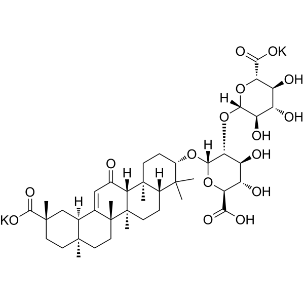
C[C@]12CC[C@](C[C@H]1C3=CC(=O)[C@@H]4[C@]5(CC[C@@H](C([C@@H]5CC[C@]4([C@@]3(CC2)C)C)(C)C)O[C@@H]6[C@@H]([C@H]([C@@H]([C@H](O6)C(=O)[O-])O)O)O[C@H]7[C@@H]([C@H]([C@@H]([C@H](O7)C(=O)[O-])O)O)O)C)(C)C(=O)O.[K+].[K+]
|
| InChi Key |
BIVBRWYINDPWKA-VLQRKCJKSA-L
|
| InChi Code |
InChI=1S/C42H62O16.2K/c1-37(2)21-8-11-42(7)31(20(43)16-18-19-17-39(4,36(53)54)13-12-38(19,3)14-15-41(18,42)6)40(21,5)10-9-22(37)55-35-30(26(47)25(46)29(57-35)33(51)52)58-34-27(48)23(44)24(45)28(56-34)32(49)50;;/h16,19,21-31,34-35,44-48H,8-15,17H2,1-7H3,(H,49,50)(H,51,52)(H,53,54);;/q;2*+1/p-2/t19-,21-,22-,23-,24-,25-,26-,27+,28-,29-,30+,31+,34-,35-,38+,39-,40-,41+,42+;;/m0../s1
|
| 化学名 |
dipotassium;(2S,3S,4S,5R,6R)-6-[(2S,3R,4S,5S,6S)-2-[[(3S,4aR,6aR,6bS,8aS,11S,12aR,14aR,14bS)-11-carboxy-4,4,6a,6b,8a,11,14b-heptamethyl-14-oxo-2,3,4a,5,6,7,8,9,10,12,12a,14a-dodecahydro-1H-picen-3-yl]oxy]-6-carboxylato-4,5-dihydroxyoxan-3-yl]oxy-3,4,5-trihydroxyoxane-2-carboxylate
|
| 别名 |
Dipotassium glycyrrhizinate; Glycyrrhizinate dipotassium; 68797-35-3; Dipotassium glycyrrhizate; CHEBI:79402; CA2Y0FE3FX; Neubormitin; Glycyrrhizic acid dipotassium;
|
| HS Tariff Code |
2934.99.9001
|
| 存储方式 |
Powder -20°C 3 years 4°C 2 years In solvent -80°C 6 months -20°C 1 month 注意: 请将本产品存放在密封且受保护的环境中,避免吸湿/受潮。 |
| 运输条件 |
Room temperature (This product is stable at ambient temperature for a few days during ordinary shipping and time spent in Customs)
|
| 溶解度 (体外实验) |
H2O : ~50 mg/mL (~55.61 mM)
DMSO : ~20.83 mg/mL (~23.17 mM) |
|---|---|
| 溶解度 (体内实验) |
配方 1 中的溶解度: 100 mg/mL (111.22 mM) in PBS (这些助溶剂从左到右依次添加,逐一添加), 澄清溶液; 超声助溶。
请根据您的实验动物和给药方式选择适当的溶解配方/方案: 1、请先配制澄清的储备液(如:用DMSO配置50 或 100 mg/mL母液(储备液)); 2、取适量母液,按从左到右的顺序依次添加助溶剂,澄清后再加入下一助溶剂。以 下列配方为例说明 (注意此配方只用于说明,并不一定代表此产品 的实际溶解配方): 10% DMSO → 40% PEG300 → 5% Tween-80 → 45% ddH2O (或 saline); 假设最终工作液的体积为 1 mL, 浓度为5 mg/mL: 取 100 μL 50 mg/mL 的澄清 DMSO 储备液加到 400 μL PEG300 中,混合均匀/澄清;向上述体系中加入50 μL Tween-80,混合均匀/澄清;然后继续加入450 μL ddH2O (或 saline)定容至 1 mL; 3、溶剂前显示的百分比是指该溶剂在最终溶液/工作液中的体积所占比例; 4、 如产品在配制过程中出现沉淀/析出,可通过加热(≤50℃)或超声的方式助溶; 5、为保证最佳实验结果,工作液请现配现用! 6、如不确定怎么将母液配置成体内动物实验的工作液,请查看说明书或联系我们; 7、 以上所有助溶剂都可在 Invivochem.cn网站购买。 |
| 制备储备液 | 1 mg | 5 mg | 10 mg | |
| 1 mM | 1.1122 mL | 5.5611 mL | 11.1221 mL | |
| 5 mM | 0.2224 mL | 1.1122 mL | 2.2244 mL | |
| 10 mM | 0.1112 mL | 0.5561 mL | 1.1122 mL |
1、根据实验需要选择合适的溶剂配制储备液 (母液):对于大多数产品,InvivoChem推荐用DMSO配置母液 (比如:5、10、20mM或者10、20、50 mg/mL浓度),个别水溶性高的产品可直接溶于水。产品在DMSO 、水或其他溶剂中的具体溶解度详见上”溶解度 (体外)”部分;
2、如果您找不到您想要的溶解度信息,或者很难将产品溶解在溶液中,请联系我们;
3、建议使用下列计算器进行相关计算(摩尔浓度计算器、稀释计算器、分子量计算器、重组计算器等);
4、母液配好之后,将其分装到常规用量,并储存在-20°C或-80°C,尽量减少反复冻融循环。
计算结果:
工作液浓度: mg/mL;
DMSO母液配制方法: mg 药物溶于 μL DMSO溶液(母液浓度 mg/mL)。如该浓度超过该批次药物DMSO溶解度,请首先与我们联系。
体内配方配制方法:取 μL DMSO母液,加入 μL PEG300,混匀澄清后加入μL Tween 80,混匀澄清后加入 μL ddH2O,混匀澄清。
(1) 请确保溶液澄清之后,再加入下一种溶剂 (助溶剂) 。可利用涡旋、超声或水浴加热等方法助溶;
(2) 一定要按顺序加入溶剂 (助溶剂) 。
| NCT Number | Recruitment | interventions | Conditions | Sponsor/Collaborators | Start Date | Phases |
| NCT04028869 | Completed | Drug: Glycyrrhizic acid preparation | Autoimmune Liver Disease | Beijing Ditan Hospital | March 1, 2018 | |
| NCT04742660 | Completed | Drug: Ammonium Glycyrrhizinate | Postoperative Nausea | Konkuk University Medical Center | May 11, 2021 | Not Applicable |
| NCT05895773 | Completed | Drug: Povidone-Iodine Drug: Saline spray |
Ventilator Associated Pneumonia | Menoufia University | June 24, 2023 | Phase 2 Phase 3 |
| NCT05788705 | Not yet recruiting | Dietary Supplement: "apigenin" and "glycyrrhizin" |
Rheumatoid Arthritis | Adel A.Gomaa | July 2023 | Not Applicable |
InvivoChem的所有产品仅用于作科学研究,不面向患者销售
Copyright 2020 InvivoChem LLC | All Rights Reserved 粤ICP备20063088号-1


 463611831
