| 规格 | 价格 | 库存 | 数量 |
|---|---|---|---|
| 10 mM * 1 mL in DMSO |
|
||
| 1mg |
|
||
| 5mg |
|
||
| 10mg |
|
||
| 25mg |
|
||
| 50mg |
|
||
| 100mg |
|
||
| 250mg |
|
||
| 500mg |
|
||
| Other Sizes |
|

| 靶点 |
Ferroptosis (EC50 = 60 nM)
Ferrostatin-1 (Fer-1) targets ferroptosis, a non-apoptotic cell death pathway driven by iron-dependent lipid peroxidation. It inhibits lipid radical propagation, with an EC50 of 60 nM for protecting HT-1080 cells from erastin-induced ferroptosis [2] - Ferrostatin-1 (Fer-1) exhibits antifungal activity by inhibiting fungal lipid peroxidation, with minimum inhibitory concentrations (MICs) of 2 μg/mL against Candida albicans and 4 μg/mL against Aspergillus fumigatus [3] |
|---|---|
| 体外研究 (In Vitro) |
Ferrostatin-1 抑制由erastin 产生的脂质和胞质ROS 的积累。在器官型大鼠脑切片中,ferrostatin-1 可抑制谷氨酸引起的神经毒性 [1]。 Ferrostatin-1(2 μM;24 小时)可保护大鼠器官型海马切片培养物 (OHSC) 免受谷氨酸 (5 mM) 诱导的神经毒性 [2]。 Ferrostatin-1 抑制脂质过氧化,但不抑制溶酶体膜的通透性或线粒体中活性氧的产生 [2]。在肾衰竭、脑室周围白质软化 (PVL) 和亨廷顿病 (HD) 的细胞模型中,ferrostatin-1 可减少细胞死亡 [2]。在 HT-1080 细胞中,ferrostatin-1(1 μM;6 小时)可防止不饱和脂肪酸氧化降解,从而增加健康中型多棘神经元 (MSN) 的数量 [3]。
HT-1080纤维肉瘤细胞铁死亡抑制:erastin(10 μM,铁死亡诱导剂)处理前1小时加入Fer-1(10-1000 nM),可显著提高细胞活力。60 nM时,细胞活力从erastin单独处理组的22%升至85%(MTT法);C11-BODIPY染色显示,100 nM Fer-1使erastin诱导的脂质过氧化减少72%(流式细胞术检测)[2] - 小鼠胚胎成纤维细胞(MEF)实验:Fer-1(50-500 nM)保护MEF免受RSL3(GPX4抑制剂)诱导的铁死亡。200 nM时,丙二醛(MDA,脂质过氧化标志物)水平降低65%(TBARS法),谷胱甘肽(GSH)水平得以维持(仅降低30%,而RSL3单独组降低75%)[2] - 抗真菌活性:Fer-1(0.5-16 μg/mL)抑制病原真菌生长。对白色念珠菌(ATCC 90028)的MIC为2 μg/mL,烟曲霉(ATCC 204305)为4 μg/mL,新型隐球菌(ATCC 24067)为8 μg/mL;4 μg/mL Fer-1使白色念珠菌的MDA水平降低58%[3] - LPS诱导BEAS-2B(人支气管上皮细胞)损伤:Fer-1(1-10 μM)预处理减少LPS(1 μg/mL)诱导的细胞死亡。5 μM时,细胞活力从52%升至83%,TNF-α mRNA表达降低62%(qPCR),脂质活性氧(ROS)水平降低55%(DCFH-DA染色)[4] - 缺氧缺血(HI)诱导新生大鼠原代皮质神经元损伤:Fer-1(0.1-1 μM)提高神经元活力。0.5 μM时,乳酸脱氢酶(LDH)释放减少48%(LDH法),GPX4蛋白表达升高2.1倍(Western blot)[6] |
| 体内研究 (In Vivo) |
在横纹肌溶解症小鼠中,Ferrostatin-1(5 mg/kg;腹膜内注射;单剂量,注射甘油前 30 分钟给药)可改善肾功能;然而,这种益处在缺乏泛半胱天冬酶抑制剂 zVAD 或 RIPK3 的小鼠中并未显示出来。 Frostatin-1(0.8 mg/kg;尾静脉注射)可以有效治疗 LPS 引起的急性肺损伤(ALI)[4]。 Ferrostatin-1(5 mg/kg;腹腔注射;C57BL/6J 小鼠)可改善横纹肌溶解症小鼠的肾功能 [5]。
研究结果显示,Ferrostatin-1(Fer-1)显著减轻了由缺氧和缺血引发的脑损伤。Fer-1显著增加SLC3A2、SLC7A11、ACSL3、GSS和GPX4的表达(P<0.05),显著降低GFAP、ACSL4、TFRC、FHC、FLC、4-HNE、HIF-1α和ROS的表达(P<0.05)。 结论:Fer-1通过潜在靶向GPX4/ACL3/ACSL4轴抑制铁下垂并缓解HIBD;然而,其具体机制值得进一步探索。[6] 绝经后口干症的机制尚未完全阐明。本研究旨在探讨绝经后动物模型中口干症的机制以及铁蛋白抑制剂去铁胺(DFO)和Ferrostatin-1(FER)对唾液腺功能障碍的影响。24只雌性Sprague-Dawley大鼠随机分为四组:SHAM组(n=6,假手术大鼠)、OVX组(n=5,去卵巢大鼠),FER组(n=7,切除卵巢大鼠腹腔注射FER)和DFO组(n=8,切除卵巢的大鼠腹腔内注射DFO)。分析GPX4活性、铁积累、脂质过氧化、炎症、纤维化和唾液腺功能。DFO组GPX4活性恢复,铁积累和细胞质MDA+HAE减少。此外,与OVX组相比,DFO组的I型胶原、III型胶原、TGF-β、IL-6、TNF-α和TGF-β水平降低。FER组还观察到GPX4活性和线粒体形态的恢复,以及细胞质MDA+HAE的减少。此外,在DFO和FER组中观察到炎性细胞因子和纤维化标志物的表达降低,AQP5的表达增加。绝经后唾液腺功能障碍与铁下垂有关,DFO和FER可能逆转绝经后的唾液腺功能障碍。因此,DFO和FER被认为是治疗绝经后口干症的有前景的方法。[7] 小鼠LPS诱导急性肺损伤(ALI)模型:6-8周龄雄性C57BL/6小鼠气管滴注LPS(5 mg/kg)诱导ALI,LPS处理前1小时腹腔注射Fer-1(5 mg/kg)可显著缓解损伤:肺湿重/干重比(水肿指标)从5.8降至3.2,支气管肺泡灌洗液(BALF)中TNF-α和IL-6水平分别降低55%和60%,肺组织MDA水平降低48%;组织病理学显示肺泡出血和炎症细胞浸润减少[4] - 新生大鼠缺氧缺血性脑损伤(HIBD)模型:7日龄(P7)SD大鼠行右侧颈总动脉结扎+2小时缺氧(8% O₂)处理,缺氧后立即腹腔注射Fer-1(10 mg/kg),72小时后脑梗死体积减少42%(TTC染色);神经行为评分改善(转棒实验:120秒 vs. 65秒;握力:假手术组的85% vs. 52%),脑脂质ROS水平降低58%[6] - 去卵巢(OVX)大鼠唾液腺功能障碍模型:8周龄雌性SD大鼠行双侧卵巢切除术,Fer-1(5 mg/kg/天,皮下注射)处理4周,唾液流量增加45%(从0.5 mL/10分钟升至0.72 mL/10分钟),唾液腺MDA水平降低52%,超氧化物歧化酶(SOD)活性升高2.3倍;组织病理学显示腺泡细胞结构保留(萎缩率从40%降至15%)[7] |
| 酶活实验 |
蛋白质印迹[4]
在我们的研究中,使用放射免疫沉淀分析裂解缓冲液裂解细胞样品,并使用Pierce BCA蛋白质分析试剂盒检测不同组的总蛋白质浓度。在我们的研究中,细胞裂解物(20 μg/泳道)用10%SDS-PAGE凝胶分离,然后转移到硝化纤维膜上。用在PBS中稀释的5%脱脂奶粉封闭膜,并进一步与一级抗体在4 °C。在此,使用的不同一级抗体是:抗SLC7A11(1:3000;细胞信号,类别号:12691)、抗GPX4(1:1000)、抗FTH(1:2000)和抗GAPDH(1:3000)。所用的第二抗体是:抗小鼠IgG(HRP缀合;1:5000)和抗兔IgG(HRP-缀合;1:10000)。最后,使用SuperSignal West Femto Maximum Sensitivity Substrate和ChemiDoc Images对每条泳道中的蛋白质带进行可视化。最后使用ImageJ1.x软件对结果进行量化。整个论文中图像的所有未切割的原始印迹如补充图所示。1。[4] 丙二醛(MDA)、4-羟基壬烯醛(4-HNE)和铁水平的评估[4] 在我们的研究中,为了评估不同组的脱铁水平,检测各组的MDA、4-HNE和铁水平。细胞裂解物中的MDA浓度、4-HNE浓度和铁浓度根据制造商的说明使用脂质过氧化(MDA)测定试剂盒、脂质过氧化测定试剂盒和铁测定试剂盒进行评估。 脂质过氧化抑制实验(TBARS法):取100 μL细胞或组织匀浆,与200 μL硫代巴比妥酸(TBA)试剂(0.67% TBA溶于50%冰醋酸)混合,95°C加热30分钟,冰浴冷却后3000×g离心10分钟,检测上清液532 nm吸光度。以1,1,3,3-四甲氧基丙烷为标准品制作标准曲线,计算MDA浓度。实验中在加入TBA试剂前加入Fer-1(100 nM-10 μM),评估其对脂质过氧化的抑制作用[2,4,6] - GPX4活性实验:取50 μg组织匀浆蛋白,与反应缓冲液(50 mM Tris-HCl pH 7.6、1 mM GSH、0.2 mM H₂O₂、0.1 mM NADPH)混合,检测340 nm吸光度变化(反映NADPH氧化)5分钟,GPX4活性以每毫克蛋白每分钟氧化的NADPH纳摩尔数计算。实验中Fer-1(0.5-5 μM)与匀浆预孵育30分钟,评估其对GPX4活性的影响[6] |
| 细胞实验 |
细胞活力测定[4]
为了评估细胞活力,我们的研究中使用CCK-8方法作为参考。简而言之,将BEAS-2B细胞以5的浓度接种到96孔板中 × 104个细胞/孔。细胞培养24小时 h、 然后用不同浓度的LPS和Fer-1处理16 h,然后添加20 μl CCK-8溶液直接加入培养基(200 μl/孔)并在37 °C 4 h.在450时检测不同组的吸光度(Abs) nm(n= 3.在空白组中,孔仅包含培养基,未经任何处理的细胞用作对照组。这里,细胞活力 = (实验组Abs空白组Abs)/(对照组Abs × 100%. HT-1080细胞铁死亡保护实验:HT-1080细胞以5×10³细胞/孔接种于96孔板,含10% FBS的DMEM培养24小时,erastin(10 μM)处理前1小时加入Fer-1(10-1000 nM)。24小时后加入20 μL MTT(5 mg/mL),孵育4小时后DMSO溶解甲瓒,检测570 nm吸光度计算细胞活力[2] - C11-BODIPY脂质过氧化染色:6孔板中2×10⁵细胞/孔的HT-1080细胞,用Fer-1(100 nM)+erastin(10 μM)处理12小时,37°C下5 μM C11-BODIPY(脂质ROS探针)染色30分钟,PBS洗涤后流式细胞术分析(激发488 nm,非氧化探针发射515 nm,氧化探针发射580 nm)[2] - 真菌MIC实验:真菌在含2%葡萄糖的RPMI 1640培养基中培养至对数期,96孔板中加入Fer-1(0.5-16 μg/mL),再加入真菌悬液(1×10⁴ CFU/孔),35°C孵育(念珠菌24-48小时,曲霉菌72小时)。MIC定义为抑制≥90%真菌生长的最低Fer-1浓度(600 nm吸光度检测)[3] - 新生大鼠原代皮质神经元HI损伤实验:从P1-P3 SD大鼠分离皮质神经元,含B27添加剂的Neurobasal培养基培养7天,氧糖剥夺(OGD:1% O₂、无糖培养基)2小时后复氧,复氧时加入Fer-1(0.1-1 μM)。24小时后LDH法检测LDH释放,Western blot检测GPX4蛋白[6] |
| 动物实验 |
动物/疾病模型:雄性C57BL/6小鼠(LPS诱导的ALI)[4]
剂量:0.8 mg/kg 给药途径:尾静脉注射 实验结果:对LPS诱导的ALI具有治疗作用。 在本研究中,雄性C57BL/6小鼠被随机分为4组(每组n=4,8-10周龄,体重=23-25 g):对照组接受0.9% NaCl(含0.1% DMSO),LPS组接受LPS+0.9% NaCl(含0.1% DMSO),Fer-1组仅接受Fer-1,LPS+Fer-1组同时接受Fer-1和LPS。通过气管内滴注50 μl LPS溶液(0.2 g/L)建立LPS诱导的急性肺损伤(ALI)模型,随后经尾静脉注射Fer-1(0.8 mg/kg)。Fer-1先溶于DMSO,再用0.9% NaCl溶液稀释,最终Fer-1和DMSO的浓度分别为0.2 mg/ml和0.1%。治疗16小时后,处死各组小鼠,并通过肺灌洗收集支气管肺泡灌洗液(BALF)。为分析BALF细胞分类计数,使用Cytospin 4浓缩细胞,并使用Shandon Kwik-Diff试剂盒进行细胞染色。此外,根据制造商的说明,使用Pierce BCA蛋白测定试剂盒、IL-6 ELISA试剂盒和TNF-α ELISA试剂盒检测各样本中的总蛋白浓度以及IL-6和TNF-α的水平。收集不同组的肺组织进行qPCR和Western blot检测,部分肺组织用10%中性缓冲福尔马林固定,然后包埋于石蜡中进行组织学分析,作为参考。本研究采用0-4分的评分系统评估肺损伤程度。 小鼠LPS诱导的急性肺损伤模型:雄性C57BL/6小鼠(6-8周龄,20-22 g)饲养于SPF级条件下(22±2℃,12小时光照/黑暗循环)。小鼠被随机分为3组(每组n=8): 1. 假手术组:气管内滴注生理盐水+腹腔注射生理盐水(10 mL/kg); 2. 仅LPS组:气管内滴注LPS(5 mg/kg,溶于生理盐水)+腹腔注射生理盐水; 3. LPS+Fer-1组:LPS滴注前1小时腹腔注射Fer-1(5 mg/kg,溶于0.1% DMSO + 生理盐水)。 LPS滴注24小时后,处死小鼠。切除肺脏:取一个肺叶测定湿重/干重比,另一个肺叶匀浆用于MDA测定,收集支气管肺泡灌洗液(BALF)用于检测TNF-α/IL-6水平(ELISA)[4] - 新生大鼠缺氧缺血性脑病(HIBD)模型:将P7 Sprague-Dawley大鼠(10-12 g)用异氟烷麻醉。用6-0丝线结扎右侧颈总动脉,然后将大鼠置于低氧舱(8% O₂,92% N₂)中2小时。大鼠被随机分为3组(每组n=10): 1. 假手术组:假结扎+常氧+腹腔注射生理盐水; 2. 仅缺氧组:结扎+缺氧+腹腔注射生理盐水; 3. 缺氧+Fer-1组:缺氧后立即腹腔注射Fer-1(10 mg/kg,溶于0.1% DMSO + 生理盐水)。 缺氧72小时后,处死大鼠:取脑组织进行TTC染色(梗死体积)和脂质ROS测定;在安乐死前 24 小时进行神经行为学测试(转棒测试、握力测试)[6] - OVX 大鼠唾液腺功能障碍模型:雌性 Sprague-Dawley 大鼠(8 周龄,220-250 克)用戊巴比妥钠麻醉。进行双侧卵巢切除术(假手术组:仅开腹)。大鼠被随机分为3组(每组n=6): 1. 假手术组:假手术+皮下注射生理盐水; 2. 卵巢切除组:卵巢切除+皮下注射生理盐水; 3. 卵巢切除+Fer-1组:卵巢切除+皮下注射Fer-1(5 mg/kg/天,溶于0.1% DMSO + 生理盐水)。 4周后,测量唾液流率(腹腔注射毛果芸香碱5 mg/kg刺激)。切除唾液腺:一部分用于MDA/SOD测定,另一部分用4%多聚甲醛固定,用于组织病理学检查(H&E染色)[7] |
| 毒性/毒理 (Toxicokinetics/TK) |
急性体外毒性:在HT-1080、MEF和BEAS-2B细胞中,用Fer-1(0.1-10 μM)处理48小时后,未观察到细胞毒性——细胞活力保持在90%以上(MTT/CCK-8检测)[2,4]
- 急性体内毒性:用Fer-1(5-10 mg/kg,腹腔/皮下注射)处理小鼠/大鼠1-4周后,未观察到异常行为(例如嗜睡、腹泻)、体重减轻(<5%的基线水平)或血清ALT、AST、BUN或肌酐水平的变化。肝脏、肾脏和靶器官(肺、脑、唾液腺)的组织病理学检查未发现组织损伤[4,6,7] |
| 参考文献 | |
| 其他信息 |
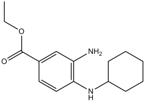
Ferrostatin-1 是一种乙酯,由 3-氨基-4-(环己基氨基)苯甲酸的羧基与乙醇缩合而成。它是一种强效的铁死亡抑制剂,铁死亡是一种独特的非凋亡性细胞死亡形式,由脂质过氧化引起。它也是一种自由基清除抗氧化剂,能够减少脂质过氧化物和链式过氧自由基的积累。它具有多种功能,包括作为铁死亡抑制剂、辐射防护剂、抗氧化剂、自由基清除剂、抗真菌剂和神经保护剂。它是一种取代苯胺、乙酯和伯芳胺。
背景:铁死亡是一种新近发现的细胞死亡类型,不同于传统的坏死、凋亡或自噬性细胞死亡。然而,铁死亡在脂多糖(LPS)诱导的急性肺损伤(ALI)中的作用尚未得到深入研究。本研究主要分析了铁死亡与LPS诱导的ALI之间的关系。方法:本研究采用LPS和铁死亡抑制剂ferrostatin-1(Fer-1)处理人支气管上皮细胞系BEAS-2B,并使用CCK-8法检测细胞活力。此外,还检测了不同组别中丙二醛(MDA)、4-羟基壬烯醛(4-HNE)和铁的含量,以及SLC7A11和GPX4的蛋白表达水平。为了进一步验证体外实验结果,我们构建了LPS诱导的小鼠ALI模型,并评估了Fer-1的治疗效果以及对肺组织中铁死亡水平的影响。结果:LPS处理可下调BEAS-2B细胞的活力,同时降低铁死亡标志物SLC7A11和GPX4的表达;LPS处理呈剂量依赖性地增加MDA、4-HNE和总铁的水平,而Fer-1可逆转这些变化。体内实验结果也表明,Fer-1对LPS诱导的急性肺损伤(ALI)具有治疗作用,并可下调肺组织中的铁死亡水平。结论:本研究表明,铁死亡在LPS诱导的ALI进展中起着重要作用,铁死亡可能成为ALI患者治疗的新靶点。[4] 背景:缺氧缺血性脑损伤(HIBD)是由围产期窒息引起的脑损伤,严重损害中枢神经系统。目前尚无有效药物治疗该疾病,且HIBD的发病机制仍不清楚。虽然研究表明铁死亡在缺氧缺血性脑损伤(HIBD)中发挥重要作用,但其在HIBD中的作用和机制尚未完全阐明。方法:采用Rice-Vannucci法建立新生大鼠HIBD模型。将PC12细胞的完全培养基调整为低糖培养基,连续缺氧12小时后建立氧糖剥夺模型。采用激光多普勒血流成像技术检测造模后的血流强度。采用2,3,5-氯化三苯基四氮唑染色检测大鼠脑组织中的缺血性脑梗死,并采用苏木精-伊红染色和透射电镜观察脑损伤和线粒体损伤。采用免疫荧光法监测GFAP的表达。采用实时定量PCR、Western blot和免疫荧光法检测mRNA和蛋白质的表达。使用活性氧(ROS)检测试剂盒检测细胞内ROS水平。结果:结果表明,铁死亡抑制剂-1(Fer-1)显著减轻了缺氧缺血引起的脑损伤。Fer-1显著上调了SLC3A2、SLC7A11、ACSL3、GSS和GPX4的表达(P<0.05),并显著下调了GFAP、ACSL4、TFRC、FHC、FLC、4-HNE、HIF-1α和ROS的表达(P<0.05)。结论:Fer-1可能通过靶向GPX4/ACSL3/ACSL4轴抑制铁死亡并减轻缺氧缺血性脑损伤(HIBD);然而,其具体机制尚需进一步研究。[6] 绝经后口干症的机制尚未完全阐明。本研究旨在探讨绝经后动物模型中口干症的机制以及铁死亡抑制剂去铁胺(DFO)和铁死亡抑制剂-1(FER)对唾液腺功能障碍的影响。24只雌性Sprague-Dawley大鼠被随机分为四组:假手术组(n = 6,假手术大鼠)、卵巢切除组(n = 6,卵巢切除大鼠)、FER组(n = 6,卵巢切除后腹腔注射FER)和DFO组(n = 6,卵巢切除后腹腔注射DFO)。分析了GPX4活性、铁积累、脂质过氧化、炎症、纤维化和唾液腺功能。结果显示,DFO组大鼠的GPX4活性恢复,铁积累和胞质MDA+HAE水平降低。此外,与卵巢切除组(OVX组)相比,去铁胺组(DFO组)中I型胶原蛋白、III型胶原蛋白、TGF-β、IL-6、TNF-α和TGF-β水平降低。铁胺治疗组(FER组)中还观察到GPX4活性和线粒体形态的恢复,以及胞质MDA+HAE的减少。此外,DFO组和FER组均观察到炎症细胞因子和纤维化标志物的表达降低,以及AQP5表达升高。绝经后唾液腺功能障碍与铁死亡相关,DFO和FER可能逆转绝经后唾液腺功能障碍。因此,DFO 和 FER 被认为是治疗绝经后口干症的有效方法。[7] 作用机制:铁抑制剂-1 (Fer-1)通过清除脂质过氧自由基 (LOO•) 并阻止铁依赖性脂质过氧化链式反应来抑制铁死亡。它不直接螯合铁或激活 GPX4,而是通过降低脂质 ROS 水平来维持 GPX4 的功能。[2,4,6] - 研究应用:Fer-1是一种广泛用于体外和体内研究铁死亡的工具化合物。它已被应用于研究铁死亡相关疾病,包括急性器官损伤(肺、脑)、神经退行性疾病和癌症(作为正常细胞的潜在化学保护剂)[2,4,6] - 抗真菌机制:与传统抗真菌药物(例如唑类)不同,Fer-1通过抑制脂质过氧化作用靶向真菌脂质代谢,从而降低真菌细胞膜的完整性并抑制其生长。它与氟康唑联用对耐氟康唑的白色念珠菌具有协同作用(FICI = 0.5)[3] - 局限性:Fer-1水溶性差(需要DMSO溶解),主要用于临床前研究;尚未在临床试验中进行评估,也未获得FDA批准用于治疗用途[2,4,6,7] |
| 分子式 |
C15H22N2O2
|
|
|---|---|---|
| 分子量 |
262.35
|
|
| 精确质量 |
262.168
|
|
| 元素分析 |
C, 68.67; H, 8.45; N, 10.68; O, 12.20
|
|
| CAS号 |
347174-05-4
|
|
| 相关CAS号 |
|
|
| PubChem CID |
4068248
|
|
| 外观&性状 |
Gray to gray purple solid
|
|
| 密度 |
1.1±0.1 g/cm3
|
|
| 沸点 |
437.3±35.0 °C at 760 mmHg
|
|
| 闪点 |
218.3±25.9 °C
|
|
| 蒸汽压 |
0.0±1.1 mmHg at 25°C
|
|
| 折射率 |
1.595
|
|
| LogP |
3.9
|
|
| tPSA |
64.35
|
|
| 氢键供体(HBD)数目 |
2
|
|
| 氢键受体(HBA)数目 |
4
|
|
| 可旋转键数目(RBC) |
5
|
|
| 重原子数目 |
19
|
|
| 分子复杂度/Complexity |
290
|
|
| 定义原子立体中心数目 |
0
|
|
| SMILES |
O(C([H])([H])C([H])([H])[H])C(C1C([H])=C([H])C(=C(C=1[H])N([H])[H])N([H])C1([H])C([H])([H])C([H])([H])C([H])([H])C([H])([H])C1([H])[H])=O
|
|
| InChi Key |
UJHBVMHOBZBWMX-UHFFFAOYSA-N
|
|
| InChi Code |
InChI=1S/C15H22N2O2/c1-2-19-15(18)11-8-9-14(13(16)10-11)17-12-6-4-3-5-7-12/h8-10,12,17H,2-7,16H2,1H3
|
|
| 化学名 |
3-amino-4-(cyclohexylamino)-benzoic acid, ethyl ester
|
|
| 别名 |
Frer-1; 3-amino-4-(cyclohexylamino)-benzoic acid, ethyl ester; Ferrostatin-1; 347174-05-4; Ethyl 3-amino-4-(cyclohexylamino)benzoate; Fer-1; Ferrostatin-1 (Fer-1); Ferrostatin 1; ferrrostatin 1; MFCD08072959;
|
|
| HS Tariff Code |
2934.99.9001
|
|
| 存储方式 |
Powder -20°C 3 years 4°C 2 years In solvent -80°C 6 months -20°C 1 month 注意: 本产品在运输和储存过程中需避光。 |
|
| 运输条件 |
Room temperature (This product is stable at ambient temperature for a few days during ordinary shipping and time spent in Customs)
|
| 溶解度 (体外实验) |
|
|||
|---|---|---|---|---|
| 溶解度 (体内实验) |
配方 1 中的溶解度: ≥ 2.5 mg/mL (9.53 mM) (饱和度未知) in 10% DMSO + 40% PEG300 + 5% Tween80 + 45% Saline (这些助溶剂从左到右依次添加,逐一添加), 澄清溶液。
例如,若需制备1 mL的工作液,可将100 μL 25.0 mg/mL澄清DMSO储备液加入到400 μL PEG300中,混匀;然后向上述溶液中加入50 μL Tween-80,混匀;加入450 μL生理盐水定容至1 mL。 *生理盐水的制备:将 0.9 g 氯化钠溶解在 100 mL ddH₂O中,得到澄清溶液。 配方 2 中的溶解度: 2.5 mg/mL (9.53 mM) in 10% DMSO + 90% Corn Oil (这些助溶剂从左到右依次添加,逐一添加), 悬浊液; 超声助溶。 例如,若需制备1 mL的工作液,可将 100 μL 25.0 mg/mL 澄清 DMSO 储备液加入到 900 μL 玉米油中并混合均匀。 View More
配方 3 中的溶解度: ≥ 2.08 mg/mL (7.93 mM) (饱和度未知) in 10% DMSO + 90% (20% SBE-β-CD in Saline) (这些助溶剂从左到右依次添加,逐一添加), 澄清溶液。 配方 4 中的溶解度: 0.2 mg/mL (0.76 mM) in 10% DMSO + 90% Saline (这些助溶剂从左到右依次添加,逐一添加), 澄清溶液; 超声助溶. *生理盐水的制备:将 0.9 g 氯化钠溶解在 100 mL ddH₂O中,得到澄清溶液。 配方 5 中的溶解度: 2% DMSO+50% PEG 300+5% Tween 80+ddH2O: 5mg/mL 1、请先配制澄清的储备液(如:用DMSO配置50 或 100 mg/mL母液(储备液)); 2、取适量母液,按从左到右的顺序依次添加助溶剂,澄清后再加入下一助溶剂。以 下列配方为例说明 (注意此配方只用于说明,并不一定代表此产品 的实际溶解配方): 10% DMSO → 40% PEG300 → 5% Tween-80 → 45% ddH2O (或 saline); 假设最终工作液的体积为 1 mL, 浓度为5 mg/mL: 取 100 μL 50 mg/mL 的澄清 DMSO 储备液加到 400 μL PEG300 中,混合均匀/澄清;向上述体系中加入50 μL Tween-80,混合均匀/澄清;然后继续加入450 μL ddH2O (或 saline)定容至 1 mL; 3、溶剂前显示的百分比是指该溶剂在最终溶液/工作液中的体积所占比例; 4、 如产品在配制过程中出现沉淀/析出,可通过加热(≤50℃)或超声的方式助溶; 5、为保证最佳实验结果,工作液请现配现用! 6、如不确定怎么将母液配置成体内动物实验的工作液,请查看说明书或联系我们; 7、 以上所有助溶剂都可在 Invivochem.cn网站购买。 |
| 制备储备液 | 1 mg | 5 mg | 10 mg | |
| 1 mM | 3.8117 mL | 19.0585 mL | 38.1170 mL | |
| 5 mM | 0.7623 mL | 3.8117 mL | 7.6234 mL | |
| 10 mM | 0.3812 mL | 1.9059 mL | 3.8117 mL |
1、根据实验需要选择合适的溶剂配制储备液 (母液):对于大多数产品,InvivoChem推荐用DMSO配置母液 (比如:5、10、20mM或者10、20、50 mg/mL浓度),个别水溶性高的产品可直接溶于水。产品在DMSO 、水或其他溶剂中的具体溶解度详见上”溶解度 (体外)”部分;
2、如果您找不到您想要的溶解度信息,或者很难将产品溶解在溶液中,请联系我们;
3、建议使用下列计算器进行相关计算(摩尔浓度计算器、稀释计算器、分子量计算器、重组计算器等);
4、母液配好之后,将其分装到常规用量,并储存在-20°C或-80°C,尽量减少反复冻融循环。
计算结果:
工作液浓度: mg/mL;
DMSO母液配制方法: mg 药物溶于 μL DMSO溶液(母液浓度 mg/mL)。如该浓度超过该批次药物DMSO溶解度,请首先与我们联系。
体内配方配制方法:取 μL DMSO母液,加入 μL PEG300,混匀澄清后加入μL Tween 80,混匀澄清后加入 μL ddH2O,混匀澄清。
(1) 请确保溶液澄清之后,再加入下一种溶剂 (助溶剂) 。可利用涡旋、超声或水浴加热等方法助溶;
(2) 一定要按顺序加入溶剂 (助溶剂) 。
|
|
|
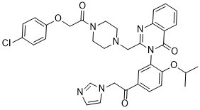 Imidazole ketone erastin
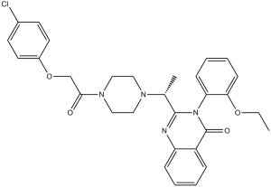 爱拉斯汀
爱拉斯汀
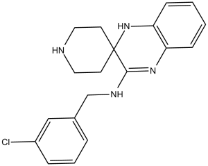 利普司他丁-1
InvivoChem的所有产品仅用于作科学研究,不面向患者销售
Copyright 2020 InvivoChem LLC | All Rights Reserved 粤ICP备20063088号-1


 COA
COA


 463611831
