| 规格 | 价格 | 库存 | 数量 |
|---|---|---|---|
| 10 mM * 1 mL in DMSO |
|
||
| 5mg |
|
||
| 10mg |
|
||
| 50mg |
|
||
| 100mg |
|
||
| 500mg |
|
||
| 1g |
|
||
| 5g |
|
||
| 10g |
|
||
| Other Sizes |
|

| 靶点 |
Tyr1173 (IC50 = 26 nM); Tyr1173 (IC50 = 37 nM); Tyr992 (IC50 = 37 nM); Tyr992 (IC50 = 57 nM)
|
|---|---|
| 体外研究 (In Vitro) |
体外活性:吉非替尼可有效抑制高表达和低表达 EGFR 的细胞系(包括 NR6、NR6M 和 NR6W 细胞系)中 EGFR 上的所有酪氨酸磷酸化位点。磷酸化位点 Tyr1173 和 Tyr992 不太敏感,需要更高浓度的吉非替尼进行抑制。吉非替尼在 NR6W 细胞中有效阻断 PLC-γ 的磷酸化,IC50 为 27nM。 NR6wtEGFR 和 NR6M 细胞系具有低水平的 PLC-γ 磷酸化,但 NR6M 细胞系中的水平对吉非替尼的抑制具有更强的抵抗力,IC50 分别为 43 nM 和 369 nM。 Gefitinib 在低 EGFR 和 EGFRvIII 表达细胞系中抑制 Akt 磷酸化,IC50 分别为 220 和 263nM。剂量范围为 0.1 至 0.5μM 的吉非替尼显着促进而非消除 NR6M 细胞的集落形成。然而,浓度为 2 μM 的吉非替尼完全阻断 NR6M 集落形成。在高 EGFR 表达细胞系和低 EGFR 表达细胞系中,吉非替尼在 EGF 刺激后 72 小时内以剂量依赖性方式快速抑制 EGFR 和 ERK 磷酸化。吉非替尼是这些 EGF 驱动的未转化 MCF10A 细胞的单层生长,IC50 为 20 NM。与单独放疗相比,吉非替尼(0.2 μM 和 0.5 μM)与放疗组合可显着抑制 LoVo 细胞的生长。激酶测定:盐酸吉非替尼是一种特异性结合并抑制 EGFR 酪氨酸激酶的抑制剂,在 NR6wtEGFR 细胞中的 IC50 值为 2-37 nM。吉非替尼在 NR6W 细胞中有效阻断 PLC-γ 的磷酸化,IC50 为 27nM。 NR6wtEGFR 和 NR6M 细胞系具有低水平的 PLC-γ 磷酸化,但 NR6M 细胞系中的水平对吉非替尼的抑制具有更强的抵抗力,IC50 分别为 43 nM 和 369 nM。 Gefitinib 在低 EGFR 和 EGFRvIII 表达细胞系中抑制 Akt 磷酸化,IC50 分别为 220 和 263nM。细胞测定:将指数生长的细胞(包括 NR6、NR6M、NR6M 和 NR6W 细胞)以 2000 个细胞/孔的浓度接种在 96 孔板中,使其粘附,随后在 PBS 中洗涤,并在含有 0.5% FCS 的培养基中孵育过夜。然后用不同浓度 (0-2 μM) 的吉非替尼或溶质对照 DMSO 和 EGF 处理细胞。诱导 NR6wtEGFR 和 NR6W 细胞增殖的最佳 EGF 浓度先前已确定,因此 NR6wtEGFR 和 NR6W 细胞分别补充有 10 nM 和 0.1 nM EGF。 NR6 和 NR6M 细胞中未添加 EGF。 72小时后,通过进行MTT增殖测定来测量细胞数量。
表皮生长因子受体(EGFR)在许多人类肿瘤中经常被扩增和/或突变,并且来自该受体的异常信号被认为是导致这些肿瘤中出现恶性表型的原因。吉非替尼是一种特异性结合和抑制EGFR酪氨酸激酶的小分子抑制剂,已被证明可以抑制一系列过表达EGFR的肿瘤细胞的生长、增殖、存活和侵袭。然而,吉非替尼的临床反应与EGFR水平和活性没有相关性,这表明下游信号传导和突变等其他分子机制在预测临床反应方面可能很重要。因此,我们研究了特异性EGFR抑制剂吉非替尼对表达天然存在的组成型活性EGFR变体EGFRvIII、低非转化水平EGFR和高转化水平EGFR的细胞的磷酸化水平、信号传导和生长的影响。结果显示,在表达EGFRvIII的细胞中,吉非替尼足以抑制EGFR磷酸化、EGFR介导的增殖和EGFR-介导的锚定非依赖性生长的水平不足以抑制这些特征。此外,数据表明,表达EGFRvIII的细胞长期暴露于低浓度吉非替尼(0.01-0.1微M)会导致受体磷酸酪氨酸负荷增加,ERK信号传导增加,并刺激增殖和锚定非依赖性生长,可能是通过诱导EGFRvⅢ二聚化。另一方面,较高浓度的吉非替尼(1-2微M)显著降低了EGFRvIII磷酸酪氨酸负荷、EGFRvⅢ介导的增殖和锚定非依赖性生长。需要进一步的研究来调查这些重要发现在临床环境中的意义。[1] 表皮生长因子受体(EGFR)在许多人类肿瘤中普遍过表达,为抗癌药物开发提供了新的靶点。ZD1839(“易瑞沙”)是一种对EGFR具有选择性的喹唑啉酪氨酸激酶抑制剂,在临床前研究和临床试验的早期阶段显示出良好的活性。然而,由于尚不清楚哪种肿瘤类型是该药物治疗的最佳靶点,因此已经研究了与ZD1839肿瘤敏感性相关的分子特征。在一组人类乳腺癌症和其他上皮肿瘤细胞系中,HER2-过表达肿瘤对ZD1839特别敏感。这些肿瘤细胞系的生长抑制与EGFR、HER2和HER3的去磷酸化有关,同时HER3与磷脂酰肌醇3-激酶的结合丧失,Akt活性下调。这些研究表明,HER2过表达的肿瘤特别容易受到HER家族酪氨酸激酶信号传导的抑制,并提出了治疗这些特别侵袭性肿瘤的新策略。[2] 转化生长因子α(TGF-α)是人类癌症的一种自分泌生长因子。TGF-α及其特异性受体表皮生长因子受体(EGFR)的过表达与侵袭性疾病和预后不良有关。EGFR已被提出作为抗癌治疗的靶点。已经开发出阻断配体诱导的EGFR激活的化合物。ZD-1839(易瑞沙)是一种口服活性喹唑啉衍生物,选择性抑制EGFR酪氨酸激酶,目前正在癌症患者中进行临床开发。在共表达EGFR和TGF-α的人卵巢(OVCAR-3)、乳腺(ZR-75-1,MCF-10A ras)和癌症(GEO)细胞中,评价了ZD-1839单独或与作用机制不同的细胞毒性药物(如顺铂、卡铂、奥沙利铂、紫杉醇、多烯紫杉醇、阿霉素、依托泊苷、拓扑替康和雷替曲塞)组合的抗增殖活性。ZD-1839在所有癌症细胞系中以剂量依赖性方式抑制软琼脂中的菌落形成。抗增殖作用主要是抑制细胞生长。然而,高剂量治疗导致细胞凋亡增加2-4倍。当用每种细胞毒性药物和ZD-1839治疗癌症细胞时,观察到生长抑制的剂量依赖性超加性增加。联合治疗显著增强了单药治疗诱导的凋亡细胞死亡。[4] 生长因子受体EGFR和erbB2的过表达在几种人类癌症中经常发生,并与侵袭性肿瘤行为和患者预后不良有关。我们研究了新型EGFR酪氨酸激酶抑制剂ZD1839(Iressa)在体外和体内对表达不同水平EGFR和erbB2的人癌症细胞系生长的影响。ZD1839以低纳摩尔范围内的一半最大有效剂量有效抑制了EGFR过表达的A431和MDA-MB-231细胞在体外的增殖(50%-70%)。同时,ZD1839阻断了EGFR的自磷酸化,并阻止了EGF激活PLCγ1、ERK MAP激酶和PKB/Akt。它还抑制过表达erbB2的EGFR(+)癌症细胞系(SKBr3,SKOV3,BT474)增殖20%至80%,这种作用与抑制EGF-依赖性erbB2磷酸化和激活SKOV3细胞中ERK-MAP激酶和PKB/Akt有关。 总的来说,这些结果表明,在所研究的剂量下,ZD1839不仅在过表达EGFR的细胞中,而且在过表达erbB2的EGFR(+)细胞中都是一种强效的增殖抑制剂[6]。 |
| 体内研究 (In Vivo) |
吉非替尼 (100 mg/kg) 可改善 LoVo 肿瘤异种移植物放疗的抗肿瘤效果。吉非替尼对携带已建立的人 GEO 结肠癌异种移植物的裸鼠进行治疗,显示出对肿瘤生长的可逆剂量依赖性抑制,因为 GEO 肿瘤在治疗结束时恢复了对照的生长速度。
在体外和体内评估了ZD1839('ressa')对人肿瘤细胞(LoVo结直肠癌)辐射反应的影响,ZD1839是表皮生长因子受体酪氨酸激酶活性的特异性抑制剂。ZD1839(0.5μM,孵育第1-5天)显著增加了分次放射治疗(2 Gy,第1-3天)对体外生长的LoVo细胞的抗增殖作用(P=0.002)。与单独使用任何一种治疗方式相比,ZD1839在携带LoVo肿瘤异种移植物的小鼠中联合单次或分次放疗也显著增加了肿瘤生长抑制(Pс0.001)。当以分级方案给药时,ZD1839的放射性增强作用更为明显。这种现象可能归因于ZD1839对放射治疗组分之间肿瘤细胞再增殖的抗增殖作用。这些数据表明,使用ZD1839佐剂进行放射治疗可以增强治疗反应。因此,ZD1839联合放疗的临床研究是有必要的。[3] ZD-1839/吉非替尼对携带已建立的人GEO结肠癌癌症异种移植物的裸鼠的治疗显示了对肿瘤生长的可逆剂量依赖性抑制,因为GEO肿瘤在治疗结束时恢复了对照的生长速率。相比之下,使用细胞毒性药物(如拓扑替康、拉曲肽或紫杉醇)和ZD-1839的联合治疗在所有小鼠中都产生了肿瘤生长停滞。治疗结束后,肿瘤缓慢生长约4-8周,最终恢复到与对照组相似的生长速度。在注射GEO细胞后4-6周内,所有对照小鼠的GEO肿瘤达到与正常生活不相容的大小,在所有单药治疗的小鼠中,GEO肿瘤在6-8周内达到与正常生命不相容的尺寸。相反,在癌症细胞注射后10、12和15周,分别有50%的ZD-1839加拓扑替康、雷曲曲塞或紫杉醇治疗的小鼠仍然存活。这些结果证明了这种EGFR选择性酪氨酸激酶抑制剂的抗肿瘤作用,并为其与细胞毒性药物联合使用的临床评估提供了理论基础。[4] 单克隆抗体阻断表皮生长因子受体(EGFR)功能在体内对人类肿瘤具有主要的抗增殖作用。EGFR相关酪氨酸激酶的特异性抑制剂也观察到对其中一些相同肿瘤的类似抗增殖作用。其中一种抑制剂,口服活性吉非替尼/ZD1839(易瑞沙),对人类肿瘤异种移植物具有明显的抗增殖活性。我们现在表明,与抗EGFR一样,ZD1839的联合给药将增强细胞毒性药物对人类外阴(A431)、肺(A549和SK-LC-16 NSCL和LX-1)和前列腺(PC-3和TSU-PR1)肿瘤的疗效。对患有成熟肿瘤的小鼠口服ZD1839(每日五次x2)和细胞毒性药物(每3-4天i.p.x4),持续2周。按照这个时间表,ZD1839的最大耐受剂量(150mg/kg)诱导A431的部分消退,A431是一种表达高水平EGFR的肿瘤,在EGFR表达水平低但变化很大的肿瘤(A549、SKLC-16、TSU-PR1和PC-3)中抑制70-80%,对表达极低水平EGFR的LX-1肿瘤抑制50-55%。ZD1839在增强大多数细胞毒性药物对所有这些肿瘤的联合治疗中非常有效,无论EGFR状态如何,但需要将ZD1839的剂量减少到低于其单药最大耐受剂量,以获得最佳耐受性。当添加ZD1839时,铂、顺铂和卡铂作为单一药物对A431外阴、A549和LX-1肺以及TSU-PR1和PC-3前列腺肿瘤的明显生长抑制作用增加了几倍,A431和PC-3肿瘤有所消退。尽管紫杉烷、紫杉醇或多西他赛作为单一药物显著抑制了A431、LX-1、SK-LC-16、TSU-PR1和PC-3的生长,但当与ZD1839联合使用时,通常会看到部分或完全的消退。ZD1839对A549的生长抑制作用增加了10倍(>99%)。叶酸类似物依达拉特对A549、LX-1和TSU-PR1具有高度的生长抑制作用,而依达拉特与ZD1839联合使用可导致这些肿瘤的部分或完全消退。对于A431肿瘤,单独使用紫杉醇要么具有高度的生长抑制作用,要么诱导了一些消退,但当与ZD1839联合使用时,可以获得明显的消退。与吉西他滨联合使用既不会增加也不会降低基线细胞毒性疗效,而ZD1839与长春瑞滨联合使用的耐受性较差。总体而言,这些结果表明,ZD1839增强细胞毒性治疗不需要靶肿瘤中高水平的EGFR表达。他们还表明,ZD1839与各种广泛使用的细胞毒性药物联合使用具有显著的临床益处[5]。 |
| 酶活实验 |
盐酸吉非替尼是一种抑制剂,在 NR6wtEGFR 细胞中 IC50 值为 2-37 nM,选择性结合并抑制 EGFR 酪氨酸激酶。吉非替尼的 IC50 为 27 nM,可有效防止 NR6W 细胞中的 PLC-γ 磷酸化。 PLC-γ 磷酸化在 NR6wtEGFR 和 NR6M 细胞系中较低,但后者对吉非替尼抑制更具有抵抗力,IC50 值分别为 43 nM 和 369 nM。吉非替尼抑制低 EGFR 和 EGFRvIII 表达细胞系中的 Akt 磷酸化,IC50 值分别为 220 和 263 nM。
交联试验[1] 受体的交联如所述进行(Montgomery,2002)。简而言之,将经吉非替尼处理的细胞在冰冷的磷酸盐缓冲盐水(PBS)中洗涤两次,并在4°C下溶解在含有蛋白酶和磷酸酶抑制剂、10%甘油和1 mM双(磺基琥珀酰亚胺基)琥珀酸盐(BS3)的RIPA缓冲液中20分钟。随后加入终浓度为250 mM的甘氨酸5分钟,然后在14 000 g下离心10分钟。通过SDS-PAGE分离等量的蛋白质,并将其电印迹到硝化纤维膜上。如上所述,使用抗EGFR和抗磷酸酪氨酸抗体进行印迹和抗体孵育。 |
| 细胞实验 |
将呈指数生长的细胞(例如 NR6、NR6M、NR6M 和 NR6W 细胞)以 2000 个细胞/孔的密度接种到 96 孔板中,使其粘附,然后用 PBS 清洗,然后在含有 0.5% 的培养基中孵育过夜。燃料电池系统。之后,将细胞暴露于不同浓度 (0–2 μM) 的吉非替尼或溶质对照、DMSO 和 EGF。由于 NR6wtEGFR 和 NR6W 细胞在已知的 EGF 浓度下可以最佳增殖,因此将 10 nM 和 0.1 nM EGF 分别添加到 NR6wtEGFR 和 NR6W 细胞中。 NR6 和 NR6M 细胞不接受额外的 EGF。使用 MTT 增殖测定来量化 72 小时后的细胞数量。
增殖试验[1] 将指数增长的细胞以2000个细胞/孔的浓度在96孔板中以六倍接种,使其粘附,随后在PBS中洗涤,并在含有0.5%FCS的培养基中孵育过夜。然后用不同浓度的易瑞沙或溶质对照DMSO和EGF处理细胞。先前已经确定了诱导NR6wtEGFR和NR6W细胞增殖的最佳EGF浓度,因此分别向NR6wtEGFR和NR6W细胞中添加了10和0.1 nM EGF(Pedersen等人,未发表的观察结果)。NR6和NR6M细胞未添加EGF。72小时后,通过进行3-[4,5-二甲基噻唑-2-基]-2,5-二苯基溴化四唑(MTT)增殖试验来测量细胞量。 非锚定生长的软琼脂试验[1] 将指数生长的细胞(1×10⁵)悬浮在溶解在DMEM+0.5%FCS中的3 ml 0.5%(w/v)NuSieve低熔点琼脂中,并将其铺在覆盖有溶解在DMEM+0.5%FCS中的0.5%琼脂的六孔板上。然后用不同浓度的易瑞沙或溶质对照DMSO处理细胞。先前已经确定了诱导NR6wtEGFR和NR6W细胞锚定非依赖性生长的EGF的最佳浓度,因此分别添加了10和0.1 nM EGF(Pedersen等人,未发表的观察结果)。NR6和NR6M未受到EGF的刺激。每种情况下,每周三次用新鲜培养基补充培养物。3周后,用结晶紫对平板进行染色,并计数>50个细胞的菌落。 体外增殖分析[6] 将细胞(5×10⁴)在正常生长培养基中一式三份铺成24孔细胞培养簇。24小时后,细胞用吉非替尼或DMSO载体再处理48小时。然后使用血细胞计数器对细胞进行计数。通过台盼蓝染色评估的细胞存活率始终≥95%,并且不随药物治疗而改变。增殖计算为48小时治疗期间细胞数量的增加。ZD1839的作用相对于对照培养物中观察到的细胞数量的增加而表现出来。所有实验均在每个细胞系的3个不同场合进行。 体外细胞凋亡分析[6] 用吉非替尼处理后,通过胰蛋白酶收集贴壁细胞,并与非贴壁细胞结合。洗涤细胞并将其重新悬浮在PBS中。使用Cytospin 2将细胞悬浮液(50μl)施加到显微镜载玻片上。然后将细胞固定在多聚甲醛中,用赫斯特染色处理2分钟。然后通过确定含有具有凋亡形态的细胞核的细胞比例来定量凋亡。每种处理对100个细胞进行三次评估。 |
| 动物实验 |
将8-10周龄的雌性裸鼠(cba nu/nu)皮内注射LoVo细胞。
100 mg/kg 每日一次口服给药(0.1 mL/10 g体重),持续14天。 无胸腺小鼠肿瘤生长及药物治疗[6] 将MDA-MB-231或SKOV3细胞(2 × 10⁶)悬浮于200 μl含10%胎牛血清和50% (v/v) Matrigel的溶液中,皮下注射至每只小鼠(n = 15)两侧(每只小鼠2个肿瘤),并使其在21天内形成肿瘤。之后,小鼠每日灌胃给予75 mg/kg吉非替尼/ZD1839或载体,持续14天。每周两次使用游标卡尺测量肿瘤直径,并根据以下公式计算肿瘤体积:肿瘤体积 = (宽度)² × 长度/2。实验结束时,将肿瘤从小鼠体内取出,进行分割,一部分用于免疫组织化学染色,另一部分保存在液氮中。吉非替尼/ZD1839 的生长抑制效果根据以下公式计算:公式如下,其中 d 为 ZD1839 治疗后的最终肿瘤体积,c 为 ZD1839 治疗前的肿瘤体积,b 为对照治疗后的最终肿瘤体积,a 为对照治疗前的肿瘤体积。 |
| 药代性质 (ADME/PK) |
吸收、分布和排泄
口服后吸收缓慢,平均生物利用度为 60%。血浆峰浓度出现在给药后 3-7 小时。食物不影响吉非替尼的生物利用度。 消除途径主要是代谢(主要通过 CYP3A4)和粪便排泄。主要通过粪便排泄 (86%),药物及其代谢物的肾脏排泄量不到给药剂量的 4%。 1400 L [静脉给药] 595 mL/min [静脉给药] 代谢/代谢物 主要通过肝脏 CYP3A4 代谢。已确定三个生物转化位点:N-丙氧基吗啉基团的代谢、喹唑啉上甲氧基取代基的去甲基化以及卤代苯基的氧化脱氟化。 吉非替尼已知的代谢产物包括O-去甲基吉非替尼和4-去氟-4-羟基吉非替尼。 生物半衰期 48小时[静脉给药] |
| 毒性/毒理 (Toxicokinetics/TK) |
肝毒性
在早期的大型临床试验中,接受标准剂量吉非替尼治疗的患者中,9%至13%出现血清转氨酶水平升高,2%至4%的患者因转氨酶水平超过正常值上限5倍而不得不停止治疗。血清酶升高通常在治疗4至12周后出现,呈肝细胞型。尚未有免疫过敏和自身免疫特征的报道,但接受吉非替尼治疗的患者中皮疹较为常见。文献报道的大多数吉非替尼引起的肝损伤病例症状轻微或无症状,停药后1至2个月内即可恢复。重新开始治疗后,通常(但不总是)会出现血清酶水平快速升高,而皮质类固醇治疗似乎并不能预防这种复发。在某些情况下,患者可以耐受较低剂量,且ALT升高程度轻微或无升高。建议在治疗期间定期监测肝功能。尽管吉非替尼治疗期间血清转氨酶升高较为常见,但出现临床上明显的肝损伤并伴有黄疸的病例却很少见。申办方已收到严重甚至致命性肝毒性病例报告,因此建议在治疗期间监测肝功能。 可能性评分:B(可能导致临床上明显的肝损伤)。 妊娠和哺乳期影响 ◉ 哺乳期用药概述 目前尚无吉非替尼在哺乳期临床应用的信息。由于吉非替尼与血浆蛋白的结合率高达90%,因此其在乳汁中的含量可能很低。然而,其半衰期约为48小时,因此可能会在婴儿体内蓄积。制造商建议在吉非替尼治疗期间停止母乳喂养。 ◉ 对母乳喂养婴儿的影响 截至修订日期,未找到相关的已发表信息。 ◉ 对泌乳和母乳的影响 截至修订日期,未找到相关的已发表信息。 蛋白质结合 90% 主要与血清白蛋白和α1-酸性糖蛋白结合(与药物浓度无关)。 |
| 参考文献 | |
| 其他信息 |
吉非替尼属于喹唑啉类化合物,其结构为喹唑啉,在4、6和7位分别被(3-氯-4-氟苯基)亚硝基、3-(吗啉-4-基)丙氧基和甲氧基取代。它是一种表皮生长因子受体(EGFR)激酶抑制剂,用于治疗非小细胞肺癌。它具有表皮生长因子受体拮抗剂和抗肿瘤药物的作用。吉非替尼是一种芳香醚,属于单氯苯类、单氟苯类、仲胺类、叔胺类、喹唑啉类和吗啉类化合物。
吉非替尼(原编码为ZD1839)是一种用于治疗某些类型癌症的药物。吉非替尼的作用机制与厄洛替尼(商品名:特罗凯)类似,它能选择性地靶向恶性细胞中的突变蛋白。吉非替尼由阿斯利康公司以易瑞沙(Iressa)的商品名销售。 吉非替尼是一种激酶抑制剂。吉非替尼的作用机制是作为蛋白激酶抑制剂。 吉非替尼是一种选择性酪氨酸激酶受体抑制剂,用于治疗非小细胞肺癌。吉非替尼治疗与血清转氨酶水平的短暂升高以及罕见的临床表现明显的急性肝损伤有关。 已有报道称吉非替尼可感染布罗卡青霉(Penicillium brocae),并有相关数据。 吉非替尼是一种具有抗肿瘤活性的苯胺基喹唑啉类化合物。吉非替尼可抑制多种酪氨酸激酶的催化活性,包括表皮生长因子受体(EGFR),这可能导致酪氨酸激酶依赖性肿瘤生长的抑制。具体而言,该药物与 ATP 竞争结合 EGFR 的酪氨酸激酶结构域,从而抑制受体自身磷酸化,最终抑制信号转导。吉非替尼还可能诱导细胞周期阻滞并抑制血管生成。 (NCI04) 一种选择性表皮生长因子受体 (EGFR) 酪氨酸激酶抑制剂,用于治疗局部晚期或转移性非小细胞肺癌。 药物适应症 用于铂类或多西他赛化疗失败后局部晚期或转移性非小细胞肺癌患者的持续治疗。 FDA 标签 吉非替尼 (Gefitinib Mylan) 适用于单药治疗携带 EGFR-TK 激活突变的局部晚期或转移性非小细胞肺癌 (NSCLC) 成人患者。 易瑞沙 (Iressa) 适用于治疗携带表皮生长因子受体酪氨酸激酶激活突变的局部晚期或转移性非小细胞肺癌成人患者。 作用机制作用机制 吉非替尼是一种表皮生长因子受体 (EGFR) 酪氨酸激酶抑制剂,它与该酶的腺苷三磷酸 (ATP) 结合位点结合。EGFR 在某些人类癌细胞(例如肺癌和乳腺癌细胞)中经常过表达。过表达会导致抗凋亡 Ras 信号转导级联的激活增强,进而导致癌细胞存活率增加和细胞不受控制地增殖。吉非替尼是首个选择性 EGFR 酪氨酸激酶抑制剂,EGFR 也被称为 Her1 或 ErbB-1。通过抑制 EGFR 酪氨酸激酶,下游信号级联反应也受到抑制,从而抑制恶性细胞增殖。 药效学 吉非替尼抑制多种与跨膜细胞表面受体相关的酪氨酸激酶的细胞内磷酸化,包括与表皮生长因子受体 (EGFR) 相关的酪氨酸激酶 (EGFR-TK)。EGFR 在许多正常细胞和癌细胞的细胞表面表达。 表皮生长因子受体 (EGFR) 在多种人类肿瘤中经常发生扩增和/或突变,该受体的异常信号传导被认为与这些肿瘤中观察到的恶性表型有关。吉非替尼是一种小分子抑制剂,可特异性结合并抑制EGFR酪氨酸激酶,已被证实能够抑制多种EGFR过表达肿瘤细胞的生长、增殖、存活和侵袭。然而,吉非替尼的临床疗效与EGFR水平和活性之间缺乏相关性,表明其他分子机制,例如下游信号通路和基因突变,可能在预测临床疗效方面发挥重要作用。因此,我们研究了特异性EGFR抑制剂吉非替尼对表达天然存在的组成型活性EGFR变体EGFRvIII、低水平非转化EGFR和高水平转化EGFR的细胞的磷酸化水平、信号通路和生长的影响。结果表明,足以抑制EGFR磷酸化、EGFR介导的增殖和EGFR介导的非锚定依赖性生长的吉非替尼剂量,不足以抑制表达EGFRvIII细胞的这些特征。此外,数据表明,EGFRvIII 表达细胞长期暴露于低浓度吉非替尼(0.01-0.1 μM)会导致受体磷酸酪氨酸水平升高,ERK 信号通路增强,并刺激细胞增殖和非锚定依赖性生长,这可能是通过诱导 EGFRvIII 二聚化实现的。另一方面,较高浓度的吉非替尼(1-2 μM)则显著降低 EGFRvIII 磷酸酪氨酸水平,并抑制 EGFRvIII 介导的细胞增殖和非锚定依赖性生长。需要进一步研究以探讨这些重要发现的临床意义。[1] 表皮生长因子受体 (EGFR) 在许多人类肿瘤中普遍过表达,为抗癌药物的研发提供了一个新的靶点。 ZD1839(易瑞沙)是一种选择性靶向EGFR的喹唑啉酪氨酸激酶抑制剂,在临床前研究和早期临床试验中均显示出良好的活性。然而,由于目前尚不清楚哪些肿瘤类型是该药物的最佳治疗靶点,因此研究人员对与肿瘤对ZD1839敏感性相关的分子特征进行了研究。在一组人乳腺癌和其他上皮肿瘤细胞系中,HER2过表达的肿瘤对ZD1839尤为敏感。这些肿瘤细胞系的生长抑制与EGFR、HER2和HER3的去磷酸化相关,同时伴随着HER3与磷脂酰肌醇3-激酶的结合丧失以及Akt活性的下调。这些研究表明,HER2过表达肿瘤对HER家族酪氨酸激酶信号通路的抑制尤为敏感,并提示了治疗这些侵袭性极强的肿瘤的新策略。[2]转化生长因子α (TGF-α) 是人类癌症的一种自分泌生长因子。TGF-α及其特异性受体表皮生长因子受体 (EGFR) 的过表达与侵袭性疾病和不良预后相关。EGFR已被提出作为抗癌治疗的靶点。目前已开发出一些能够阻断配体诱导的EGFR激活的化合物。ZD-1839(易瑞沙)是一种多效性喹唑啉衍生物,可选择性抑制EGFR酪氨酸激酶,目前正在癌症患者中进行临床开发。本研究评估了ZD-1839单独或与作用机制不同的细胞毒性药物(如顺铂、卡铂、奥沙利铂、紫杉醇、多西他赛、阿霉素、依托泊苷、拓扑替康和雷替曲塞)联合用药对人卵巢癌(OVCAR-3)、乳腺癌(ZR-75-1、MCF-10A ras)和结肠癌(GEO)细胞的抗增殖活性,这些细胞均共表达EGFR和TGF-α。ZD-1839以剂量依赖的方式抑制所有癌细胞系在软琼脂中的克隆形成。其抗增殖作用主要表现为细胞抑制。然而,较高剂量处理可使细胞凋亡增加2-4倍。当癌细胞与每种细胞毒性药物和ZD-1839联合处理时,观察到生长抑制作用呈剂量依赖性的超加性增强。联合治疗显著增强了单药治疗诱导的细胞凋亡。[4] 生长因子受体EGFR和erbB2的过表达在多种人类癌症中频繁发生,并与肿瘤侵袭性行为和患者预后不良相关。我们研究了新型EGFR酪氨酸激酶抑制剂ZD1839(易瑞沙)对表达不同水平EGFR和erbB2的人类癌细胞系在体外和体内生长的影响。ZD1839在低纳摩尔浓度范围内即可有效抑制EGFR过表达的A431和MDA-MB-231细胞的体外增殖(抑制率达50%-70%)。同时,ZD1839阻断了EGFR的自身磷酸化,并抑制了EGF激活PLC-γ1、ERK MAP激酶和PKB/Akt。 ZD1839还能抑制EGFR(+)癌细胞系(SKBr3、SKOV3、BT474)中erbB2过表达细胞的增殖,抑制率在20%至80%之间。这种抑制作用与SKOV3细胞中EGF依赖性erbB2磷酸化以及ERK MAP激酶和PKB/Akt激活的抑制相关。口服ZD1839可分别抑制MDA-MB-231和SKOV3肿瘤(在无胸腺小鼠体内建立的异种移植瘤)的生长71%和32%。生长抑制与增殖减少同时发生,但凋亡指数没有变化。综上所述,这些结果表明,在所研究的剂量下,ZD1839不仅能有效抑制EGFR过表达细胞的增殖,还能有效抑制erbB2过表达的EGFR(+)细胞的增殖[6]。 |
| 分子式 |
C22H24CLFN4O3
|
|---|---|
| 分子量 |
446.90
|
| 精确质量 |
446.152
|
| 元素分析 |
C, 59.13; H, 5.41; Cl, 7.93; F, 4.25; N, 12.54; O, 10.74
|
| CAS号 |
184475-35-2
|
| 相关CAS号 |
184475-35-2;857091-32-8; 184475-56-7; 184475-55-6; 1173976-40-3; 1173976-40-3; 1228664-49-0
|
| PubChem CID |
123631
|
| 外观&性状 |
White solid powder
|
| 密度 |
1.3±0.1 g/cm3
|
| 沸点 |
586.8±50.0 °C at 760 mmHg
|
| 熔点 |
119-1200C
|
| 闪点 |
308.7±30.1 °C
|
| 蒸汽压 |
0.0±1.6 mmHg at 25°C
|
| 折射率 |
1.621
|
| LogP |
4.11
|
| tPSA |
68.74
|
| 氢键供体(HBD)数目 |
1
|
| 氢键受体(HBA)数目 |
8
|
| 可旋转键数目(RBC) |
8
|
| 重原子数目 |
31
|
| 分子复杂度/Complexity |
545
|
| 定义原子立体中心数目 |
0
|
| SMILES |
ClC1=C(C([H])=C([H])C(=C1[H])N([H])C1C2C(=C([H])C(=C(C=2[H])OC([H])([H])C([H])([H])C([H])([H])N2C([H])([H])C([H])([H])OC([H])([H])C2([H])[H])OC([H])([H])[H])N=C([H])N=1)F
|
| InChi Key |
XGALLCVXEZPNRQ-UHFFFAOYSA-N
|
| InChi Code |
InChI=1S/C22H24ClFN4O3/c1-29-20-13-19-16(12-21(20)31-8-2-5-28-6-9-30-10-7-28)22(26-14-25-19)27-15-3-4-18(24)17(23)11-15/h3-4,11-14H,2,5-10H2,1H3,(H,25,26,27)
|
| 化学名 |
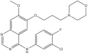
N-(3-chloro-4-fluorophenyl)-7-methoxy-6-(3-morpholin-4-ylpropoxy)quinazolin-4-amine
|
| 别名 |
Gefitinib; ZD-1839; ZD1839; ZD 1839; Brand name: Iressa
|
| HS Tariff Code |
2934.99.9001
|
| 存储方式 |
Powder -20°C 3 years 4°C 2 years In solvent -80°C 6 months -20°C 1 month |
| 运输条件 |
Room temperature (This product is stable at ambient temperature for a few days during ordinary shipping and time spent in Customs)
|
| 溶解度 (体外实验) |
|
|||
|---|---|---|---|---|
| 溶解度 (体内实验) |
1、请先配制澄清的储备液(如:用DMSO配置50 或 100 mg/mL母液(储备液)); 2、取适量母液,按从左到右的顺序依次添加助溶剂,澄清后再加入下一助溶剂。以 下列配方为例说明 (注意此配方只用于说明,并不一定代表此产品 的实际溶解配方): 10% DMSO → 40% PEG300 → 5% Tween-80 → 45% ddH2O (或 saline); 假设最终工作液的体积为 1 mL, 浓度为5 mg/mL: 取 100 μL 50 mg/mL 的澄清 DMSO 储备液加到 400 μL PEG300 中,混合均匀/澄清;向上述体系中加入50 μL Tween-80,混合均匀/澄清;然后继续加入450 μL ddH2O (或 saline)定容至 1 mL; 3、溶剂前显示的百分比是指该溶剂在最终溶液/工作液中的体积所占比例; 4、 如产品在配制过程中出现沉淀/析出,可通过加热(≤50℃)或超声的方式助溶; 5、为保证最佳实验结果,工作液请现配现用! 6、如不确定怎么将母液配置成体内动物实验的工作液,请查看说明书或联系我们; 7、 以上所有助溶剂都可在 Invivochem.cn网站购买。 |
| 制备储备液 | 1 mg | 5 mg | 10 mg | |
| 1 mM | 2.2376 mL | 11.1882 mL | 22.3764 mL | |
| 5 mM | 0.4475 mL | 2.2376 mL | 4.4753 mL | |
| 10 mM | 0.2238 mL | 1.1188 mL | 2.2376 mL |
1、根据实验需要选择合适的溶剂配制储备液 (母液):对于大多数产品,InvivoChem推荐用DMSO配置母液 (比如:5、10、20mM或者10、20、50 mg/mL浓度),个别水溶性高的产品可直接溶于水。产品在DMSO 、水或其他溶剂中的具体溶解度详见上”溶解度 (体外)”部分;
2、如果您找不到您想要的溶解度信息,或者很难将产品溶解在溶液中,请联系我们;
3、建议使用下列计算器进行相关计算(摩尔浓度计算器、稀释计算器、分子量计算器、重组计算器等);
4、母液配好之后,将其分装到常规用量,并储存在-20°C或-80°C,尽量减少反复冻融循环。
计算结果:
工作液浓度: mg/mL;
DMSO母液配制方法: mg 药物溶于 μL DMSO溶液(母液浓度 mg/mL)。如该浓度超过该批次药物DMSO溶解度,请首先与我们联系。
体内配方配制方法:取 μL DMSO母液,加入 μL PEG300,混匀澄清后加入μL Tween 80,混匀澄清后加入 μL ddH2O,混匀澄清。
(1) 请确保溶液澄清之后,再加入下一种溶剂 (助溶剂) 。可利用涡旋、超声或水浴加热等方法助溶;
(2) 一定要按顺序加入溶剂 (助溶剂) 。
Study of EGF816 in Combination With Selected Targeted Agents in EGFR-mutant NSCLC
CTID: NCT03333343
Phase: Phase 1 Status: Active, not recruiting
Date: 2024-09-19
A, effect of metformin (MET) alone and in combination with gefitinib (GEF) on cell proliferation, on anchorage-independent growth ability of NSCLC cell lines, and on the induction of apoptosis in CALU-3, CALU-3 GEF-R, and H1299 cell lines.Clin Cancer Res.2013 Jul 1;19(13):3508-19. |
Effects on the downstream pathway by combined treatment of metformin and gefitinib. Western blotting of EGFR, MAPK, AKT p70S6K, and S6 activation following treatment with the indicated concentration of metformin and gefitinib in CALU-3 and CALU-3 GEF-R cell lines. β-Actin was included as a loading control.Clin Cancer Res.2013 Jul 1;19(13):3508-19. |
Effects of the combination treatment of metformin and gefitinib on NSCLC tumor xenografts.Clin Cancer Res.2013 Jul 1;19(13):3508-19. |
") |
") |
 EGFR Ligand-Linker Conjugates 1
 PROTAC Her3-binding moiety 2
 EGFR ligand-14
 EGFR ligand-14-PEG3-Boc
InvivoChem的所有产品仅用于作科学研究,不面向患者销售
Copyright 2020 InvivoChem LLC | All Rights Reserved 粤ICP备20063088号-1


 COA
COA
")
")
")
 463611831
