| 规格 | 价格 | 库存 | 数量 |
|---|---|---|---|
| 10 mM * 1 mL in DMSO |
|
||
| 1mg |
|
||
| 5mg |
|
||
| 10mg |
|
||
| 25mg |
|
||
| 50mg |
|
||
| 100mg |
|
||
| 250mg |
|
||
| 500mg |
|
||
| Other Sizes |
|
| 靶点 |
PKA (Ki = 48 nM); S6K1 (IC50 = 80 nM); PKG (Ki = 0.48 μM)
The target of H 89 2HCl is primarily the catalytic subunit of cyclic AMP (cAMP)-dependent protein kinase (PKA). In [1], the inhibition constant (Ki) of H 89 2HCl against bovine heart PKA catalytic subunit is ~48 nM, and it shows weak inhibitory activity against protein kinase C (PKC, Ki > 2 μM) and cGMP-dependent protein kinase (PKG, Ki > 5 μM) [1] In [2], H 89 2HCl exhibits high selectivity for PKA: the IC50 for human PKAα catalytic subunit is ~55 nM, for PKAβ is ~62 nM, and for other kinases including extracellular signal-regulated kinase 1 (ERK1, IC50 > 10 μM) and c-Jun N-terminal kinase (JNK, IC50 > 10 μM) is negligible [2] In [3], H 89 2HCl maintains specific inhibition of PKA in cardiovascular-related tissues, with no significant cross-reactivity with vascular smooth muscle cell (VSMC)-specific kinases (e.g., Rho kinase, IC50 > 8 μM) [3] |
|---|---|
| 体外研究 (In Vitro) |
在添加福司可林前 1 小时用 H-89 (30 M) 预处理细胞,可显着且剂量依赖性地抑制 orskolin 诱导的蛋白质磷酸化。 [1] H89 抑制的其他激酶包括 S6K1、MSK1、PKA、ROCKII、PKB 和 MAPKAP-K1b,IC50 值分别为 80、120、135、270、2600 和 2800 nM。 [2] [3] 一些细胞受体和离子通道,包括 Kv1.3 K+ 通道、1AR 和 2AR,对 H89 也有活性。 [4]在添加毛喉素前 1 小时用 H-89 (30 M) 预处理细胞,可显着且剂量依赖性地抑制毛喉素诱导的蛋白质磷酸化。 [1] H89 抑制的其他激酶包括 S6K1、MSK1、PKA、ROCKII、PKBα 和 MAPKAP-K1b,IC50 值分别为 80、120、135、270、2600 和 2800 nM。 [2] [3] 一些细胞受体和离子通道,包括 Kv1.3 K+ 通道、β1AR 和 β2AR,对 H89 也具有活性。 [4]
1. 抑制PKA介导的磷酸化(来自[1]):无细胞实验中,H 89 2HCl剂量依赖性抑制PKA催化的特异性底物kemptide(序列:LRRASLG)磷酸化。100 nM时抑制约90%的PKA活性,10 nM时抑制约35%,与Ki值一致;即使在5 μM浓度下,对PKC介导的组蛋白H1磷酸化也无显著影响[1] 2. 调控胰岛β细胞胰岛素分泌(来自[2]):用H 89 2HCl(0.1 μM、0.5 μM、1 μM)处理INS-1大鼠胰岛素瘤细胞2小时,可浓度依赖性降低葡萄糖(16.7 mM)刺激的胰岛素分泌。具体而言,1 μM H 89 2HCl使胰岛素分泌减少约60%(放射免疫法检测),而基础胰岛素分泌(2.8 mM葡萄糖)无变化。Western blot显示,0.5 μM H 89 2HCl可降低PKA下游底物CREB(Ser133)的磷酸化水平[2] 3. 抑制血管平滑肌细胞(VSMC)增殖(来自[3]):H 89 2HCl(0.2 μM、1 μM、5 μM)可抑制血小板衍生生长因子(PDGF)诱导的大鼠主动脉VSMC增殖(MTT法),72小时增殖抑制的IC50约为1.2 μM。5 μM时还可减少PDGF诱导的VSMC迁移约45%(Transwell法),Western blot显示增殖相关蛋白(cyclin D1、PCNA)表达下调[3] |
| 体内研究 (In Vivo) |
H89 的蛋白质磷酸化以多种方式发生改变,但果糖 1,6-二磷酸酶、异质核核糖核蛋白 (hnRNP) 和 NSFL1 辅因子 p47 表现出最强的磷酸化变化。这些蛋白可能都与cAMP/PKA有调节关系。
在经过 PTZ 治疗的动物中,H-89(0.2 mg/100g,腹腔注射)显着增加了癫痫潜伏期和阈值。 H-89 显着提高癫痫潜伏期和癫痫阈值,并抑制布克拉地辛 (300 nM) 的致癫痫作用,剂量为 0.05 和 0.2 mg/100 g,腹膜内注射 [Eur J Pharmacol. 2011 Nov 30;670(2-3):464-70.]。 H-89预处理对PTZ诱发癫痫的影响[Eur J Pharmacol. 2011 Nov 30;670(2-3):464-70.] 图2A和B显示了用不同剂量的H-89(0.05、0.1和0.2 mg/100 g,i.p.,30分钟)预处理对PTZ(0.5%w/v i.v)诱导的癫痫发作的影响。与对照组相比,以0.2 mg/100克的剂量给药H-89显著增加了癫痫发作的潜伏期和阈值(***p<0.001)。与对照组动物相比,其他两种剂量的H-89(0.05和0.1 mg/100 g)在癫痫发作潜伏期和阈值方面没有观察到显著差异(图2A和B)。 己酮可可碱和H-89联合预处理对PTZ诱导的小鼠癫痫发作的影响[Eur J Pharmacol. 2011 Nov 30;670(2-3):464-70.] 属于该组合组的所有动物在PTZ输注前45分钟接受PTX作为第一组分,30分钟接受H-89作为第二组分。与对照组相比,接受PTX 50 mg/kg和H-89 0.2 mg/kg以及PTX 100 mg/kg和H-890.2 mg/100 g的组在癫痫发作潜伏期和阈值方面存在显著差异(***P<0.001)(图4A和B)。PTX(50和100 mg/kg)给药显著减弱了H-89(0.2 mg/100 g)对癫痫发作阈值和潜伏期的影响(*P<0.05)(图4A和B)。 1. 调节大鼠糖代谢(来自[2]):250~300 g雄性SD大鼠禁食12小时后,腹腔注射H 89 2HCl(10 mg/kg、20 mg/kg)或溶剂。30分钟后给予口服葡萄糖负荷(2 g/kg)。葡萄糖负荷后60分钟,10 mg/kg组和20 mg/kg组血糖分别比溶剂组降低约18%和32%;30分钟时血清胰岛素水平分别降低约25%和40%,与体外胰岛素分泌抑制结果一致[2] 2. 自发性高血压大鼠(SHR)的降压作用(来自[3]):12~14周龄SHR大鼠静脉注射H 89 2HCl(0.3 mg/kg、1 mg/kg、3 mg/kg)或溶剂,持续监测平均动脉压(MAP)4小时。3 mg/kg组在给药后30分钟MAP最大降低约22 mmHg,效应持续约2小时;各剂量组均未观察到心率显著变化。3 mg/kg组主动脉组织Western blot显示,p-CREB(Ser133)水平比溶剂组降低约50%[3] |
| 酶活实验 |
cAMP 依赖性蛋白激酶活性在最终体积为 0.2 mL 的反应混合物中进行测定,其中包含 50 mM Tris-HCl (pH 7.0)、10 mM 醋酸镁、2 mM EGTA、1 μM cAMP 或不含 cAMP,3.3 -20 μM [γ-32P]ATP (4 × 10⁵ cpm)、0.5 μg 酶、100 μg 组蛋白 H2B 和每种化合物,如所示。
1. PKA催化活性实验(来自[1]): - 试剂制备:制备纯化的牛心PKA催化亚基;将特异性底物kemptide溶于反应缓冲液(50 mM Tris-HCl pH7.5、10 mM MgCl₂、1 mM二硫苏糖醇(DTT)),终浓度200 μM;将[γ-³²P]ATP稀释至10 μM(比活度~3000 cpm/pmol)[1] - 实验设置:将H 89 2HCl用DMSO系列稀释为7个浓度(1 nM、10 nM、30 nM、100 nM、300 nM、1 μM、5 μM),加入反应混合物(DMSO终浓度≤1%)。反应混合物含反应缓冲液、kemptide和[γ-³²P]ATP。加入PKA催化亚基(终浓度5 nM)启动反应,30°C孵育30分钟。设置溶剂(DMSO)和阳性对照(PKI肽,100 nM)组,每组3个重复[1] - 检测与分析:取25 μL反应混合物点样到P81磷酸纤维素滤纸上,用1%磷酸洗涤3次(每次5分钟)去除未结合的ATP,丙酮漂洗后风干。通过液体闪烁计数测量放射性。抑制率=[(对照放射性-样品放射性)/对照放射性]×100%,采用双倒数作图法(Lineweaver-Burk plot)计算Ki值[1] 2. PKA亚型及脱靶激酶选择性实验(来自[2]): - 试剂制备:纯化重组人PKAα、PKAβ、PKCα、ERK1和JNK1;使用荧光PKA底物(FAM-Kemptide-K(BHQ1)-NH₂),激发波长485 nm,发射波长520 nm[2] - 实验设置:将H 89 2HCl(0.01 μM~20 μM)与各激酶(10 nM)及对应底物(100 μM)在激酶特异性缓冲液中孵育(如PKC缓冲液含20 mM Tris-HCl pH7.4、5 mM CaCl₂、100 μg/mL磷脂酰丝氨酸)。37°C反应40分钟,每5分钟测量一次荧光强度[2] - 分析:计算初始反应速率以确定各激酶的IC50,证实H 89 2HCl对PKA亚型的选择性[2] 3. VSMC相关激酶抑制实验(来自[3]): - 试剂制备:使用重组Rho激酶和PKA催化亚基;Rho激酶反应缓冲液含25 mM Tris-HCl pH7.5、10 mM MgCl₂、0.1 mM ATP[3] - 实验设置:将H 89 2HCl(0.1 μM~10 μM)与Rho激酶或PKA及其特异性底物(Rho激酶用MYPT1肽,PKA用kemptide)在30°C孵育30分钟,通过ADP-Glo™实验(检测ADP生成)测定活性[3] - 分析:比较对Rho激酶和PKA的抑制率,证实H 89 2HCl对Rho激酶无显著抑制[3] |
| 细胞实验 |
测定细胞内cAMP 的水平。培养 48 小时后,PC12D 细胞在含有 30 μM H-89 的测试培养基中生长 1 小时,然后暴露于含有 10 μM 毛喉素和 30 μM H-89 的全新培养基中。添加 0.5 ml 6% 三氯乙酸,同时用橡胶警察刮下细胞并进行超声处理。加入2ml石油醚,混合,3000rpm离心10分钟,提取三氯乙酸。吸出顶层后,残留样品溶液用于分析。
1. PKA介导的酪氨酸羟化酶磷酸化实验(来自[1]): - 细胞制备:牛肾上腺皮质细胞在含10% FBS的DMEM中培养,以2×10⁵个细胞/孔接种到6孔板,37°C、5% CO₂孵育至70%汇合度[1] - 药物处理:细胞用H 89 2HCl(0.1 μM、1 μM、10 μM)预处理1小时,再用毛喉素(forskolin,10 μM,cAMP激活剂)刺激30分钟。设置溶剂对照(1% DMSO)和单独毛喉素组[1] - 检测:用含蛋白酶/磷酸酶抑制剂的RIPA缓冲液裂解细胞,30 μg蛋白经10% SDS-PAGE分离后转移至PVDF膜。膜用抗磷酸化酪氨酸羟化酶(Ser40)和抗总酪氨酸羟化酶抗体孵育,定量条带强度显示,1 μM H 89 2HCl抑制毛喉素诱导的酪氨酸羟化酶磷酸化约75%[1] 2. INS-1细胞胰岛素分泌实验(来自[2]): - 细胞接种:INS-1细胞在含11 mM葡萄糖、10% FBS的RPMI-1640培养基中培养,以1×10⁵个细胞/孔接种到24孔板,过夜孵育[2] - 葡萄糖与药物处理:细胞用含2.8 mM葡萄糖的克雷布斯-林格碳酸氢盐缓冲液(KRBB)洗涤,预孵育1小时。随后用含H 89 2HCl(0.1 μM、0.5 μM、1 μM)的KRBB(含2.8 mM基础葡萄糖或16.7 mM刺激葡萄糖)处理2小时[2] - 胰岛素检测:收集培养上清,通过放射免疫法测定胰岛素浓度;细胞裂解液用于BCA法测定蛋白浓度以标准化结果,显示H 89 2HCl浓度依赖性抑制刺激态胰岛素分泌[2] 3. VSMC增殖与迁移实验(来自[3]): - 增殖实验(MTT法):大鼠主动脉VSMC以5×10³个细胞/孔接种到96孔板,孵育24小时。细胞用H 89 2HCl(0.2 μM、1 μM、5 μM)+PDGF-BB(20 ng/mL)或单独PDGF-BB处理。72小时后加入20 μL MTT(5 mg/mL),孵育4小时,DMSO溶解甲瓒结晶,测量570 nm吸光度,通过逻辑回归计算IC50[3] - 迁移实验(Transwell法):VSMC血清饥饿24小时,用H 89 2HCl(1 μM、5 μM)处理1小时,以1×10⁴个细胞/室接种到Transwell小室(8 μm孔径)上室,下室含PDGF-BB(20 ng/mL)。24小时后固定下室膜上细胞,结晶紫染色并计数,5 μM H 89 2HCl使迁移减少约45%[3] |
| 动物实验 |
大鼠;小鼠
20 或 200 mg/kg(大鼠);0-5 mg/kg(小鼠) 皮下注射(大鼠);腹腔注射(小鼠) 在静脉注射戊四唑 (PTZ) 前 30 分钟,腹腔注射己酮可可碱 (25、50、100 mg/kg)、布克拉地辛 (50、100、300 nM/只小鼠) 和 H-89 (0.05、0.1、0.2 mg/100 g)。在联合用药组中,第一种和第二种成分分别在 PTZ 注射前 45 分钟和 30 分钟注射。所有组的相应对照动物均接受等体积的溶剂。静脉输注时,将针头插入尾侧静脉,用一小段胶带固定于尾静脉上,并允许动物自由活动(Gholipour et al., 2008, 2009)。戊四唑溶液以1 ml/min的浓度速率输注。[Eur J Pharmacol. 2011 Nov 30;670(2-3):464-70.] 1. SD大鼠葡萄糖耐量试验(引自[2]):- 动物准备:雄性SD大鼠(250–300 g)饲养于12小时光照/12小时黑暗循环环境中,可自由获取食物和水。实验前大鼠禁食12小时[2] - 药物配制和给药:将H 89 2HCl溶解于5% DMSO + 95%生理盐水中,配制成浓度分别为10 mg/mL和20 mg/mL的溶液。大鼠随机分为3组(每组n=6):溶剂组(5% DMSO + 生理盐水,1 mL/kg,腹腔注射)、H 89 2HCl 10 mg/kg组(1 mL/kg,腹腔注射)、H 89 2HCl 20 mg/kg组(1 mL/kg,腹腔注射)[2] - 葡萄糖负荷和取样:给药30分钟后,所有大鼠均口服葡萄糖(2 g/kg,溶于水)。分别于葡萄糖负荷后0、30、60、90和120分钟从尾静脉采集血样。使用血糖仪测量血糖,并使用放射免疫分析法检测血清胰岛素[2]。 2. 高血压大鼠血压监测(引自[3]): - 动物模型:使用自发性高血压大鼠(SHR,12-14周龄,雄性,300-350 g),以正常血压的Wistar-Kyoto(WKY)大鼠作为对照。实验前,动物适应环境7天[3]。 - 药物配制和给药:将H 89 2HCl溶解于2% DMSO + 98%生理盐水中,配制成浓度分别为0.3 mg/mL、1 mg/mL和3 mg/mL的溶液。大鼠用异氟烷麻醉,并植入颈动脉导管进行连续平均动脉压(MAP)监测。大鼠分为4组(每组n=5):赋形剂组(2% DMSO + 生理盐水,1 mL/kg,静脉注射)、H 89 2HCl 0.3 mg/kg、1 mg/kg、3 mg/kg(1 mL/kg,静脉注射)[3] - 数据采集:给药后每10分钟记录一次MAP和心率,持续4小时。实验结束时,处死大鼠,并采集主动脉组织进行p-CREB的Western blot分析[3] |
| 药代性质 (ADME/PK) |
1. 大鼠药代动力学参数(引自[2]):雄性SD大鼠分别经口灌胃(20 mg/kg)或静脉注射(5 mg/kg)给予H 89 2HCl。分别于给药后0.083、0.25、0.5、1、2、4、6、8和12小时采集血样。采用LC-MS/MS法测定血浆中H 89 2HCl的浓度。主要参数:(1)口服生物利用度:约25%;(2)半衰期(t1/2):口服约2.5小时,静脉注射约1.8小时;(3)峰浓度(Cmax):口服1小时约1.2 μg/mL。 (4) 曲线下面积 (AUC₀-∞):口服约为 9.2 μg·h/mL,静脉注射约为 8.5 μg·h/mL [2]
2. SHR 中的组织分布(引自 [3]):SHR 大鼠静脉注射 H 89 2HCl 1 mg/kg。给药后 0.5、1 和 2 小时,处死大鼠,并收集心脏、主动脉、肝脏、肾脏和血浆。采用 LC-MS/MS 法测定 H 89 2HCl 的浓度。主动脉浓度在 0.5 小时约为 0.8 μg/g,1 小时约为 0.5 μg/g,2 小时约为 0.2 μg/g,均高于血浆浓度(相应时间点分别为 0.6 μg/mL、0.3 μg/mL 和 0.1 μg/mL)。肝脏浓度最高(0.5 小时约为 1.5 μg/g),而肾脏浓度约为 0.6 μg/g [3] |
| 毒性/毒理 (Toxicokinetics/TK) |
1. 小鼠急性毒性试验(引自[2]):雌性ICR小鼠腹腔注射单剂量H 89 2HCl(50 mg/kg、100 mg/kg、150 mg/kg、200 mg/kg)。对小鼠进行7天的观察。50 mg/kg和100 mg/kg剂量组未观察到死亡;150 mg/kg剂量组导致20%的死亡率(1/5只小鼠);200 mg/kg剂量组导致60%的死亡率(3/5只小鼠)。计算得出半数致死量(LD50)约为150 mg/kg。 100 mg/kg剂量下出现的轻度毒性症状(嗜睡、食欲减少)在48小时内消退[2]
2. 大鼠亚慢性毒性(引自[2]):SD大鼠每日一次灌胃给予H 89 2HCl(10 mg/kg、20 mg/kg),持续28天。未观察到体重、食欲或器官重量(心脏、肝脏、肾脏)的显著变化。血清丙氨酸氨基转移酶(ALT)、天冬氨酸氨基转移酶(AST)、血尿素氮(BUN)和肌酐(Cr)水平均在正常范围内,表明未发生肝肾损伤[2] 3. 血浆蛋白结合率(引自[3]):采用超滤法测定H 89 2HCl的血浆蛋白结合率。将H 89 2HCl(0.1 μM、1 μM、10 μM)加入人、大鼠和小鼠血浆中。经超滤(30 kDa截留分子量)后,采用液相色谱-串联质谱法(LC-MS/MS)测定滤液和血浆中的浓度。在所有浓度下,结合率分别为~88%(人)、~85%(大鼠)和~83%(小鼠)[3] |
| 参考文献 | |
| 其他信息 |
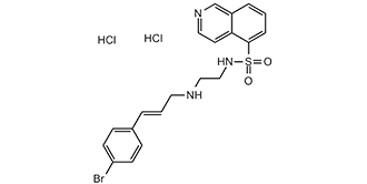
N-[2-(4-溴肉桂基氨基)乙基]异喹啉-5-磺酰胺二盐酸盐是由N-[2-(4-溴肉桂基氨基)乙基]异喹啉-5-磺酰胺与两当量氯化氢制备的盐酸盐。它是一种EC 2.7.11.11(cAMP依赖性蛋白激酶)抑制剂。它含有N-[2-(4-溴肉桂基氨基)乙基]异喹啉-5-磺酰胺(2+)。
一种新合成的异喹啉磺酰胺H-89(N-[2-(对溴肉桂基氨基)乙基]-5-异喹啉磺酰胺)被证实对环磷酸腺苷依赖性蛋白激酶(蛋白激酶A)具有强效且选择性的抑制作用,其抑制常数为0.048 ± 0.008 μM。 H-89 对其他激酶的抑制作用较弱,其对这些激酶的 Ki 值分别为:cGMP 依赖性蛋白激酶(蛋白激酶 G)、Ca2+/磷脂依赖性蛋白激酶(蛋白激酶 C)、酪蛋白激酶 I 和 II、肌球蛋白轻链激酶以及 Ca2+/钙调蛋白依赖性蛋白激酶 II,分别为 0.48 ± 0.13 μM、31.7 ± 15.9 μM、38.3 ± 6.0 μM、136.7 ± 17.0 μM、28.3 ± 17.5 μM 和 29.7 ± 8.1 μM。动力学分析表明,H-89 以与 ATP 竞争的方式抑制蛋白激酶 A。为了研究蛋白激酶A在PC12细胞神经突生长中的作用,我们分别使用H-89、神经生长因子(NGF)、福斯克林或二丁酰环磷酸腺苷(dibutyryl cAMP)处理PC12细胞。H-89预处理可剂量依赖性地抑制福斯克林诱导的蛋白磷酸化,且PC12D细胞内环磷酸腺苷(cAMP)水平未见降低;而NGF诱导的蛋白磷酸化则未被抑制。H-89还显著抑制了福斯克林诱导的PC12D细胞神经突生长。当在添加二丁酰环磷酸腺苷之前添加H-89时,也观察到了这种抑制作用。用H-89(30 μM)预处理PC12D细胞可显著抑制细胞裂解液中cAMP依赖的组蛋白IIb磷酸化活性,但对其他蛋白质磷酸化活性无影响,例如cGMP依赖的组蛋白IIb磷酸化活性、Ca2+/磷脂依赖的组蛋白IIIs磷酸化活性、Ca2+/钙调蛋白依赖的肌球蛋白轻链磷酸化活性以及α-酪蛋白磷酸化活性。然而,这种蛋白激酶A抑制剂并不抑制NGF诱导的PC12D细胞神经突生长。因此,福斯克林和二丁酰环磷酸腺苷(dibutyryl cAMP)诱导的神经突生长显然是由蛋白激酶A介导的,而NGF诱导的神经突生长则是由一条不依赖于蛋白激酶A的通路介导的。[1] 我们检测了28种市售化合物的特异性,这些化合物据报道是特定丝氨酸/苏氨酸特异性蛋白激酶的相对选择性抑制剂,并针对大量蛋白激酶进行了测试。结果发现,化合物KT 5720、Rottlerin和槲皮素能够抑制多种蛋白激酶,有时其抑制效力甚至远超其假定的靶标,因此基于这些化合物在细胞实验中得出的结论可能存在误差。Ro 318220及其相关的双吲哚马来酰亚胺类化合物,以及H89、HA1077和Y 27632是更具选择性的抑制剂,但它们仍然能够以相似的效力抑制两种或两种以上的蛋白激酶。研究发现,LY 294002 对酪蛋白激酶-2的抑制效力与对磷脂酰肌醇3-激酶的抑制效力相当。选择性最显著的化合物包括 KN62、PD 98059、U0126、PD 184352、雷帕霉素、沃特曼宁、SB 203580 和 SB 202190。与 PD 98059 类似,U0126 和 PD 184352 在细胞实验中通过抑制丝裂原活化蛋白激酶 (MAPK) 的活化而非直接抑制 MKK1 的活性来阻断丝裂原活化蛋白激酶 (MAPK) 级联反应。除雷帕霉素和 PD 184352 外,即使是选择性最高的抑制剂也至少会影响一种其他蛋白激酶。我们的研究结果表明,仅通过研究蛋白激酶抑制剂对一级结构密切相关的激酶的影响,无法评估其特异性。我们提出了在基于细胞的实验中使用蛋白激酶抑制剂的指导原则。[2] H89 是一种选择性强效的蛋白激酶 A (PKA) 抑制剂。自发现以来,它已被广泛用于评估 PKA 在心脏、成骨细胞、肝细胞、平滑肌细胞、神经组织、上皮细胞等组织中的作用。尽管 H89 应用广泛,但其特异性抑制 PKA 的机制仍未完全阐明。研究还表明,H89 至少抑制 8 种其他激酶,同时还具有相当多的 PKA 非依赖性效应,这可能会严重影响数据的解读。因此,尽管 H89 具有激酶抑制特性,但建议不要将其作为 PKA 参与的唯一证据来源。 H-89 应与其他 PKA 抑制剂(例如 Rp-cAMPS 或 PKA 类似物)联合使用。[3] 1. 作用机制(引自[1]):H 89 2HCl 是一种 PKA 竞争性抑制剂,可与 PKA 催化亚基的 ATP 结合口袋结合。这种结合阻断了 ATP 的进入,抑制了 PKA 介导的下游底物(例如 CREB、酪氨酸羟化酶)的磷酸化,从而调节 cAMP 依赖性信号通路。[1] 2. 研究工具应用(引自[2]):由于其对 PKA 的高选择性,H 89 2HCl 被广泛用作研究 PKA 在细胞过程中作用的工具化合物,包括胰岛素分泌、神经递质合成和细胞增殖。它有助于验证 PKA 作为代谢和神经系统疾病治疗靶点的有效性。[2] 3.心血管治疗潜力(引自[3]):H 89 2HCl可抑制高血压大鼠的血管平滑肌细胞增殖/迁移并降低血压,提示其具有治疗高血压和血管重塑(例如动脉粥样硬化、再狭窄)的潜力。然而,由于担心全身性PKA抑制会影响其他组织(例如胰腺、脑),目前仍处于临床前研究阶段[3]。 4. 研发历程(引自[1]):H 89 2HCl于1990年首次报道,是最早的选择性PKA抑制剂之一,克服了早期PKA抑制剂(例如H-8)的非特异性。它的研发使得在体外和体内精确调控PKA活性成为可能[1]。 |
| 分子式 |
C20H24BRCL2N3O3S
|
|---|---|
| 分子量 |
519.28
|
| 精确质量 |
516.9993
|
| 元素分析 |
C, 46.26; H, 4.27; Br, 15.39; Cl, 13.65; N, 8.09; O, 6.16; S, 6.17
|
| CAS号 |
130964-39-5
|
| 相关CAS号 |
H-89;127243-85-0
|
| PubChem CID |
5702541
|
| 外观&性状 |
White to light yellow solid powder
|
| 熔点 |
195-200ºC
|
| LogP |
6.98
|
| tPSA |
88.7
|
| 氢键供体(HBD)数目 |
4
|
| 氢键受体(HBA)数目 |
5
|
| 可旋转键数目(RBC) |
8
|
| 重原子数目 |
29
|
| 分子复杂度/Complexity |
570
|
| 定义原子立体中心数目 |
0
|
| SMILES |
O=S(C1=CC=CC2=C1C=CN=C2)(NCCNC/C=C/C3=CC=C(Br)C=C3)=O.Cl.Cl
|
| InChi Key |
GELOGQJVGPIKAM-WTVBWJGASA-N
|
| InChi Code |
InChI=1S/C20H20BrN3O2S.2ClH/c21-18-8-6-16(7-9-18)3-2-11-22-13-14-24-27(25,26)20-5-1-4-17-15-23-12-10-19(17)20;;/h1-10,12,15,22,24H,11,13-14H2;2*1H/b3-2+;;
|
| 化学名 |
N-[2-[[(E)-3-(4-bromophenyl)prop-2-enyl]amino]ethyl]isoquinoline-5-sulfonamide;dihydrochloride
|
| 别名 |
H-89; H 89 HCl; 30964-39-5; H-89 DIHYDROCHLORIDE; H-89 dihydrochloride hydrate; H 89 2HCl; H 89 dihydrochloride; H-89 (dihydrochloride); N-(2-((3-(4-Bromophenyl)allyl)amino)ethyl)isoquinoline-5-sulfonamide dihydrochloride; H-89 2HCL; H-89 Dihydrochloride; H89
|
| HS Tariff Code |
2934.99.9001
|
| 存储方式 |
Powder -20°C 3 years 4°C 2 years In solvent -80°C 6 months -20°C 1 month 注意: 请将本产品存放在密封且受保护的环境中,避免吸湿/受潮。 |
| 运输条件 |
Room temperature (This product is stable at ambient temperature for a few days during ordinary shipping and time spent in Customs)
|
| 溶解度 (体外实验) |
DMSO: ~104 mg/mL (~200.3 mM)
Water: ~6 mg/mL (~11.6 mM) Ethanol: <1 mg/mL |
|---|---|
| 溶解度 (体内实验) |
配方 1 中的溶解度: 5 mg/mL (9.63 mM) in 10% DMSO + 90% Saline (这些助溶剂从左到右依次添加,逐一添加), 悬浮液;超声助溶。
*生理盐水的制备:将 0.9 g 氯化钠溶解在 100 mL ddH₂O中,得到澄清溶液。 配方 2 中的溶解度: ≥ 2.75 mg/mL (5.30 mM) (饱和度未知) in 5% DMSO + 40% PEG300 + 5% Tween80 + 50% Saline (这些助溶剂从左到右依次添加,逐一添加), 澄清溶液。 *生理盐水的制备:将 0.9 g 氯化钠溶解在 100 mL ddH₂O中,得到澄清溶液。 View More
配方 3 中的溶解度: ≥ 2.75 mg/mL (5.30 mM) (饱和度未知) in 5% DMSO + 95% (20% SBE-β-CD in Saline) (这些助溶剂从左到右依次添加,逐一添加), 澄清溶液。 配方 4 中的溶解度: ≥ 2.5 mg/mL (4.81 mM) (饱和度未知) in 10% DMSO + 40% PEG300 + 5% Tween80 + 45% Saline (这些助溶剂从左到右依次添加,逐一添加), 澄清溶液。 例如,若需制备1 mL的工作液,可将100 μL 25.0 mg/mL 澄清的 DMSO 储备液加入到400 μL PEG300中,混匀;再向上述溶液中加入50 μL Tween-80,混匀;然后加入450 μL 生理盐水定容至1 mL。 *生理盐水的制备:将 0.9 g 氯化钠溶解在 100 mL ddH₂O中,得到澄清溶液。 配方 5 中的溶解度: ≥ 2.5 mg/mL (4.81 mM) (饱和度未知) in 10% DMSO + 90% (20% SBE-β-CD in Saline) (这些助溶剂从左到右依次添加,逐一添加), 澄清溶液。 例如,若需制备1 mL的工作液,可将100μL 25.0mg/mL澄清的DMSO储备液加入到900μL 20%SBE-β-CD生理盐水中,混匀。 *20% SBE-β-CD 生理盐水溶液的制备(4°C,1 周):将 2 g SBE-β-CD 溶解于 10 mL 生理盐水中,得到澄清溶液。 配方 6 中的溶解度: ≥ 2.5 mg/mL (4.81 mM) (饱和度未知) in 10% DMSO + 90% Corn Oil (这些助溶剂从左到右依次添加,逐一添加), 澄清溶液。 例如,若需制备1 mL的工作液,可将 100 μL 25.0 mg/mL 澄清 DMSO 储备液加入到 900 μL 玉米油中并混合均匀。 配方 7 中的溶解度: ≥ 0.55 mg/mL (1.06 mM) (饱和度未知) in 1% DMSO 99% Saline (这些助溶剂从左到右依次添加,逐一添加), 澄清溶液。 *生理盐水的制备:将 0.9 g 氯化钠溶解在 100 mL ddH₂O中,得到澄清溶液。 配方 8 中的溶解度: 1% DMSO+30% polyethylene glycol+1% Tween 80: 30mg/mL 1、请先配制澄清的储备液(如:用DMSO配置50 或 100 mg/mL母液(储备液)); 2、取适量母液,按从左到右的顺序依次添加助溶剂,澄清后再加入下一助溶剂。以 下列配方为例说明 (注意此配方只用于说明,并不一定代表此产品 的实际溶解配方): 10% DMSO → 40% PEG300 → 5% Tween-80 → 45% ddH2O (或 saline); 假设最终工作液的体积为 1 mL, 浓度为5 mg/mL: 取 100 μL 50 mg/mL 的澄清 DMSO 储备液加到 400 μL PEG300 中,混合均匀/澄清;向上述体系中加入50 μL Tween-80,混合均匀/澄清;然后继续加入450 μL ddH2O (或 saline)定容至 1 mL; 3、溶剂前显示的百分比是指该溶剂在最终溶液/工作液中的体积所占比例; 4、 如产品在配制过程中出现沉淀/析出,可通过加热(≤50℃)或超声的方式助溶; 5、为保证最佳实验结果,工作液请现配现用! 6、如不确定怎么将母液配置成体内动物实验的工作液,请查看说明书或联系我们; 7、 以上所有助溶剂都可在 Invivochem.cn网站购买。 |
| 制备储备液 | 1 mg | 5 mg | 10 mg | |
| 1 mM | 1.9257 mL | 9.6287 mL | 19.2574 mL | |
| 5 mM | 0.3851 mL | 1.9257 mL | 3.8515 mL | |
| 10 mM | 0.1926 mL | 0.9629 mL | 1.9257 mL |
1、根据实验需要选择合适的溶剂配制储备液 (母液):对于大多数产品,InvivoChem推荐用DMSO配置母液 (比如:5、10、20mM或者10、20、50 mg/mL浓度),个别水溶性高的产品可直接溶于水。产品在DMSO 、水或其他溶剂中的具体溶解度详见上”溶解度 (体外)”部分;
2、如果您找不到您想要的溶解度信息,或者很难将产品溶解在溶液中,请联系我们;
3、建议使用下列计算器进行相关计算(摩尔浓度计算器、稀释计算器、分子量计算器、重组计算器等);
4、母液配好之后,将其分装到常规用量,并储存在-20°C或-80°C,尽量减少反复冻融循环。
计算结果:
工作液浓度: mg/mL;
DMSO母液配制方法: mg 药物溶于 μL DMSO溶液(母液浓度 mg/mL)。如该浓度超过该批次药物DMSO溶解度,请首先与我们联系。
体内配方配制方法:取 μL DMSO母液,加入 μL PEG300,混匀澄清后加入μL Tween 80,混匀澄清后加入 μL ddH2O,混匀澄清。
(1) 请确保溶液澄清之后,再加入下一种溶剂 (助溶剂) 。可利用涡旋、超声或水浴加热等方法助溶;
(2) 一定要按顺序加入溶剂 (助溶剂) 。
 LJI308
 LY2584702
 PF-4708671
 BI-D1870
InvivoChem的所有产品仅用于作科学研究,不面向患者销售
Copyright 2020 InvivoChem LLC | All Rights Reserved 粤ICP备20063088号-1


 COA
COA
 463611831
