| 规格 | 价格 | 库存 | 数量 |
|---|---|---|---|
| 10 mM * 1 mL in DMSO |
|
||
| 1mg |
|
||
| 5mg |
|
||
| 10mg |
|
||
| 25mg |
|
||
| 50mg |
|
||
| 100mg |
|
||
| 250mg |
|
||
| Other Sizes |
|
| 靶点 |
β-catenin-responsive transcription (CRT; IC50 = 40.3 nM)
β-catenin/TCF4 transcription complex iCRT14 is an inhibitor of catenin responsive transcription (CRT). It disrupts the interaction between β-catenin and TCF4, and may also interfere with TCF binding to DNA.[1] |
|---|---|
| 体外研究 (In Vitro) |
除了影响 TCF-β-cat 反应外,iCRT 14 还能够阻碍 TCF 的 DNA 结合 [1]。尽管仍低于 iCRT 3,但 iCRT 14(10、25、50 μM)可有效且呈时间和剂量依赖性地减少 BT-549 细胞的生长 [2]。
在果蝇Cl8细胞中,当通过dsRNA介导的Axin敲低激活Wnt信号时,iCRT14抑制Wnt响应性荧光素酶报告基因dTF12的活性。 在HEK293细胞中,iCRT14抑制Wnt响应性STF16荧光素酶报告基因的活性,其IC50在纳摩尔到微摩尔范围内(具体数值未提供)。 iCRT14在体外降低了纯化的重组His标记β-连环蛋白与GST标记的TCF4 N端结构域之间的相互作用。 在转染了抗降解β-连环蛋白(S37Aβ-cat)的HEK293细胞中,iCRT14处理显著抑制了免疫共沉淀实验中β-连环蛋白与TCF的相互作用。 热熔分析显示,iCRT14使β-连环蛋白-TCF4复合物的熔解温度改变高达2°C,表明其直接结合该复合物。 用50 µM iCRT14处理HCT116结肠癌细胞(携带β-连环蛋白S45缺失)导致Axin2和细胞周期蛋白D1 mRNA水平显著降低,效果与β-连环蛋白 siRNA相似。 Western印迹分析显示,经iCRT14处理的HCT116细胞中细胞周期蛋白D1蛋白水平相应降低。 流式细胞术分析显示,用50 µM iCRT14处理HCT116细胞导致细胞周期显著停滞在G0/G1期。 在用50 µM iCRT14处理的HT29结肠癌细胞(具有APC突变)中也观察到类似的G0/G1期阻滞。 这种细胞周期阻滞与HCT116细胞中磷酸化组蛋白H3阳性细胞数量和BrdU掺入的急剧减少相关。 在基质胶包被的Boyden小室实验中,iCRT14降低了稳定表达S37Aβ-cat-HA的MCF7乳腺癌细胞的侵袭能力。 iCRT14抑制了MCF7-S37Aβ-cat-HA细胞中膜性E-钙粘蛋白的下调。 在C57MG小鼠乳腺上皮细胞中,iCRT14抑制了Wnt3a诱导的形态学转化,并降低了β-连环蛋白靶基因WISP1的mRNA水平。[1] |
| 体内研究 (In Vivo) |
在 HCT116 异种移植物中,iCRT 14(50 mg/kg,腹腔注射)显着降低 CycD1(肿瘤肿胀)[1]。
在无胸腺裸鼠中建立的HCT116和HT29异种移植模型中,腹腔注射iCRT14(50 mg/kg,每周三次,持续3周)导致与DMSO对照组相比,肿瘤切片中细胞周期蛋白D1染色显著减少,磷酸化组蛋白H3阳性细胞数量减少。 这与治疗最初3周(约第19天)内肿瘤初始生长率降低约50%相吻合。第19天后,生长率与对照组相当。 通过微型泵给予较低浓度(20 mg/kg)的iCRT14在HCT116异种移植模型中产生了相同的效果。 在整个研究过程中,未观察到小鼠出现全身毒性迹象或体重减轻。[1] |
| 酶活实验 |
热稳定性分析。[1]
在室温下,在不同浓度的iCRTs(如iCRT-14)和0.5×SYPRO Orange的存在下,将纯化的β-cat-His和TCF4-N-GST以1:2的摩尔比在1×PBS中混合。使用LightCycler 480系统在96孔板中以0.06°C/s的升温速率将样品从20°C加热到95°C。在加热阶段,以0.1s的间隔采集荧光读数。通过绘制荧光强度读数的负导数与温度的关系来计算熔化温度。负导数曲线上的拐点被认为是熔化温度(Tm)。 |
| 细胞实验 |
对于每种细胞系的细胞增殖测定,细胞用DMSO作为载体或不同浓度的每种Wnt抑制剂处理:iCRT-3(25、50、75μM)、iCRT-5(50、100、200μM),iCRT-14(10、25、50μM);IWP-4(1、2.5、5μM)和XAV-939(5、10μM)。对于SOX4敲除的BT549细胞的细胞增殖、迁移和侵袭试验,用DMSO或25μM iCRT-3处理细胞。CIM板16的上腔涂有Matrigel(1:40稀释),用于细胞侵袭试验。此外,在实验时用50μM染料木素和25μM iCRT-3处理6天的SOX4敲除的BT-549细胞中测量了细胞增殖。每个样品都进行了三次分析,并进行了三个独立的实验。细胞增殖试验进行48小时,细胞迁移和侵袭实验进行24小时。RTCA软件包1.2计算了每个样本的细胞指数值,用于测量电阻抗的相对变化,以表示细胞形态、粘附或存活率。[2]
将细胞以20000个细胞/孔的速度接种到96孔板中。孵育过夜后,用DMSO或每种Wnt抑制剂(iCRT-3,75μM;iCRT-5,200μM;iCRT-14,50μM;IWP-4,5μM和XAV-939,10μM)处理细胞48小时。根据制造商的说明,使用Cell Titer Glo发光细胞存活率测定试剂盒测定细胞存活率。使用FLUOstar微孔板读数器测量发光。所有治疗均进行三次,每次实验重复三次[2]。 初筛(Wnt报告基因实验): 将果蝇Cl8细胞转染Wnt响应性dTF12-荧光素酶报告基因和靶向Axin的dsRNA以激活信号通路。4天后,加入小分子化合物库中的化合物(终浓度9 ng/mL)。约16小时后,使用双荧光素酶系统测量荧光素酶活性。该实验的Z因子为0.77。 次级上位性分析: 将Cl8细胞转染靶向Slimb/βTrCP(位于Axin/APC/GSK-3β复合物下游)的dsRNA和dTF12报告基因。评估化合物对报告基因活性的影响。 β-连环蛋白-TCF相互作用实验(免疫共沉淀): 将HEK293细胞转染S37Aβ-cat,并用iCRT14处理过夜。用抗β-连环蛋白抗体对全细胞裂解液进行免疫沉淀,沉淀物用内源性TCF4抗体进行免疫印迹分析。 体外蛋白质结合实验: 将纯化的重组His标记β-连环蛋白与不同浓度的iCRT14预孵育,评估其与纯化的GST标记TCF4 N端结构域的结合能力。 热熔分析: 将纯化的β-连环蛋白-His和TCF4-N-GST以1:2的摩尔比在PBS中与iCRT14和SYPRO Orange染料混合。样品在热循环仪中从20°C加热到95°C,升温速率为每秒0.06°C。监测荧光,并根据荧光曲线的负导数计算熔解温度。 定量RT-PCR(qRT-PCR): 用iCRT14处理细胞指定时间(MCF7/HCT116细胞1天,C57MG细胞5天),裂解细胞并合成cDNA。使用SYBR Green化学法和特异性引物分析基因表达(如Axin2、Cyclin D1、WISP1、c-Myc、GAPDH)。 细胞周期分析(流式细胞术): 固定处理后的细胞,用碘化丙啶染色,通过流式细胞术分析DNA含量和细胞周期分布。 侵袭实验(Boyden小室): 将MCF7-S37Aβ-cat-HA细胞接种于基质胶包被的Transwell上室。下室含有含或不含iCRT14的培养基。孵育后,计数穿过基质胶迁移到膜下侧的细胞。 免疫荧光/免疫组织化学: 固定细胞或肿瘤切片,透化,封闭,用一抗(如抗E-钙粘蛋白、抗磷酸化组蛋白H3、抗细胞周期蛋白D1)染色,然后用荧光或酶标二抗染色并成像。[1] |
| 动物实验 |
异种移植模型:将HCT116或HT29结肠癌细胞皮下植入无胸腺裸鼠体内。当肿瘤体积达到80-120 mm³时,用溶于DMSO的iCRT14治疗小鼠。
给药:iCRT14以50 mg/kg的剂量腹腔注射,每周三次,持续3周。在另一项实验中,采用微型泵以较低剂量20 mg/kg给药。 对照:对照组动物注射DMSO溶剂。 监测:定期测量肿瘤体积。监测小鼠的全身毒性反应和体重变化。[1] |
| 毒性/毒理 (Toxicokinetics/TK) |
在小鼠异种移植研究中,以 50 mg/kg 的剂量(腹腔注射,每周 3 次,持续 3 周)给予 iCRT14,未引起动物出现任何全身毒性或体重减轻的迹象。[1]
|
| 参考文献 |
|
| 其他信息 |
β-catenin 反应性转录 (CRT) 的异常调控与多种恶性肿瘤的发生密切相关,包括结直肠癌,并且是多种癌症治疗的关键靶点。尽管人们已付出巨大努力,但 CRT 抑制剂疗法的成功临床应用仍然面临挑战。部分原因是难以找到能够特异性调节 β-catenin 核转录活性,同时又不影响其在细胞膜黏附连接稳定中发挥细胞骨架功能的抑制剂。本文报道了一种基于 RNAi 的修饰筛选策略,用于鉴定 CRT 抑制剂。我们的数据支持这些抑制剂能够特异性地拮抗核 β-catenin 的转录功能。我们发现,这些抑制剂能够有效阻断多种哺乳动物和癌细胞系中 Wnt/β-catenin 信号通路诱导的靶基因表达和表型。重要的是,这些Wnt抑制剂对人结肠肿瘤活检培养物以及表现出Wnt信号通路失调的结肠癌细胞系具有特异性细胞毒性。
[1] 背景:三阴性乳腺癌(TNBC)是一种侵袭性乳腺癌临床亚型,其特征是缺乏雌激素受体(ER)和孕激素受体(PR)表达以及人表皮生长因子受体2(HER2)过表达。TNBC亚型约占所有乳腺癌的10%-20%,但目前尚无有效的分子靶向治疗方法。先前对来自21项研究的587例TNBC病例的基因表达谱进行的荟萃分析表明,TNBC的基底样2型和间质型亚型中Wnt信号通路相关基因高表达。本研究探讨了Wnt信号通路抑制剂在三阴性乳腺癌(TNBC)有效治疗中的潜力。 方法:采用共聚焦显微镜和Western blot分析检测四种TNBC细胞系(BT-549、MDA-MB-231、HCC-1143和HCC-1937)以及ER阳性细胞系MCF-7中Wnt信号通路的激活情况。体外实验检测了五种不同的Wnt信号通路抑制剂(iCRT-3、iCRT-5、iCRT-14、IWP-4和XAV-939)对细胞增殖和凋亡的影响。通过定量实时RT-PCR分析Axin2表达和双荧光素酶报告基因检测,评估了iCRT-3对TNBC中经典Wnt信号通路的抑制作用。本研究还评估了shRNA敲低SOX4联合iCRT-3和/或染料木素处理对BT-549细胞增殖、迁移和侵袭的影响。 结果:TNBC细胞系中β-catenin的免疫荧光染色显示其定位于细胞核和细胞质,表明TNBC细胞中Wnt信号通路被激活。iCRT-3是抑制TNBC细胞增殖和拮抗Wnt信号通路最有效的化合物。此外,iCRT-3处理可增加体外细胞凋亡。在三阴性 BT-549 细胞中敲低 Wnt 通路转录因子 SOX4 可降低细胞增殖和迁移能力,而 iCRT-3 与 SOX4 敲低联合治疗对抑制细胞增殖和诱导细胞凋亡具有协同作用。 结论:这些数据表明,靶向 SOX4 和/或 Wnt 通路可能对三阴性乳腺癌 (TNBC) 患者具有治疗益处。[2] iCRT14 是一种噻唑烷二酮类小分子,通过基于 RNAi 的化学遗传筛选鉴定为 catenin 反应性转录 (CRT) 抑制剂。 它通过破坏 β-catenin/TCF4 转录复合物发挥作用,也可能干扰 TCF 与 DNA 的结合。 它对具有组成型激活的 Wnt/β-catenin 信号通路的癌细胞系(例如 HCT116、HT29)具有特异性细胞毒性,但对正常细胞或不具有此类信号通路的细胞系无毒性。成瘾性。 在异种移植模型中,它表现出适度但稳定的抗肿瘤疗效,且无明显毒性,凸显了其作为靶向Wnt驱动型癌症的先导化合物的潜力。[1] |
| 分子式 |
C21H17N3O2S
|
|---|---|
| 分子量 |
375.44358
|
| 精确质量 |
375.104
|
| 元素分析 |
C, 67.18; H, 4.56; N, 11.19; O, 8.52; S, 8.54
|
| CAS号 |
677331-12-3
|
| 相关CAS号 |
901751-47-1(iCRT3); 18623-44-4 (iCRT5)
|
| PubChem CID |
5967294
|
| 外观&性状 |
Light yellow to yellow solid powder
|
| LogP |
4.795
|
| tPSA |
80.5
|
| 氢键供体(HBD)数目 |
0
|
| 氢键受体(HBA)数目 |
4
|
| 可旋转键数目(RBC) |
3
|
| 重原子数目 |
27
|
| 分子复杂度/Complexity |
617
|
| 定义原子立体中心数目 |
0
|
| SMILES |
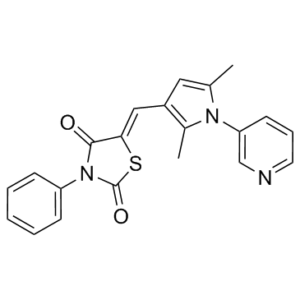
CC1=CC(=C(N1C2=CN=CC=C2)C)/C=C\3/C(=O)N(C(=O)S3)C4=CC=CC=C4
|
| InChi Key |
NCSHZXNGQYSKLR-XDHOZWIPSA-N
|
| InChi Code |
InChI=1S/C21H17N3O2S/c1-14-11-16(15(2)23(14)18-9-6-10-22-13-18)12-19-20(25)24(21(26)27-19)17-7-4-3-5-8-17/h3-13H,1-2H3/b19-12+
|
| 化学名 |
5-[[2,5-dimethyl-1-(3-pyridinyl)-1H-pyrrol-3-yl]methylene]-3-phenyl-2,4-thiazolidinedione
|
| 别名 |
iCRT14; iCRT-14; 677331-12-3; iCRT-14; iCRT14; (5Z)-5-[(2,5-dimethyl-1-pyridin-3-ylpyrrol-3-yl)methylidene]-3-phenyl-1,3-thiazolidine-2,4-dione; CHEMBL3589010; (5Z)-5-{[2,5-Dimethyl-1-(3-pyridinyl)-1H-pyrrol-3-yl]methylene}-3-phenyl-1,3-thiazolidine-2,4-dione; (5Z)-5-{[2,5-dimethyl-1-(pyridin-3-yl)-1H-pyrrol-3-yl]methylidene}-3-phenyl-1,3-thiazolidine-2,4-dione; iCRT 14
|
| HS Tariff Code |
2934.99.9001
|
| 存储方式 |
Powder -20°C 3 years 4°C 2 years In solvent -80°C 6 months -20°C 1 month |
| 运输条件 |
Room temperature (This product is stable at ambient temperature for a few days during ordinary shipping and time spent in Customs)
|
| 溶解度 (体外实验) |
DMSO : ≥ 29 mg/mL (~77.24 mM)
|
|---|---|
| 溶解度 (体内实验) |
配方 1 中的溶解度: ≥ 2.5 mg/mL (6.66 mM) (饱和度未知) in 10% DMSO + 40% PEG300 +5% Tween-80 + 45% Saline (这些助溶剂从左到右依次添加,逐一添加), 澄清溶液。
例如,若需制备1 mL的工作液,可将100 μL 25.0 mg/mL澄清DMSO储备液加入到400 μL PEG300中,混匀;然后向上述溶液中加入50 μL Tween-80+,混匀;加入450 μL生理盐水定容至1 mL。 *生理盐水的制备:将 0.9 g 氯化钠溶解在 100 mL ddH₂O中,得到澄清溶液。 请根据您的实验动物和给药方式选择适当的溶解配方/方案: 1、请先配制澄清的储备液(如:用DMSO配置50 或 100 mg/mL母液(储备液)); 2、取适量母液,按从左到右的顺序依次添加助溶剂,澄清后再加入下一助溶剂。以 下列配方为例说明 (注意此配方只用于说明,并不一定代表此产品 的实际溶解配方): 10% DMSO → 40% PEG300 → 5% Tween-80 → 45% ddH2O (或 saline); 假设最终工作液的体积为 1 mL, 浓度为5 mg/mL: 取 100 μL 50 mg/mL 的澄清 DMSO 储备液加到 400 μL PEG300 中,混合均匀/澄清;向上述体系中加入50 μL Tween-80,混合均匀/澄清;然后继续加入450 μL ddH2O (或 saline)定容至 1 mL; 3、溶剂前显示的百分比是指该溶剂在最终溶液/工作液中的体积所占比例; 4、 如产品在配制过程中出现沉淀/析出,可通过加热(≤50℃)或超声的方式助溶; 5、为保证最佳实验结果,工作液请现配现用! 6、如不确定怎么将母液配置成体内动物实验的工作液,请查看说明书或联系我们; 7、 以上所有助溶剂都可在 Invivochem.cn网站购买。 |
| 制备储备液 | 1 mg | 5 mg | 10 mg | |
| 1 mM | 2.6635 mL | 13.3177 mL | 26.6354 mL | |
| 5 mM | 0.5327 mL | 2.6635 mL | 5.3271 mL | |
| 10 mM | 0.2664 mL | 1.3318 mL | 2.6635 mL |
1、根据实验需要选择合适的溶剂配制储备液 (母液):对于大多数产品,InvivoChem推荐用DMSO配置母液 (比如:5、10、20mM或者10、20、50 mg/mL浓度),个别水溶性高的产品可直接溶于水。产品在DMSO 、水或其他溶剂中的具体溶解度详见上”溶解度 (体外)”部分;
2、如果您找不到您想要的溶解度信息,或者很难将产品溶解在溶液中,请联系我们;
3、建议使用下列计算器进行相关计算(摩尔浓度计算器、稀释计算器、分子量计算器、重组计算器等);
4、母液配好之后,将其分装到常规用量,并储存在-20°C或-80°C,尽量减少反复冻融循环。
计算结果:
工作液浓度: mg/mL;
DMSO母液配制方法: mg 药物溶于 μL DMSO溶液(母液浓度 mg/mL)。如该浓度超过该批次药物DMSO溶解度,请首先与我们联系。
体内配方配制方法:取 μL DMSO母液,加入 μL PEG300,混匀澄清后加入μL Tween 80,混匀澄清后加入 μL ddH2O,混匀澄清。
(1) 请确保溶液澄清之后,再加入下一种溶剂 (助溶剂) 。可利用涡旋、超声或水浴加热等方法助溶;
(2) 一定要按顺序加入溶剂 (助溶剂) 。
 iCRT-14 effectively inhibits cell proliferation in BT-549 cells in a dose- and time-dependent manner.J Transl Med.2013 Nov 4;11:280. |
|---|
 (A) Primary screen. dsRNA-mediated knockdown of Axin results in cytoplasmic stabilization of β-cat, which, on translocation to the nucleus, results in activation of the β-cat responsive dTF12 reporter.Proc Natl Acad Sci U S A.2011 Apr 12;108(15):5954-63. |
 (A) Effect of candidate compounds on the interaction of purified β-cat-His and GST-TCF4. iCRT3, -5, and -14 show a significant inhibitory effect on these interactions compared with nontreated (NT) and DMSO-treated binding reactions.Bottomshows comparable amounts of GST-TCF4 being pulled down.Proc Natl Acad Sci U S A.2011 Apr 12;108(15):5954-63 |
 hsBCL9CT-24
 Cardiogenol C HCl
 PNU-74654
 HLY78
HLY78
InvivoChem的所有产品仅用于作科学研究,不面向患者销售
Copyright 2020 InvivoChem LLC | All Rights Reserved 粤ICP备20063088号-1


 COA
COA
 463611831
