| 规格 | 价格 | 库存 | 数量 |
|---|---|---|---|
| 10 mM * 1 mL in DMSO |
|
||
| 1mg |
|
||
| 5mg |
|
||
| 10mg |
|
||
| 25mg |
|
||
| 50mg |
|
||
| 100mg |
|
||
| 250mg |
|
||
| 500mg |
|
||
| Other Sizes |
|

| 靶点 |
USP4(EC50= 3.93 μM);USP8(EC50= 4.9 μM);USP7(EC50= 6.86 μM);USP2(EC50= 7.2 μM);USP5(EC50= 8.61 μM)
PR-619 is a pan-inhibitor of deubiquitinating enzymes (DUBs), with IC50 values for key subtypes: USP1 (0.3 μM), USP2 (0.5 μM), USP5 (0.7 μM), USP7 (1.2 μM), USP14 (0.9 μM), and UCH-L1 (1.5 μM) [1] PR-619 shows no significant inhibition of proteasomal catalytic subunits (IC50 > 50 μM) [1] |
|---|---|
| 体外研究 (In Vitro) |
PR-619是一种细胞渗透性吡啶胺类广谱DUB抑制剂,已知靶点包括ATXN3、BAP1、JOSD2、OTUD5、UCH-L1、UCH-L3、UCH-L5/UCH37、USP1、2、4 、5、7、8、9X、10、14、15、16、19、20、22、24、28、47、48、VCIP135、YOD1,以及 deISGylase PLpro、deNEDDylase DEN1 和 deSUMOlyase SENP6。 PR-619 显示以剂量和时间依赖性方式(20 至 150 μM,0.5 至 20 小时)增加 HEK293T 细胞中总体蛋白质多泛素化。 PR619 处理导致 K 48 和 K63 连接的多聚泛素链上调。 PR-619 诱导 HCT116 细胞死亡,EC50 值为 6.3 μM。激酶测定:将重组酶置于 20 mM Tris-HCl(pH 8.0)、2 mM CaCl2 和 2 mM β-巯基乙醇(DUB 测定缓冲液)中,与单剂量或剂量范围的 PR-619 或 P22077 在 96 孔中预孵育 30 分钟添加 Ub-PLA2 和 NBD C6-HPC 之前的板。在室温下使用荧光板读数器监测测定线性范围内荧光产物的释放。载体 (2%(v/v) DMSO) 和 10 mM N-乙基马来酰亚胺作为对照。当观察到 ≥60% 的抑制时,使用 S 形剂量反应方程确定 EC50 值。细胞测定:72小时后,将在磷酸盐缓冲盐水中制备的0.2mg/mL刃天青添加到每个孔中,并将细胞再孵育3-6小时。使用荧光计上的 Ex=535 nm 和 Em=590 nm 滤光片测量刃天青还原产物的荧光。 EC50 值在 Prism 中计算。
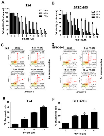
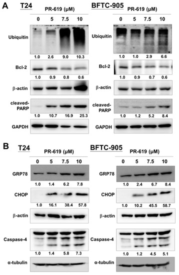
PR-619 是一种细胞可透过性广谱去泛素化酶(DBUs)抑制剂。PR-619 在不直接影响蛋白酶体活性的情况下,诱导细胞中多泛素化蛋白质的积聚。 在重组DUB酶实验中,PR-619 剂量依赖性抑制多种USP家族成员(USP1、USP2、USP5、USP7、USP14)和UCH-L1的去泛素化活性,IC50值范围为0.3-1.5 μM。浓度高达50 μM时,对蛋白酶体β5亚基活性无影响 [1] - 在一组人癌细胞系(HCT116、HeLa、A549、MDA-MB-231、U2OS)中,PR-619 表现出抗增殖活性,IC50值范围为2-8 μM。处理72小时后,5 μM浓度使不同细胞系的细胞活力降低55-70% [3] - 在HCT116结肠癌细胞中,PR-619(4 μM)处理24小时后,多聚泛素化蛋白积累(较对照组增加3.2倍)并诱导内质网应激,表现为CHOP(增加2.9倍)和BIP(增加2.5倍)蛋白水平上调 [1] - 在HeLa细胞中,PR-619(6 μM)处理18小时后诱导G2/M期细胞周期阻滞(G2/M期细胞比例从11%升至36%),处理48小时后诱导凋亡(膜联蛋白V阳性细胞比例从4%升至33%),胱天蛋白酶-3/7活性提高2.8倍 [3] - 在转染泛素-GFP报告基因的人胚肾(HEK293T)细胞中,PR-619(3 μM)处理12小时后,GFP阳性斑点增加4.1倍(较对照组),证实其抑制DUB介导的去泛素化作用 [2] |
| 体内研究 (In Vivo) |
PR-619(10 mg/kg/天)在顺铂未耐受和顺铂耐药的 UC 异种移植裸鼠中增强了顺铂的抗肿瘤作用[2]。 PR-619增强了顺铂对裸鼠顺铂钠和顺铂耐药UC异种移植物的抗肿瘤作用[2] 研究人员使用异种移植物小鼠模型评估了顺铂、PR-619治疗或顺铂和PR-619联合治疗的体内抗肿瘤作用。T24和BFTC-905UC细胞与Matrigel混合,皮下注射到裸鼠的侧翼。正如我们在“方法”一节中所述,根据不同的治疗方法,将小鼠分为四组:DMSO(对照组,n=5)、顺铂(n=5),PR-619(10mg/kg/天,n=5。与单药(顺铂或PR-619)治疗相比,顺铂和PR-619的联合治疗对T24和BFTC-905的异种移植物肿瘤显示出最显著的抗肿瘤作用(图6A,B)。除了提高化疗疗效的药物联合治疗外,一种规避顺铂耐药性的新型药物为这一临床未解决的问题提供了其他解决方案。我们进一步研究了PR-619对顺铂耐药UC(T24/R)的体外和体内抗肿瘤作用。PR-619在处理24小时后以剂量依赖的方式诱导细胞毒性和凋亡。使用异种移植物小鼠模型显示的体内数据显示,PR-619(10mg/kg/天)在28天的治疗期间抑制了肿瘤生长。 在荷HCT116结肠癌细胞异种移植瘤的裸鼠中,腹腔注射 PR-619(15 mg/kg,每周两次,持续3周)显著抑制肿瘤生长。与溶媒处理组相比,肿瘤体积减少65%,且无显著体重下降 [3] - 在同一异种移植模型中,PR-619(15 mg/kg)处理导致肿瘤组织中多聚泛素化蛋白积累(较溶媒组增加2.7倍),胱天蛋白酶-3激活(裂解型胱天蛋白酶-3水平增加2.3倍),证实了靶向DUB的抑制作用和凋亡诱导效果 [3] |
| 酶活实验 |
将重组酶置于 20 mM Tris-HCl、pH 8.0、2 mM CaCl2 和 2 mM β-巯基乙醇(DUB 检测缓冲液)中,与单剂量或剂量范围的PR-619或 P22077 在 96 孔板中预孵育 30 分钟,然后再进行实验。添加 Ub-PLA2 和 NBD C6-HPC。在室温下使用荧光板读数器监测测定线性范围内荧光产物的释放。载体 (2%(v/v) DMSO) 和 10 mM N-乙基马来酰亚胺作为对照。当观察到 ≥60% 的抑制时,使用 S 形剂量反应方程确定 EC50 值。
KCa3.1质膜降解速率的测定[3] 测定了内吞膜KCa3.1的降解速率。简而言之,极化MDCK、Caco-2或FRT细胞中的KCa3.1使用BirA进行特异性生物素化,并用非共轭链霉抗生物素蛋白标记,然后将细胞在37°C下孵育不同时间,如图所示。在某些实验中,在37°C孵育步骤之前,将溶酶体蛋白酶抑制剂亮肽(100μM)和蛋白胨(1μg/ml;Leu/Pep)、蛋白酶体抑制剂乳胱氨酸(10μM)或通用去泛素酶(DUB)抑制剂PR-619(50μM)添加到顶膜和BL膜中。然后裂解细胞,通过SDS-PAGE分离等量的总蛋白,然后通过IB分离链霉抗生物素蛋白。使用ImageJ软件通过密度测定对条带进行定量。将获得的各个时间点的带强度相对于时间0(T=0)的强度进行归一化,并进行报告。还检测了印迹中的α-微管蛋白和β-肌动蛋白作为蛋白质负载对照。 重组DUB活性实验:将纯化的重组USP1、USP2、USP5、USP7、USP14或UCH-L1与泛素-AMC(荧光底物)和 PR-619(0.01-20 μM)在实验缓冲液中于37°C孵育60分钟。检测荧光强度(激发光360 nm,发射光460 nm)以评估去泛素化活性,从剂量-效应抑制曲线计算IC50值 [1] - 蛋白酶体选择性实验:将纯化的20S蛋白酶体与Suc-LLVY-AMC(蛋白酶体β5亚基底物)和 PR-619(0.1-50 μM)在37°C孵育60分钟。检测荧光强度以评估蛋白酶体活性,若存在抑制则确定IC50值 [1] - DUB特异性实验:将20种重组DUB与各自的荧光底物和 PR-619(1 μM)在最适条件下孵育,定量去泛素化活性以评估泛抑制谱 [1] |
| 细胞实验 |
PR-619和顺铂的联合作用[2]
使用CalcuSyn软件确定PR-619和顺铂的联合作用。以1:2的比例用PR-619和顺铂治疗评估联合效果。如前所述,对中值效应和组合指数(CI)进行了分析。CI值小于1、等于1和大于1分别被定义为协同、相加和拮抗。 细胞凋亡检测[2] 根据制造商的方案,使用Muse®膜联蛋白V和死细胞检测试剂盒进行凋亡检测。然后,使用Muse®细胞分析仪并配备Muse分析软件(版本1.6.0.0)对染色的凋亡细胞进行检查和定量。 流式细胞术细胞周期分析[2] 接种细胞直至达到40%融合。然后用DMSO(对照)或PR-169处理细胞24小时。将细胞置于Muse®细胞周期分析试剂盒中,使用Muse®Cell Analyzer进行细胞周期分析,并配备Muse analysis软件。 72小时后,将0.2 mg/mL在磷酸盐缓冲盐水中制备的刃天青加入每个孔中,并将细胞再孵育3-6小时。使用荧光计上的Ex=535 nm和Em=590 nm滤光片测量刃天青还原产物的荧光。EC50值用Prism计算。 抗增殖实验:将癌细胞系(HCT116、HeLa、A549、MDA-MB-231、U2OS)以3×10³个/孔接种到96孔板中,培养24小时。加入浓度为0.5-50 μM的 PR-619,孵育72小时。MTT法评估细胞活力,推导IC50值 [3] - 多聚泛素化和内质网应激实验:将HCT116细胞以2×10⁵个/孔接种到6孔板中,用 PR-619(4 μM)处理24小时。裂解细胞后,通过特异性抗体Western blot分析多聚泛素化蛋白、CHOP和BIP水平 [1] - 细胞周期和凋亡实验:用 PR-619(6 μM)处理HeLa细胞18小时(细胞周期检测)或48小时(凋亡检测)。细胞周期分析采用固定细胞后碘化丙啶染色,流式细胞术检测;凋亡检测采用膜联蛋白V-FITC/PI染色,荧光素酶试剂盒检测胱天蛋白酶-3/7活性 [3] - 去泛素化报告基因实验:HEK293T细胞转染泛素-GFP质粒,培养24小时。用 PR-619(3 μM)处理12小时后固定细胞并DAPI染色,荧光显微镜计数GFP阳性斑点(泛素聚集物)[2] |
| 动物实验 |
裸鼠(HCT116异种移植模型):将6-8周龄的裸鼠皮下接种HCT116结肠癌细胞(5×10⁶个细胞/只)。当肿瘤体积达到约120 mm³时,将小鼠随机分为载体组和PR-619组。PR-619溶于DMSO,并用生理盐水稀释(最终DMSO浓度≤5%),以15 mg/kg的剂量腹腔注射,每周两次,持续3周。载体组小鼠注射DMSO/生理盐水混合液。每3天测量一次肿瘤体积,每周监测一次体重。切除肿瘤组织进行多聚泛素化蛋白和裂解型caspase-3的Western blot分析[3]
|
| 毒性/毒理 (Toxicokinetics/TK) |
在体内异种移植研究中,PR-619(15 mg/kg,腹腔注射,每周两次,持续3周)未引起裸鼠显著的体重下降(与基线相比变化≤5%)或明显的毒性[3]
- 在体外,PR-619对正常人成纤维细胞的毒性降低(IC50 > 30 μM),表明其在癌细胞和正常细胞之间存在治疗窗口[3] - 与载体对照组相比,PR-619治疗组小鼠的肝功能(ALT、AST)或肾功能(肌酐、BUN)均未观察到显著变化[3] |
| 参考文献 | |
| 其他信息 |
PR-619 的用途包括:
1) 作为裂解缓冲液的成分,以及用于处理 SILAC 标记的 Jurkat 细胞来源蛋白质的去泛素化酶抑制剂。 2) 作为去泛素化酶抑制剂,用于研究其对腺相关病毒 (AAV) 转导的影响。 3) 用于泛素化检测的放射免疫沉淀分析缓冲液 (RIPA)。 将先导化合物转化为候选药物是药物开发的关键步骤,需要尽早评估其效力、选择性和脱靶效应。我们利用基于活性的化学蛋白质组学方法,在细胞培养模型中测定了去泛素化酶 (DUB) 抑制剂的效力和选择性。重要的是,我们鉴定出小分子 PR-619 是一种广谱 DUB 抑制剂,而 P22077 是一种 USP7 抑制剂,具有进一步开发为癌症化疗药物的潜力。在不直接损害蛋白酶体蛋白水解的情况下,选择性或普遍抑制细胞内DUB活性后,观察到多聚泛素化蛋白的显著积累。通过串联质谱分析泛素化底物谱,鉴定出DUB普遍或特异性抑制的不同亚群。这使得我们能够发现USP7与DNA修复相关酶之间先前未知的功能联系。[1] 转移性膀胱尿路上皮癌(UC)患者在接受化疗后,大多数不可避免地会遇到耐药性,导致治疗失败。去泛素化酶(DUB)可去除靶蛋白上的泛素,在维持蛋白质稳态中发挥关键作用。本研究探讨了DUB抑制剂PR-619联合顺铂治疗膀胱UC的抗肿瘤效果。我们的研究结果表明,PR-619 能有效诱导人尿路上皮癌细胞(T24 和 BFTC-905)产生剂量和时间依赖性的细胞毒性、凋亡以及内质网应激相关的凋亡。此外,PR-619 与顺铂联合治疗可增强顺铂对尿路上皮癌细胞的细胞毒性,并伴有 Bcl-2 表达的抑制。我们还通过免疫组织化学(IHC)染色证实,Bcl-2 过表达与转移性尿路上皮癌患者的化疗耐药状态相关。在异种移植小鼠模型中,我们证实 PR-619 能增强顺铂对未接受过顺铂治疗和对顺铂耐药的尿路上皮癌的抗肿瘤作用。我们的研究结果表明,PR-619 通过抑制 Bcl-2 的表达水平,有效增强顺铂的抗肿瘤作用。这些发现为开发溃疡性结肠炎(UC)的治疗策略提供了有希望的见解。[2] 中等电导钙激活钾通道(KCa3.1)定位于极化上皮细胞的基底外侧膜(BL膜),并在跨上皮离子转运中发挥关键作用。然而,目前尚无研究阐明KCa3.1在极化上皮细胞中的顺行和逆行转运。本文中,我们利用生物素连接酶受体肽(BLAP)标记的KCa3.1,在MDCK、Caco-2和FRT细胞中研究了极化上皮细胞中的这些转运步骤。我们证明,当这些细胞在滤膜上培养时,KCa3.1仅定位于BL膜。内吞作用后,溶酶体/蛋白酶体途径的抑制可阻止KCa3.1的降解。此外,KCa3.1从基底外侧膜内吞后泛素化水平升高,而去泛素化酶抑制剂PR-619可阻止其降解,表明KCa3.1的降解是通过泛素化实现的。我们证实,在缺乏AP-1复合物μ1B亚基的极化LLC-PK1细胞中,KCa3.1仍能定位到基底外侧膜,表明KCa3.1的基底外侧膜定位与μ1B无关。由于Rab1、2、6和8在内质网/高尔基体输出以及蛋白质向基底外侧膜的转运中发挥作用,我们评估了这些Rab蛋白在KCa3.1转运中的作用。在显性负性Rab1或Rab8存在的情况下,KCa3.1的细胞表面表达显著降低,而Rab2和Rab6则无此作用。我们还进行了KCa3.1与Rab1和Rab8的共免疫沉淀实验。这些结果表明,这些Rab蛋白对于KCa3.1的顺行运输是必需的。最后,我们确定了KCa3.1在MDCK细胞中是直接运输到基底外侧膜(BL膜)还是通过循环内体运输。在这些研究中,我们使用了循环内体消融或显性负性RME-1构建体,并确定KCa3.1是直接运输到基底外侧膜(BL膜)而不是通过循环内体运输。这些结果首次描述了KCa3.1在极化上皮细胞中的顺行和逆行转运。[3] PR-619是一种细胞渗透性、可逆的泛泛泛素化酶(DUBs)抑制剂,对USP家族成员和UCH-L1具有优先活性[1] - 其作用机制涉及抑制DUB介导的去泛素化,导致多泛素化蛋白的积累、内质网应激、细胞周期阻滞,最终导致癌细胞凋亡[1][3] - PR-619被广泛用作研究DUBs在蛋白质稳态、细胞周期调控和癌症生物学中作用的工具化合物[1][2] - 它在体内对结肠癌异种移植瘤表现出抗肿瘤活性,支持DUB抑制作为一种潜在的癌症治疗策略[3] |
| 分子式 |
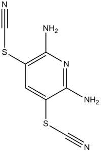
C7H5N5S2
|
|
|---|---|---|
| 分子量 |
223.28
|
|
| 精确质量 |
222.998
|
|
| 元素分析 |
C, 37.66; H, 2.26; N, 31.37; S, 28.72
|
|
| CAS号 |
2645-32-1
|
|
| 相关CAS号 |
|
|
| PubChem CID |
2817763
|
|
| 外观&性状 |
Dark mustard yellow fluffy powder
|
|
| 密度 |
1.6±0.1 g/cm3
|
|
| 沸点 |
406.0±45.0 °C at 760 mmHg
|
|
| 熔点 |
210℃
|
|
| 闪点 |
199.3±28.7 °C
|
|
| 蒸汽压 |
0.0±0.9 mmHg at 25°C
|
|
| 折射率 |
1.764
|
|
| LogP |
2.05
|
|
| tPSA |
163.84
|
|
| 氢键供体(HBD)数目 |
2
|
|
| 氢键受体(HBA)数目 |
7
|
|
| 可旋转键数目(RBC) |
2
|
|
| 重原子数目 |
14
|
|
| 分子复杂度/Complexity |
261
|
|
| 定义原子立体中心数目 |
0
|
|
| SMILES |
S(C#N)C1C(N([H])[H])=NC(=C(C=1[H])SC#N)N([H])[H]
|
|
| InChi Key |
ZXOBLNBVNROVLC-UHFFFAOYSA-N
|
|
| InChi Code |
InChI=1S/C7H5N5S2/c8-2-13-4-1-5(14-3-9)7(11)12-6(4)10/h1H,(H4,10,11,12)
|
|
| 化学名 |
(2,6-diamino-5-thiocyanatopyridin-3-yl) thiocyanate
|
|
| 别名 |
|
|
| HS Tariff Code |
2934.99.9001
|
|
| 存储方式 |
Powder -20°C 3 years 4°C 2 years In solvent -80°C 6 months -20°C 1 month |
|
| 运输条件 |
Room temperature (This product is stable at ambient temperature for a few days during ordinary shipping and time spent in Customs)
|
| 溶解度 (体外实验) |
DMSO : 3~21 mg/mL ( 13.43~94.05 mM)
|
|---|---|
| 溶解度 (体内实验) |
配方 1 中的溶解度: 2.5 mg/mL (11.20 mM) in 10% DMSO + 40% PEG300 +5% Tween-80 + 45% Saline (这些助溶剂从左到右依次添加,逐一添加), 悬浮液;超声助溶。
例如,若需制备1 mL的工作液,可将100 μL 25.0 mg/mL澄清DMSO储备液加入到400 μL PEG300中,混匀;然后向上述溶液中加入50 μL Tween-80+,混匀;加入450 μL生理盐水定容至1 mL。 *生理盐水的制备:将 0.9 g 氯化钠溶解在 100 mL ddH₂O中,得到澄清溶液。 请根据您的实验动物和给药方式选择适当的溶解配方/方案: 1、请先配制澄清的储备液(如:用DMSO配置50 或 100 mg/mL母液(储备液)); 2、取适量母液,按从左到右的顺序依次添加助溶剂,澄清后再加入下一助溶剂。以 下列配方为例说明 (注意此配方只用于说明,并不一定代表此产品 的实际溶解配方): 10% DMSO → 40% PEG300 → 5% Tween-80 → 45% ddH2O (或 saline); 假设最终工作液的体积为 1 mL, 浓度为5 mg/mL: 取 100 μL 50 mg/mL 的澄清 DMSO 储备液加到 400 μL PEG300 中,混合均匀/澄清;向上述体系中加入50 μL Tween-80,混合均匀/澄清;然后继续加入450 μL ddH2O (或 saline)定容至 1 mL; 3、溶剂前显示的百分比是指该溶剂在最终溶液/工作液中的体积所占比例; 4、 如产品在配制过程中出现沉淀/析出,可通过加热(≤50℃)或超声的方式助溶; 5、为保证最佳实验结果,工作液请现配现用! 6、如不确定怎么将母液配置成体内动物实验的工作液,请查看说明书或联系我们; 7、 以上所有助溶剂都可在 Invivochem.cn网站购买。 |
| 制备储备液 | 1 mg | 5 mg | 10 mg | |
| 1 mM | 4.4787 mL | 22.3934 mL | 44.7868 mL | |
| 5 mM | 0.8957 mL | 4.4787 mL | 8.9574 mL | |
| 10 mM | 0.4479 mL | 2.2393 mL | 4.4787 mL |
1、根据实验需要选择合适的溶剂配制储备液 (母液):对于大多数产品,InvivoChem推荐用DMSO配置母液 (比如:5、10、20mM或者10、20、50 mg/mL浓度),个别水溶性高的产品可直接溶于水。产品在DMSO 、水或其他溶剂中的具体溶解度详见上”溶解度 (体外)”部分;
2、如果您找不到您想要的溶解度信息,或者很难将产品溶解在溶液中,请联系我们;
3、建议使用下列计算器进行相关计算(摩尔浓度计算器、稀释计算器、分子量计算器、重组计算器等);
4、母液配好之后,将其分装到常规用量,并储存在-20°C或-80°C,尽量减少反复冻融循环。
计算结果:
工作液浓度: mg/mL;
DMSO母液配制方法: mg 药物溶于 μL DMSO溶液(母液浓度 mg/mL)。如该浓度超过该批次药物DMSO溶解度,请首先与我们联系。
体内配方配制方法:取 μL DMSO母液,加入 μL PEG300,混匀澄清后加入μL Tween 80,混匀澄清后加入 μL ddH2O,混匀澄清。
(1) 请确保溶液澄清之后,再加入下一种溶剂 (助溶剂) 。可利用涡旋、超声或水浴加热等方法助溶;
(2) 一定要按顺序加入溶剂 (助溶剂) 。
|
|
|
|
 Ubiquitin thiolesterase UCHL1
 USP30-IN-20
 WCY-8-67
 Huib32
InvivoChem的所有产品仅用于作科学研究,不面向患者销售
Copyright 2020 InvivoChem LLC | All Rights Reserved 粤ICP备20063088号-1


 COA
COA


 463611831
