| 规格 | 价格 | 库存 | 数量 |
|---|---|---|---|
| 10mg |
|
||
| 50mg |
|
||
| 100mg |
|
||
| 1g |
|
||
| 5g |
|
||
| 10g |
|
||
| Other Sizes |
|
| 靶点 |
Natural tetrahydroisoquinoline
Tetrahydropapaverine HCl primarily targets phosphodiesterase 1 (PDE1), with an IC50 value of 2.3 μM for rat myocardial PDE1; it shows no significant binding affinity to other PDE subtypes (e.g., PDE5, PDE6) at concentrations up to 100 μM [1] - Tetrahydropapaverine HCl indirectly modulates intracellular calcium channels (via PDE1 inhibition-mediated cGMP elevation) [2,3] |
|---|---|
| 体外研究 (In Vitro) |
四氢罂粟碱是 TIQ 之一,也是 Salsolinol 和四氢罂粟碱的类似物,据报道对多巴胺神经元具有神经毒性作用。 Tetrahydropapaverine 抑制产生血清素的小鼠肥大细胞瘤 P815 细胞中的血清素生物合成,IC50 为 7.5 μM,并降低色氨酸羟化酶活性,IC50 为 5.7 μM。
研究了四氢罂粟碱对产生血清素的小鼠肥大细胞瘤P815细胞血清素生物合成的抑制作用。四氢罂粟碱在5-20 μ m浓度范围内以浓度依赖的方式降低P815细胞血清素含量,在5.0 μ m浓度下,24小时血清素含量抑制率为42.1%。四氢罂粟碱50%抑制浓度(IC50)为6.2 μ m。在此条件下,四氢罂粟碱对P815细胞色氨酸羟化酶(EC 1.14.16.4, TPH)的抑制作用达到24-36小时(7.5 μ m时抑制49.1%)。而四氢罂粟碱对芳香族l -氨基酸脱羧酶活性没有影响。此外,四氢罂粟碱对P815细胞(P815-TPH)制备的TPH活性有抑制作用,IC50值为5.7 μ m。四氢罂粟碱与底物l-色氨酸无竞争性抑制P815-TPH,与辅因子dl -6-甲基-5,6,7,8-四氢罂粟碱无竞争性抑制P815-TPH。四氢罂粟碱与l -色氨酸的Ki值为10.1 μ m。这些数据表明,四氢罂粟碱通过抑制P815细胞的TPH活性导致血清素含量降低。[1] 研究人员报道了罂粟碱、四氢罂粟碱、二甲氧基苯基乙胺(DMPEA)和1-甲基-4-苯基吡啶离子(MPP+)对腹侧中脑-纹状体共培养的多巴胺能神经元的神经毒性作用。这些化合物已被报道为线粒体毒素,可能与帕金森病的病因和发病机制有关。酪氨酸羟化酶(TH)阳性神经元呈剂量依赖性减少。罂粟碱和MPP+对th阳性神经元的毒性最大。对th阳性神经元的毒性程度依次为罂粟碱、MPP+、四氢罂粟碱、DMPEA。这种毒性顺序与报道的这些化合物对nadh相关的线粒体呼吸和复合体I活性的抑制作用大致相同。这些发现表明,儿茶酚环中二甲氧基残基的存在增加了培养中多巴胺能神经元的毒性。[2] 在大鼠心肌组织提取物的PDE1活性检测中,盐酸四氢罂粟碱(Tetrahydropapaverine HCl)(0.1-100 μM)呈剂量依赖性抑制PDE1活性,IC50为2.3 μM。在10 μM浓度下,药物对PDE1活性的抑制率较溶媒对照组达78%[1] - 在原代培养的大鼠皮质神经元中,用盐酸四氢罂粟碱(Tetrahydropapaverine HCl)(1-50 μM)预处理1小时,可显著减轻谷氨酸诱导的神经毒性。10 μM浓度时,药物可将细胞活力从谷氨酸单独处理组的35%(MTT法检测)恢复至78%,并降低细胞内钙浓度升高(从620 nM降至310 nM,通过Fura-2 AM荧光检测)[2] - 在暴露于缺氧环境(1% O2,6小时)的原代大鼠海马神经元中,盐酸四氢罂粟碱(Tetrahydropapaverine HCl)(5 μM,缺氧前2小时加入)可将乳酸脱氢酶(LDH)释放率从缺氧单独处理组的58%降至29%,表明细胞损伤减轻[3] |
| 体内研究 (In Vivo) |
研究人员报告了3,4-二甲氧基苯乙胺(DMPEA)和四氢罂粟碱(THP)对大鼠黑纹状体系统的毒性作用;THP是一种四氢异喹啉化合物,可由DMPEA及其氧化代谢物二甲氧基苯乙醛偶联而得;这两种化合物都是线粒体复合物i的有效抑制剂。这些化合物通过200微利微渗透泵注入雄性Sprague-Dawley大鼠单侧尾壳核7天。注射侧纹状体多巴胺在注射DMPEA 16.55微mol/7 d后显著降低至非注射侧的86%,注射7.90微mol/7 d后显著降低至非注射侧的73%;由于注射thp的大鼠未注射侧多巴胺也减少,注射侧多巴胺为生理盐水对照组的55%。DMPEA注射16.55微mol/7 d后,酪氨酸羟化酶(TH)阳性的黑质神经元减少到未注射侧的76%,THP注射7.90微mol/7 d后减少到77%。二甲氧基苯基-四氢异喹啉化合物似乎是强效的神经毒素。 [3]
在心肌缺血再灌注损伤的雄性SD大鼠(缺血30分钟,再灌注2小时)中,再灌注开始时静脉注射盐酸四氢罂粟碱(Tetrahydropapaverine HCl)(3 mg/kg),可将心肌梗死面积从45%降至22%(TTC染色)。同时,血清肌酸激酶(CK)活性从2850 U/L降至1240 U/L,乳酸脱氢酶(LDH)活性从1980 U/L降至920 U/L[1] - 在局灶性脑缺血的雄性Wistar大鼠(大脑中动脉阻塞[MCAO]2小时,再灌注24小时)中,MCAO前30分钟腹腔注射盐酸四氢罂粟碱(Tetrahydropapaverine HCl)(10 mg/kg),可将脑梗死体积从38%降至18%(TTC染色),并将Bederson神经功能评分从3.5分改善至1.2分(0分=无缺损,4分=严重缺损)[3] |
| 酶活实验 |
PDE1活性抑制实验:取新鲜大鼠心肌组织,用裂解缓冲液匀浆,离心(10,000×g,15分钟,4°C)获取含PDE1的上清液。100 μL反应体系包含50 mM Tris-HCl(pH 7.5)、5 mM MgCl2、1 μM [3H]-cGMP(底物)、不同浓度的盐酸四氢罂粟碱(Tetrahydropapaverine HCl)(0.1-100 μM)及PDE1上清液。37°C孵育30分钟启动反应,随后加入100 μL 0.5 M ZnSO4和100 μL 0.5 M Ba(OH)2终止反应并沉淀蛋白。离心(3000×g,10分钟,4°C)后收集上清液,用液体闪烁计数仪检测放射性,计算PDE1活性(每分钟计数,cpm),并以溶媒对照组为基准计算抑制率,通过非线性回归分析获得IC50值[1]
|
| 细胞实验 |
四氢罂粟碱处理P815细胞显著降低细胞内血清素含量呈浓度依赖性。在5.0 μM浓度下,四氢罂粟碱可使血清素含量降低57.9%(表1),IC50值为6.2 μM(表1)。在此条件下,四氢罂粟碱可显著抑制细胞内TPH活性(7.5 μM浓度下抑制49.1%),而AADC活性未受影响。[1]
原代大鼠皮质神经元培养及谷氨酸神经毒性实验:取胚胎18天(E18)大鼠的皮质组织,用胰蛋白酶(0.25%,37°C,15分钟)消化,中和后吹打成单细胞悬液。将细胞接种于多聚赖氨酸包被的96孔板(5×10⁴细胞/孔),培养基为DMEM/F12(添加10%胎牛血清、2 mM谷氨酰胺、50 U/mL青霉素、50 μg/mL链霉素),在37°C、5% CO2条件下培养7天。实验分组:对照组(仅培养基)、谷氨酸组(100 μM)、盐酸四氢罂粟碱(Tetrahydropapaverine HCl)处理组(1-50 μM,谷氨酸处理前1小时加入)。谷氨酸暴露24小时后,每孔加入20 μL MTT(5 mg/mL),孵育4小时后加入150 μL DMSO溶解甲瓒结晶,在570 nm波长下检测吸光度,计算细胞活力(以对照组为100%)[2] - 细胞内钙浓度检测实验:培养7天的皮质神经元用2 μM Fura-2 AM的HBSS溶液孵育30分钟(37°C),随后用PBS冲洗。加入盐酸四氢罂粟碱(Tetrahydropapaverine HCl)(1-50 μM)孵育1小时,再加入100 μM谷氨酸。通过激光共聚焦显微镜检测荧光强度(激发波长:340 nm/380 nm;发射波长:510 nm),以340/380荧光比值量化细胞内钙水平[2] - 海马神经元缺氧损伤实验:取新生1天(P1)大鼠的海马组织,培养于Neurobasal培养基(含2% B27添加剂、2 mM谷氨酰胺)中10天。实验分组:正常氧组、缺氧组(1% O2、5% CO2、94% N2,6小时)、盐酸四氢罂粟碱(Tetrahydropapaverine HCl)处理组(5 μM,缺氧前2小时加入)。缺氧处理后收集培养上清液,用商品化试剂盒检测LDH活性,以正常氧组为基准计算LDH释放率(反映细胞损伤程度)[3] |
| 动物实验 |
大鼠心肌缺血再灌注模型:雄性SD大鼠(250-300 g)饲养于22±2℃、12小时光照/12小时黑暗循环的环境中,自由摄食饮水。术前大鼠禁食12小时(自由饮水),并用戊巴比妥钠(40 mg/kg,腹腔注射)麻醉。气管插管并通气后,开胸暴露心脏,用丝线结扎左前降支冠状动脉(LAD)(心肌苍白证实缺血)。缺血30分钟后,松开结扎线进行再灌注。再灌注开始时,经尾静脉注射盐酸四氢罂粟碱(溶于生理盐水,1 mg/mL),剂量为1 mg/kg或3 mg/kg。对照组接受等体积生理盐水。再灌注2小时后,经腹主动脉采集血液,测定血清CK和LDH活性。取出心脏,切成2 mm厚的切片,并用1% TTC染色(37℃,15分钟)。使用Image-Pro Plus软件[1]分析梗死面积(白色区域)与左心室面积的比值。
- 大鼠局灶性脑缺血模型:雄性Wistar大鼠(280-320 g)用水合氯醛(350 mg/kg,腹腔注射)麻醉。分离右侧颈总动脉、颈外动脉和颈内动脉。将一根硅胶涂层尼龙线(直径0.26 mm)插入颈内动脉,以阻断大脑中动脉(MCAO,插入深度:18-20 mm)。闭塞2小时后,移除线进行再灌注。在MCAO前30分钟,腹腔注射四氢罂粟碱盐酸盐(溶于5% DMSO + 生理盐水,2 mg/mL),剂量分别为5 mg/kg或10 mg/kg;对照组注射等体积的溶剂。再灌注24小时后,采用Bederson评分评估神经功能。取出脑组织,切成2 mm厚的冠状切片,并用2% TTC染色(37°C,20分钟)。使用ImageJ软件[3]分析梗死体积(白色区域)占总脑体积的比例。 |
| 药代性质 (ADME/PK) |
在大鼠静脉注射盐酸四氢罂粟碱(3 mg/kg)后,分别于 5、15、30、60 和 120 分钟时采用高效液相色谱法 (HPLC) 测定血浆药物浓度。该药物的消除半衰期较短(t1/2 = 28 分钟)。给药后 15 分钟,心肌药物浓度达到峰值,为 125 ng/g 组织 [1]。在大鼠腹腔注射盐酸四氢罂粟碱(10 mg/kg)后,于再灌注 2 小时后采用高效液相色谱法 (HPLC) 测定脑组织药物浓度。大脑皮层和海马的药物浓度分别为 89 ng/g 组织和 95 ng/g 组织,证实了该药物能够穿透血脑屏障 [3]。
|
| 毒性/毒理 (Toxicokinetics/TK) |
雄性SD大鼠急性毒性研究:单次静脉注射盐酸四氢罂粟碱(20、40、60、80 mg/kg),观察7天。LD50为58 mg/kg。剂量≤20 mg/kg时,未观察到异常行为或血清肝酶(ALT、AST)或肾功能指标(BUN、Cr)的变化。40 mg/kg剂量下,部分大鼠出现短暂性呼吸急促,但30分钟内缓解[1]。亚急性毒性研究:连续7天腹腔注射盐酸四氢罂粟碱(10 mg/kg、20 mg/kg),与对照组相比,血清ALT、AST、BUN或Cr均无显著差异。脑组织病理学检查未发现神经元变性或炎症细胞浸润[3]
|
| 参考文献 | |
| 其他信息 |
四氢异喹啉类化合物(TIQs)已被广泛研究,证实具有神经毒性(Nagatsu,1997)和抑制多巴胺生物合成的作用(Kim等,2001;Shih等,1999)。TIQs的结构与1-甲基-4-苯基-1,2,3,6-四氢吡啶(MPTP)相似,后者可导致人类和非人灵长类动物出现帕金森样综合征(McNaught等,1998)。在接受左旋多巴治疗的帕金森病患者的尿液中,已检测到萨尔索林醇和四氢罂粟碱(Sandler等,1973)(图1)。研究发现,沙索林醇能够抑制酪氨酸羟化酶(EC 1.14.16.2)的活性,酪氨酸羟化酶是儿茶酚胺生物合成途径的限速酶(Minami等,1992),以及TPH(血清素生物合成的限速酶)(Ota等,1992)。四氢罂粟碱也能抑制酪氨酸羟化酶的活性(Lee等,2001a)。
最近有报道称,四氢罂粟碱通过抑制PC12细胞中的酪氨酸羟化酶来抑制多巴胺的生物合成(Lee等,2001a),并且还能通过抑制TPH来降低小鼠肥大细胞瘤P815细胞中的血清素含量(Kim等,2003)。此外,四氢罂粟碱已被证实能以非竞争性方式抑制底物L-色氨酸的TPH活性(Kim等,2003)。四氢罂粟碱是TIQ类化合物之一,也是萨尔索林醇和四氢罂粟碱的类似物,据报道其对多巴胺能神经元具有神经毒性作用(Koshimura等,1997)(图1)。然而,四氢罂粟碱对吲哚胺生物合成或代谢的影响尚未得到评估。已知小鼠肥大细胞瘤P815细胞能产生5-羟色胺,且具有较高的TPH活性(Schindler等,1959)。 P815细胞也表达组胺和L-组氨酸脱羧酶(Schindler等,1959;Imanishi等,1987)。因此,本研究旨在探讨四氢罂粟碱对P815细胞中5-羟色胺生物合成和TPH活性的抑制作用。TPH酶源由P815细胞制备(P815-TPH)。[1] 1-甲基-4-苯基-1,2,3,6-四氢吡啶(MPTP)是一种神经毒素,可诱发灵长类动物的长期帕金森综合征。MPTP诱发的帕金森综合征的临床特征与黑质纹状体系统中多巴胺能神经元的丢失和多巴胺的耗竭相关。因此,人们推测帕金森病可能是由内源性或环境神经毒素(如MPTP)在大脑中生物活化而诱发的,这一概念引发了对潜在的黑质神经毒素(包括内源性和外源性毒素)的大量研究。在这些化合物中,四氢异喹啉(TIQ)和β-咔啉的研究最为广泛。 TIQ存在于奶酪、葡萄酒和可可等食物中,易于被运输到大脑,并且在帕金森病患者和正常人的大脑中均发现了某些TIQ化合物。在猴子身上长期服用TIQ会产生类似帕金森病的症状,并显著降低黑质中的多巴胺和酪氨酸羟化酶(TH)活性。TIQ经N-甲基转移酶代谢为N-甲基-TIQ,后者再经单胺氧化酶氧化为N-甲基-四氢异喹啉鎓离子。该离子抑制酪氨酸羟化酶(TH)和线粒体复合物I的活性,并抑制组织培养中的神经元生长。 源自吲哚胺的β-咔啉结构与MPP+密切相关,一些衍生物已在人脑中被发现,并被证实可抑制多巴胺摄取、单胺氧化酶活性、线粒体呼吸作用以及培养的PC12细胞的生长。 在我们研究这些化合物的线粒体毒性时,我们发现儿茶酚环上二甲氧基化的化合物是更有效的线粒体呼吸抑制剂。这一观察促使我们研究这些化合物对培养的多巴胺能神经元的毒性。 在本研究中,我们使用了分离的中脑-纹状体共培养模型,因为与传统的中脑培养相比,这种模型能更接近地模拟体内环境,其中包含来自靶纹状体神经元的营养因子,从而使多巴胺能神经元的树突分支更加丰富。本研究中测试的二甲氧基化合物包括四氢罂粟碱、罂粟碱、它们的前体二甲氧基苯乙胺(DMPEA)以及作为阳性对照的1-甲基-4-苯基吡啶离子(MPP+)(图1)。[2] 盐酸四氢罂粟碱是一种合成的罂粟碱衍生物,其PDE1抑制活性高于罂粟碱;它缺乏阿片类镇痛作用和成瘾性[1] - 盐酸四氢罂粟碱通过抑制PDE1、增加细胞内cGMP水平、扩张冠状动脉、改善心肌供血和抑制心肌细胞凋亡来减轻心肌缺血再灌注损伤,支持其在急性心肌梗死治疗中的潜力[1] - 体外研究表明,盐酸四氢罂粟碱通过抑制细胞内钙超载来保护皮层神经元免受谷氨酸诱导的兴奋性毒性,提示其在神经退行性疾病(例如阿尔茨海默病)或创伤性脑损伤中的应用[2] - 在局灶性脑缺血模型中,盐酸四氢罂粟碱通过血脑屏障穿透减少梗死体积并改善神经功能;其作用机制可能包括抑制PDE1(提高脑内cGMP水平)、抗炎作用和减少神经元凋亡[3] |
| 分子式 |
C20H25NO4.HCL
|
|---|---|
| 分子量 |
379.88
|
| 精确质量 |
379.155
|
| CAS号 |
6429-04-5
|
| 相关CAS号 |
(R)-Tetrahydropapaverine hydrochloride;54417-53-7
|
| PubChem CID |
16667431
|
| 外观&性状 |
Typically exists as solid at room temperature
|
| 密度 |
1.12g/cm3
|
| 沸点 |
475.8ºC at 760 mmHg
|
| 熔点 |
213-215ºC
|
| 闪点 |
202.7ºC
|
| 折射率 |
1.549
|
| LogP |
0.697
|
| tPSA |
53.53
|
| 氢键供体(HBD)数目 |
2
|
| 氢键受体(HBA)数目 |
5
|
| 可旋转键数目(RBC) |
6
|
| 重原子数目 |
26
|
| 分子复杂度/Complexity |
407
|
| 定义原子立体中心数目 |
0
|
| SMILES |
Cl[H].O(C([H])([H])[H])C1=C(C([H])=C2C([H])([H])C([H])([H])N([H])C([H])(C([H])([H])C3C([H])=C([H])C(=C(C=3[H])OC([H])([H])[H])OC([H])([H])[H])C2=C1[H])OC([H])([H])[H]
|
| InChi Key |
VMPLLPIDRGXFTQ-UHFFFAOYSA-N
|
| InChi Code |
InChI=1S/C20H25NO4.ClH/c1-22-17-6-5-13(10-18(17)23-2)9-16-15-12-20(25-4)19(24-3)11-14(15)7-8-21-16;/h5-6,10-12,16,21H,7-9H2,1-4H3;1H
|
| 化学名 |
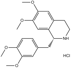
1-[(3,4-dimethoxyphenyl)methyl]-6,7-dimethoxy-1,2,3,4-tetrahydroisoquinoline;hydrochloride
|
| 别名 |
6429-04-5; Tetrahydropapaverine hydrochloride; 1-(3,4-dimethoxybenzyl)-6,7-dimethoxy-1,2,3,4-tetrahydroisoquinoline hydrochloride; Tetrahydropapaverine HCl; DL-Norlaudanosine hydrochloride; Norlaudanosine Hydrochloride; 1-[(3,4-dimethoxyphenyl)methyl]-6,7-dimethoxy-1,2,3,4-tetrahydroisoquinoline hydrochloride; 1-[(3,4-dimethoxyphenyl)methyl]-6,7-dimethoxy-1,2,3,4-tetrahydroisoquinoline;hydrochloride; Norlaudanosine HCl
|
| HS Tariff Code |
2934.99.9001
|
| 存储方式 |
Powder -20°C 3 years 4°C 2 years In solvent -80°C 6 months -20°C 1 month 注意: 请将本产品存放在密封且受保护的环境中,避免吸湿/受潮。 |
| 运输条件 |
Room temperature (This product is stable at ambient temperature for a few days during ordinary shipping and time spent in Customs)
|
| 溶解度 (体外实验) |
|
|||
|---|---|---|---|---|
| 溶解度 (体内实验) |
配方 1 中的溶解度: ≥ 2.5 mg/mL (6.58 mM) (饱和度未知) in 10% DMSO + 40% PEG300 + 5% Tween80 + 45% Saline (这些助溶剂从左到右依次添加,逐一添加), 澄清溶液。
例如,若需制备1 mL的工作液,可将100 μL 25.0 mg/mL澄清DMSO储备液加入到400 μL PEG300中,混匀;然后向上述溶液中加入50 μL Tween-80,混匀;加入450 μL生理盐水定容至1 mL。 *生理盐水的制备:将 0.9 g 氯化钠溶解在 100 mL ddH₂O中,得到澄清溶液。 配方 2 中的溶解度: ≥ 2.5 mg/mL (6.58 mM) (饱和度未知) in 10% DMSO + 90% (20% SBE-β-CD in Saline) (这些助溶剂从左到右依次添加,逐一添加), 澄清溶液。 例如,若需制备1 mL的工作液,可将 100 μL 25.0 mg/mL澄清DMSO储备液加入900 μL 20% SBE-β-CD生理盐水溶液中,混匀。 *20% SBE-β-CD 生理盐水溶液的制备(4°C,1 周):将 2 g SBE-β-CD 溶解于 10 mL 生理盐水中,得到澄清溶液。 View More
配方 3 中的溶解度: ≥ 2.5 mg/mL (6.58 mM) (饱和度未知) in 10% DMSO + 90% Corn Oil (这些助溶剂从左到右依次添加,逐一添加), 澄清溶液。 1、请先配制澄清的储备液(如:用DMSO配置50 或 100 mg/mL母液(储备液)); 2、取适量母液,按从左到右的顺序依次添加助溶剂,澄清后再加入下一助溶剂。以 下列配方为例说明 (注意此配方只用于说明,并不一定代表此产品 的实际溶解配方): 10% DMSO → 40% PEG300 → 5% Tween-80 → 45% ddH2O (或 saline); 假设最终工作液的体积为 1 mL, 浓度为5 mg/mL: 取 100 μL 50 mg/mL 的澄清 DMSO 储备液加到 400 μL PEG300 中,混合均匀/澄清;向上述体系中加入50 μL Tween-80,混合均匀/澄清;然后继续加入450 μL ddH2O (或 saline)定容至 1 mL; 3、溶剂前显示的百分比是指该溶剂在最终溶液/工作液中的体积所占比例; 4、 如产品在配制过程中出现沉淀/析出,可通过加热(≤50℃)或超声的方式助溶; 5、为保证最佳实验结果,工作液请现配现用! 6、如不确定怎么将母液配置成体内动物实验的工作液,请查看说明书或联系我们; 7、 以上所有助溶剂都可在 Invivochem.cn网站购买。 |
| 制备储备液 | 1 mg | 5 mg | 10 mg | |
| 1 mM | 2.6324 mL | 13.1621 mL | 26.3241 mL | |
| 5 mM | 0.5265 mL | 2.6324 mL | 5.2648 mL | |
| 10 mM | 0.2632 mL | 1.3162 mL | 2.6324 mL |
1、根据实验需要选择合适的溶剂配制储备液 (母液):对于大多数产品,InvivoChem推荐用DMSO配置母液 (比如:5、10、20mM或者10、20、50 mg/mL浓度),个别水溶性高的产品可直接溶于水。产品在DMSO 、水或其他溶剂中的具体溶解度详见上”溶解度 (体外)”部分;
2、如果您找不到您想要的溶解度信息,或者很难将产品溶解在溶液中,请联系我们;
3、建议使用下列计算器进行相关计算(摩尔浓度计算器、稀释计算器、分子量计算器、重组计算器等);
4、母液配好之后,将其分装到常规用量,并储存在-20°C或-80°C,尽量减少反复冻融循环。
计算结果:
工作液浓度: mg/mL;
DMSO母液配制方法: mg 药物溶于 μL DMSO溶液(母液浓度 mg/mL)。如该浓度超过该批次药物DMSO溶解度,请首先与我们联系。
体内配方配制方法:取 μL DMSO母液,加入 μL PEG300,混匀澄清后加入μL Tween 80,混匀澄清后加入 μL ddH2O,混匀澄清。
(1) 请确保溶液澄清之后,再加入下一种溶剂 (助溶剂) 。可利用涡旋、超声或水浴加热等方法助溶;
(2) 一定要按顺序加入溶剂 (助溶剂) 。
|
|---|
|
 ZnPc-O3-JQ1
 EZN-2968 sodium scrambled negative control
 Davotifan
 SK-129
InvivoChem的所有产品仅用于作科学研究,不面向患者销售
Copyright 2020 InvivoChem LLC | All Rights Reserved 粤ICP备20063088号-1


 COA
COA


 463611831
