| 规格 | 价格 | 库存 | 数量 |
|---|---|---|---|
| 10 mM * 1 mL in DMSO |
|
||
| 1mg |
|
||
| 5mg |
|
||
| 10mg |
|
||
| 25mg |
|
||
| 50mg |
|
||
| 100mg |
|
||
| 250mg |
|
||
| 500mg |
|
||
| 1g |
|
||
| Other Sizes |
|
| 靶点 |
EGFRL858R (IC50 = 0.4 nM); EGFR (wt) (IC50 = 0.5 nM); ErbB4 (IC50 = 1 nM); EGFRL858R/T790M (IC50 = 10 nM); HER2 (IC50 = 14 nM)
- EGFR (wild-type):Afatinib (BIBW2992) inhibits wild-type EGFR with an IC₅₀ of 0.5 nM. [1] - EGFR (L858R mutant):Exhibits inhibitory activity against the L858R mutant with an IC₅₀ of 0.4 nM. [1] - EGFR (exon 19 deletion mutant):Inhibits exon 19 deletion mutant EGFR with an IC₅₀ of 0.3 nM. [1] - HER2 (ErbB2):Inhibits HER2 kinase activity with an IC₅₀ of 14 nM. [1] Afatinib (BIBW2992) inhibits EGFR (IC₅₀ = 0.5 nM), HER2 (IC₅₀ = 14 nM), and HER4 (IC₅₀ = 1 nM) tyrosine kinases [1] Afatinib (BIBW2992) shows inhibitory activity against EGFR T790M mutant (IC₅₀ = 10 nM) and wild-type EGFR (IC₅₀ = 0.4 nM) [2] |
|---|---|
| 体外研究 (In Vitro) |
- 抗增殖活性:afatinib在MTT实验中抑制EGFR突变型非小细胞肺癌(NSCLC)细胞系(HCC827、PC-9),IC₅₀为10–20 nM;抑制HER2+乳腺癌细胞(SK-BR-3),IC₅₀为30 nM。[1][2]
- 信号通路抑制:在HCC827细胞中,afatinib(50 nM,4小时)通过Western blot检测显示,EGFR(Tyr1068)、AKT(Ser473)和ERK1/2(Thr202/Tyr204)的磷酸化水平分别降低90%、85%和80%,同时下调cyclin D1并上调cleaved PARP,提示诱导凋亡。[1][2] - 与放疗协同作用:在头颈部鳞状细胞癌(HNSCC)细胞(SCC-25)中,afatinib(10 nM)增强放疗诱导的细胞杀伤,辐射敏感系数提高1.5倍。[3] BIBW2992 对野生型和突变型 EGFR 和 HER2 均显示出有效的活性。它针对 L858R EGFR 的效力与吉非替尼相似,但针对吉非替尼耐药的 L858R-T790M EGFR 双突变体的活性高出约 100 倍。 BIBW2992 对体内 EGFR 和 HER2 磷酸化表现出有效的作用。在所有测试的细胞类型中,例如表达 wt EGFR 的人表皮样癌细胞系 A431、转染 wt HER2 的鼠 NIH-3T3 细胞以及乳腺癌细胞系 BT,其与参考化合物(例如 Lapatinib 等)相比均具有优势-474和胃癌细胞系NCI-N87,表达内源性HER2。激酶测定:将人 EGFR 的野生型酪氨酸激酶结构域以及 EGFR L858R/T790M 双突变体的酪氨酸激酶结构域与谷胱甘肽-S-转移酶 (GST) 融合并提取。然后在抑制剂 BIBW2992(在 50% DMSO 中连续稀释)存在的情况下测定酶活性。使用随机聚合物 pEY (4:1) 作为底物,并添加生物素化 pEY (bio-pEY) 作为示踪底物。使用杆状病毒系统克隆 HER2 的激酶结构域,并以与 EGFR 激酶类似的方式进行提取。补充信息中提供了 EGFR、HER2、SRC、BIRK 和 VEGFR2 激酶活性测定的详细信息。细胞测定:将 1 × 10⁴ 个 NSCLC 细胞转移至 96 孔板的每个孔中,并在无血清培养基中培养过夜,用于 EGFR 磷酸化测定。第二天添加 BIBW2992 后,将板在 37°C 下孵育 1 小时。 EGF 刺激使用 100 ng/mL 在室温下进行 10 分钟。用冰冷的 PBS 洗涤细胞,每孔用 120 μL HEPEX 缓冲液提取,并在室温下摇动 1 小时。每孔全部 2 × 10⁴ 个细胞用于 HER2 磷酸化测定。链霉亲和素预包被板用封闭缓冲液中 1:100 稀释的抗 EGFR-生物素和 c-erb2/HER2 癌蛋白 Ab-5(克隆 N24)-生物素包被。然后将细胞提取物转移至抗体包被的孔中并在室温下孵育1小时。消光在 450 nm 处测量。 阿法替尼(BIBW2992)剂量依赖性抑制EGFR过表达的肿瘤细胞系增殖,包括A431细胞(IC₅₀=0.07μM)、HCC827细胞(EGFR 19外显子缺失,IC₅₀=0.01μM)和NCI-N87细胞(HER2过表达,IC₅₀=0.15μM)。浓度≥0.1μM时,可阻断这些细胞中EGFR/HER2磷酸化及下游信号(ERK1/2、Akt)[1] 阿法替尼(BIBW2992)诱导HCC827细胞凋亡,EC₅₀为0.02μM,上调切割型caspase-3和PARP的表达。对NCI-H1975细胞(EGFR T790M突变体)的克隆形成能力具有抑制作用,IC₅₀为0.2μM[2] 阿法替尼(BIBW2992)在体外增强非小细胞肺癌(NSCLC)细胞(A549)的放射敏感性。0.1μM阿法替尼与2Gy放疗联合使用,相比单独放疗,细胞死亡增加约50%[3] 阿法替尼(BIBW2992)在0.2μM浓度下,可分别抑制乳腺癌细胞(SK-BR-3)的迁移和侵袭约70%和65%,其机制与下调MMP-9表达有关[4] |
| 体内研究 (In Vivo) |
- NSCLC异种移植模型肿瘤抑制:裸鼠HCC827和PC-9异种移植模型中,口服afatinib(20 mg/kg/天)21天后肿瘤体积减少70–80%,肿瘤组织中Ki-67和p-EGFR表达降低。[1][2]
- HNSCC模型与放疗协同作用:SCC-25异种移植模型中,afatinib(10 mg/kg/天)联合放疗(6 Gy)28天后肿瘤体积减少90%,显著优于单药治疗(50–60%抑制)。[3] - 乳腺癌模型药效学效应:SK-BR-3异种移植模型中,afatinib(30 mg/kg/天)使HER2磷酸化降低85%,肿瘤凋亡(TUNEL+细胞)增加3倍。[4] 每日口服 20 mg/kg 的 BIBW2992 持续 25 天可导致肿瘤显着消退,累积治疗/对照肿瘤体积比(T/C 比)为 2%。通过组织切片的免疫组织化学染色证实 EGFR 和 AKT 磷酸化的减少。因此,与拉帕替尼和来那替尼一样,BIBW2992是下一代酪氨酸激酶抑制剂(TKI),可不可逆地抑制人表皮生长因子受体2(Her2)和表皮生长因子受体(EGFR)激酶。 BIBW2992 不仅能有效对抗厄洛替尼或吉非替尼等第一代 TKI 靶向的 EGFR 突变,还能对抗那些对这些标准疗法不敏感的患者。 阿法替尼(BIBW2992)以20mg/kg/天的剂量口服给药21天,可抑制裸鼠HCC827异种移植瘤的生长。与对照组相比,肿瘤体积减少约80%,瘤内EGFR磷酸化水平显著受抑[1] 阿法替尼(BIBW2992)以40mg/kg/天的剂量口服给药28天,可延缓裸鼠NCI-H1975异种移植瘤(EGFR T790M突变体)的进展,肿瘤重量减少约60%[2] 阿法替尼(BIBW2992)在携带A549 NSCLC异种移植瘤的裸鼠中增强放疗的抗肿瘤效果。口服15mg/kg/天阿法替尼联合8Gy放疗(分4天给予),相比单独放疗,肿瘤体积减少约75%[3] 在携带EGFR突变的晚期NSCLC患者的II期临床研究中,阿法替尼(BIBW2992)(40mg口服,每日一次)的部分缓解率为56%,中位无进展生存期为11.1个月[5] |
| 酶活实验 |
- EGFR激酶活性实验:
1. 重组野生型或突变型EGFR激酶域与afatinib(0.01–100 nM)及[γ-³²P]ATP在激酶缓冲液中孵育。
2. 30°C反应30分钟后终止,磷酸化肽底物被捕获在滤膜上。
3. 测量放射性,计算各EGFR变体的IC₅₀值。[1]
- HER2激酶实验: 1. 重组HER2激酶与afatinib(1–100 nM)及荧光标记底物肽孵育。 2. 通过荧光共振能量转移(FRET)检测底物磷酸化,确定HER2抑制的IC₅₀为14 nM。[1] 将人 EGFR 野生型和 EGFR L858R/T790M 双突变酪氨酸激酶结构域与 GST 融合并提取。接下来,在使用和不使用抑制剂 BIBW2992 的情况下测量酶活性,BIBW2992 在 50% DMSO 中连续稀释。添加生物素化 pEY (bio-pEY) 作为示踪底物,并使用随机聚合物 pEY (4:1) 作为底物。利用杆状病毒系统,以类似于 EGFR 激酶的方式克隆和提取 HER2 激酶结构域。补充信息包含有关 EGFR、HER2、SRC、BIRK 和 VEGFR2 激酶活性检测的具体信息。 将重组EGFR、HER2和HER4激酶结构域分别与ATP及特异性多肽底物在系列稀释的阿法替尼(BIBW2992)存在下孵育,反应在37°C下进行60分钟,采用均相时间分辨荧光(HTRF)法检测磷酸化底物。通过与溶媒对照组的荧光强度对比计算抑制率,从量效曲线中得出IC₅₀值[1] 采用相同方案检测重组EGFR T790M突变体和野生型EGFR激酶的活性,反应混合物在30°C下孵育45分钟,通过HTRF法定量磷酸化水平,确定IC₅₀值以比较对突变体和野生型EGFR的抑制效价[2] |
| 细胞实验 |
- 增殖与信号通路实验:
1. NSCLC或乳腺癌细胞接种于96孔板,用afatinib(0.1–1,000 nM)处理72小时。
2. MTT法检测细胞活力,确定IC₅₀值。
3. 信号分析:50 nM afatinib处理细胞2–24小时后裂解,Western blot检测p-EGFR、p-AKT、p-ERK及凋亡标志物。[1][2][4]
- 放疗协同实验: 1. HNSCC细胞用afatinib(10 nM)预处理2小时,然后接受0–8 Gy辐射。 2. 14天后计数克隆形成,生存曲线计算辐射敏感性。[3] 对于 EGFR 磷酸化测试,将 1 × 10 4 NSCLC 细胞接种到 96 孔板的每个孔中,并在无血清培养基中生长整夜。第二天,添加 BIBW2992 后,将板在 37°C 下孵育一小时。 EGF 刺激在室温下使用 100 ng/mL 进行 10 分钟。在室温下振荡一小时并使用每孔 120 μL HEPEX 缓冲液提取后,用冰冷的 PBS 清洗细胞。 HER2 磷酸化检测每孔总共使用 2 × 10 4 细胞。 c-erb2/HER2 癌蛋白 Ab-5(克隆 N24)-生物素和抗 EGFR-生物素以 1:100 稀释在封闭缓冲液中包被在链霉亲和素预包被板上。一旦进入抗体包被的孔中,细胞提取物就可以在室温下静置一小时。消光测量发生在 450 nm 处。 将A431、HCC827和NCI-N87细胞以5×10³个细胞/孔接种到96孔板中,用阿法替尼(BIBW2992)(0.001-1μM)处理72小时,采用四唑盐法检测细胞活性并计算IC₅₀值。蛋白质印迹分析中,用0.05-0.5μM阿法替尼处理细胞,裂解后与抗磷酸化EGFR/HER2、ERK1/2、Akt和GAPDH的抗体孵育[1] 用阿法替尼(BIBW2992)(0.01-0.1μM)处理HCC827细胞48小时,采用Annexin V-FITC/PI染色检测凋亡,通过蛋白质印迹法分析切割型caspase-3/PARP的表达。将NCI-H1975细胞接种到6孔板中,用0.05-0.5μM阿法替尼处理14天,评估克隆形成能力[2] 用阿法替尼(BIBW2992)(0.05-0.2μM)处理A549细胞24小时后,给予0-4Gy放疗。72小时后,采用MTT法评估细胞活性,通过碘化丙啶染色检测细胞死亡[3] 用阿法替尼(BIBW2992)(0.1-0.5μM)处理SK-BR-3细胞24小时,采用Boyden小室进行迁移和侵袭实验,通过逆转录-聚合酶链反应(RT-PCR)定量MMP-9 mRNA的表达[4] |
| 动物实验 |
非小细胞肺癌(NSCLC)异种移植模型:1. 将HCC827或PC-9细胞(5×10⁶)皮下接种到裸鼠体内。
2. 当肿瘤体积达到100 mm³时,小鼠接受溶于0.5%甲基纤维素的阿法替尼(10–30 mg/kg,口服,每日一次)治疗,持续21天。 3. 每周测量两次肿瘤体积;在研究结束时,通过免疫组织化学方法分析肿瘤中p-EGFR和细胞凋亡情况。[1][2] - 头颈部鳞状细胞癌(HNSCC)放疗联合模型:1. 携带SCC-25异种移植瘤的裸鼠接受阿法替尼(10 mg/kg,口服,每日一次)和/或放疗(第7天和第14天各6 Gy)。 2. 监测肿瘤生长情况28天;对肿瘤进行DNA损伤(γ-H2AX)和增殖(Ki-67)分析。[3] 无胸腺NMRI-nu/nu雌性小鼠[1] 20 mg/kg 口服给药 四只连续喂食强力霉素超过6周的双转基因小鼠接受MRI检查(图4)以记录肺部肿瘤负荷。阿法替尼(BIBW2992)溶于0.5%甲基纤维素-0.4%聚山梨醇酯-80(吐温80)中,以20 mg/kg的剂量每日一次灌胃给药。雷帕霉素溶于100%乙醇,在治疗前用5% PEG400和5%吐温80新鲜稀释,并以2 mg/kg的剂量每日一次腹腔注射给药。每隔1或2周对小鼠进行MRI检查,以确定肿瘤体积的缩小情况,并在药物治疗后处死小鼠进行进一步的组织学和生化研究。免疫组织化学染色方面,每组选取3只荷瘤小鼠,分别接受三次治疗,每次治疗间隔24小时,治疗方案为:单独使用阿法替尼(BIBW2992)(20 mg/kg)或阿法替尼(BIBW2992)(20 mg/kg)联合雷帕霉素(2 mg/kg)。在最后一次给药后1小时处死小鼠。所有小鼠在整个实验过程中均饲喂多西环素饮食。同窝小鼠作为对照。[1] 携带HCC827异种移植瘤(100-150 mm³)的裸鼠被随机分为对照组和治疗组。将阿法替尼(BIBW2992)悬浮于 0.5% 羧甲基纤维素中,以 20 mg/kg/天的剂量口服给药,持续 21 天。每 3 天测量一次肿瘤体积,并将小鼠安乐死以收集肿瘤组织,用于 EGFR 磷酸化的蛋白质印迹分析 [1]。 携带 NCI-H1975 异种移植瘤的裸鼠接受阿法替尼(BIBW2992)口服治疗,剂量为 40 mg/kg/天,持续 28 天。治疗结束后测量肿瘤重量,并对肿瘤组织进行Ki-67(增殖标志物)免疫组化染色[2]。 携带A549非小细胞肺癌异种移植瘤的裸鼠被随机分为四组:对照组、阿法替尼单药治疗组(口服,15 mg/kg/天)、放疗单药治疗组(8 Gy,分4天,每天2 Gy)和联合治疗组。阿法替尼单药治疗持续14天(放疗前3天开始),每周记录两次肿瘤体积[3]。 |
| 药代性质 (ADME/PK) |
吸收、分布和排泄
口服给药后,达峰时间 (Tmax) 为 2 至 5 小时。在 20 至 50 mg 的剂量范围内,最大浓度 (Cmax) 和浓度-时间曲线下面积 (AUC0-∞) 的增加幅度略高于剂量比例。与口服溶液相比,20 mg 片剂的几何平均相对生物利用度为 92%。此外,与空腹服用相比,与高脂餐同服时,阿法替尼的全身暴露量分别降低 50% (Cmax) 和 39% (AUC0-∞)。基于来自各种肿瘤类型临床试验的群体药代动力学数据,在服用阿法替尼前 3 小时内或服用后 1 小时内进食,AUCss 平均降低 26%。 在人体内,阿法替尼主要通过粪便排泄。口服15 mg阿法替尼溶液后,85.4%的剂量从粪便中回收,4.3%从尿液中回收。回收剂量中88%为原药阿法替尼。 在健康男性志愿者中记录的阿法替尼分布容积为4500 L。如此高的血浆分布容积提示其组织分布可能较高。 在健康男性志愿者中记录的阿法替尼表观全身清除率几何平均值高达1530 mL/min。 代谢/代谢物 酶促代谢反应在体内对阿法替尼的作用微乎其微。蛋白质共价加合物是阿法替尼的主要循环代谢物。 生物半衰期 阿法替尼的有效半衰期约为37小时。因此,多次给药阿法替尼后,8天内即可达到稳态血浆浓度,导致药物累积量增加2.77倍(AUC0-∞)和2.11倍(Cmax)。在接受阿法替尼治疗超过6个月的患者中,估计其末端半衰期为344小时。 - 口服吸收:在小鼠中,阿法替尼(20 mg/kg,口服)在2小时后达到Cmax 1.2 μg/mL,口服生物利用度约为40%。[2] - 半衰期:小鼠的末端消除半衰期为6-8小时;在人体中,稳态时为37小时。[2][5] - 分布:在荷瘤小鼠中,阿法替尼在肿瘤中蓄积,肿瘤与血浆的浓度比为3-5:1。 [2] - 代谢:主要通过 CYP3A4 代谢;<5% 以原形经尿液排出。[5] 阿法替尼 (BIBW2992) 在小鼠单次口服 20 mg/kg 后,其生物利用度约为 83%。血浆半衰期约为 7.5 小时,给药后 2 小时达到最大血浆浓度 (Cmax) 为 5.2 μg/mL。[1] 在大鼠中,口服 40 mg/kg 的 阿法替尼 (BIBW2992) 后,其 AUC₀-24h 为 48.6 μg·h/mL。该药物广泛分布于肝脏、肺脏和肿瘤组织中,肿瘤与血浆浓度比约为3.5 [2] 在健康志愿者中,口服阿法替尼(BIBW2992)(每日一次,每次40 mg)的血药峰浓度(Cmax)为2.7 μg/mL,24小时药时曲线下面积(AUC₀-24h)为34.1 μg·h/mL,血浆半衰期为37.1小时。该药物主要经肝脏代谢,7天内85%的剂量经粪便排出,15%经尿液排出[5] |
| 毒性/毒理 (Toxicokinetics/TK) |
肝毒性
在阿法替尼治疗期间,血清转氨酶水平升高较为常见,发生率在20%至50%之间,但仅有1%至2%的患者转氨酶水平超过正常值上限5倍以上。据报道,0.2%的患者发生肝衰竭,并导致数例死亡。肝毒性似乎是EGFR2蛋白激酶抑制剂的类效应,尽管吉非替尼引起的肝损伤似乎比阿法替尼和厄洛替尼更常见且更严重。关于阿法替尼相关肝损伤的具体细节,例如潜伏期、血清酶谱、临床特征和病程,尚未发表。其他EGFR抑制剂,如厄洛替尼和吉非替尼,通常在开始治疗后数天或数周内引起肝损伤,表现为肝细胞酶升高,病程中度至重度。免疫过敏和自身免疫特征并不常见。既往存在肝硬化或因肝肿瘤负荷导致肝功能损害的患者,发生临床显著肝损伤和肝功能衰竭的风险增加。 可能性评分:D(可能导致临床明显的肝损伤)。 妊娠和哺乳期影响 ◉ 哺乳期用药概述 目前尚无阿法替尼在哺乳期临床应用的信息。由于阿法替尼与血浆蛋白的结合率约为95%,因此其在乳汁中的含量可能较低。然而,其半衰期约为37小时,因此可能在婴儿体内蓄积。制造商建议在阿法替尼治疗期间以及末次给药后 2 周内停止母乳喂养。 ◉ 对母乳喂养婴儿的影响 截至修订日期,未找到相关的已发表信息。 ◉ 对泌乳和母乳的影响 截至修订日期,未找到相关的已发表信息。 蛋白质结合 阿法替尼与人血浆蛋白的体外结合率约为 95%。阿法替尼与蛋白质的结合方式包括非共价结合(传统蛋白质结合)和共价结合。 - 临床前毒性:在大鼠中,阿法替尼(50 mg/kg,每日一次,持续 28 天)可引起轻度腹泻和皮疹,但未见明显的肝肾损伤(ALT/AST 和 BUN 均在正常范围内)。 [2][4] - 临床毒性:常见不良事件包括腹泻 (60%)、皮疹 (45%) 和口腔炎 (30%);3 级及以上不良事件罕见 (<10%)。血浆蛋白结合率 >95%。[5] 小鼠以 20 mg/kg/天的剂量接受阿法替尼 (BIBW2992) 治疗 21 天后,出现轻度体重减轻 (~10%) 和短暂性腹泻 (18% 的动物),但未出现明显的肝肾毒性。血清 ALT、AST 和肌酐水平均在正常范围内。[1] 在 II 期临床研究中,阿法替尼 (BIBW2992) 最常见的不良事件是腹泻 (90%)、皮疹 (80%) 和口腔炎 (45%)。 3/4级毒性反应包括严重腹泻(15%)和皮肤反应(10%)[5] 通过平衡透析法测定,阿法替尼(BIBW2992)在人血浆中的血浆蛋白结合率约为95%[4] |
| 参考文献 | |
| 其他信息 |
药效学
受体突变、扩增和/或受体配体过表达引发的异常ErbB信号传导会导致恶性表型。EGFR突变定义了一种独特的肺癌分子亚型。在ErbB通路失调的非临床疾病模型中,阿法替尼作为单药可有效阻断ErbB受体信号传导,从而抑制肿瘤生长或导致肿瘤消退。在非临床和临床环境中,携带常见EGFR激活突变(Del 19、L858R)以及一些较少见的EGFR突变(位于外显子18 (G719X) 和外显子21 (L861Q))的非小细胞肺癌(NSCLC)肿瘤对阿法替尼治疗尤为敏感。在携带20号外显子插入突变的非小细胞肺癌(NSCLC)肿瘤中,观察到的非临床和/或临床活性有限。获得继发性T790M突变是阿法替尼获得性耐药的主要机制,且T790M突变等位基因的基因剂量与体外耐药程度相关。在阿法替尼治疗进展的患者中,约50%的肿瘤存在T790M突变,对于此类患者,可考虑使用靶向T790M的EGFR-TKI作为二线治疗方案。临床前研究提示了其他潜在的阿法替尼耐药机制,临床上也观察到了MET基因扩增。同时,一项开放标签、单臂研究评估了多次服用阿法替尼(每日一次,每次50 mg)对复发或难治性实体瘤患者心脏电生理和QTc间期的影响。最终,该研究未检测到平均QTc间期发生显著变化(即>20 ms)。 - 作用机制:阿法替尼与EGFR、HER2和HER4的ATP结合位点不可逆结合,抑制其激酶活性,并阻断下游PI3K/AKT和MAPK通路,从而导致细胞周期阻滞和凋亡。[1][2] - 适应症:已获批用于EGFR突变型非小细胞肺癌和HER2阳性乳腺癌;正在研究其与放射治疗联合用于头颈部鳞状细胞癌的疗效。[2][3][4] - 药效学标志物:血浆CEA(癌胚抗原)水平的降低与非小细胞肺癌患者的肿瘤反应相关。 [5] 阿法替尼 (BIBW2992) 是一种不可逆的口服 EGFR、HER2 和 HER4 抑制剂,通过与这些受体的激酶结构域共价结合发挥抗肿瘤作用,从而阻断下游信号通路。[1] 阿法替尼 (BIBW2992) 对 EGFR 突变型非小细胞肺癌 (NSCLC) 有效,包括携带 T790M 耐药突变的肿瘤,使其成为对第一代 EGFR 抑制剂产生耐药性患者的潜在治疗方案。[2] 阿法替尼 (BIBW2992) 增强放射治疗反应的能力支持其用于局部晚期 NSCLC 的联合放疗。[3] |
| 分子式 |
C24H25CLFN5O3
|
|---|---|
| 分子量 |
485.94
|
| 精确质量 |
485.162
|
| 元素分析 |
C, 59.32; H, 5.19; Cl, 7.30; F, 3.91; N, 14.41; O, 9.88
|
| CAS号 |
850140-72-6
|
| 相关CAS号 |
Afatinib dimaleate;850140-73-7;Afatinib-d6;1313874-96-2;Afatinib oxalate;1398312-64-5;(R)-Afatinib;439081-17-1;Afatinib-d4
|
| PubChem CID |
10184653
|
| 外观&性状 |
White to light yellow solid powder
|
| 密度 |
1.4±0.1 g/cm3
|
| 沸点 |
676.9±55.0 °C at 760 mmHg
|
| 熔点 |
100 - 102 °C
|
| 闪点 |
363.2±31.5 °C
|
| 蒸汽压 |
0.0±2.1 mmHg at 25°C
|
| 折射率 |
1.668
|
| LogP |
3.59
|
| tPSA |
88.61
|
| 氢键供体(HBD)数目 |
2
|
| 氢键受体(HBA)数目 |
8
|
| 可旋转键数目(RBC) |
8
|
| 重原子数目 |
34
|
| 分子复杂度/Complexity |
702
|
| 定义原子立体中心数目 |
1
|
| SMILES |
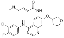
N(C1C=CC(F)=C(Cl)C=1)C1=NC=NC2=CC(=C(C=C12)NC(=O)/C=C/CN(C)C)O[C@@H]1COCC1
|
| InChi Key |
ULXXDDBFHOBEHA-CWDCEQMOSA-N
|
| InChi Code |
InChI=1S/C24H25ClFN5O3/c1-31(2)8-3-4-23(32)30-21-11-17-20(12-22(21)34-16-7-9-33-13-16)27-14-28-24(17)29-15-5-6-19(26)18(25)10-15/h3-6,10-12,14,16H,7-9,13H2,1-2H3,(H,30,32)(H,27,28,29)/b4-3+/t16-/m0/s1
|
| 化学名 |
(E)-N-[4-(3-chloro-4-fluoroanilino)-7-[(3S)-oxolan-3-yl]oxyquinazolin-6-yl]-4-(dimethylamino)but-2-enamide
|
| 别名 |
BIBW2992; Afatinib free base; BIBW 2992; BIBW 2992; Afatinib; trade name: Gilotrif, Tomtovok and Tovok
|
| HS Tariff Code |
2934.99.9001
|
| 存储方式 |
Powder -20°C 3 years 4°C 2 years In solvent -80°C 6 months -20°C 1 month |
| 运输条件 |
Room temperature (This product is stable at ambient temperature for a few days during ordinary shipping and time spent in Customs)
|
| 溶解度 (体外实验) |
|
|||
|---|---|---|---|---|
| 溶解度 (体内实验) |
配方 1 中的溶解度: ≥ 2.5 mg/mL (5.14 mM) (饱和度未知) in 10% DMSO + 40% PEG300 + 5% Tween80 + 45% Saline (这些助溶剂从左到右依次添加,逐一添加), 澄清溶液。
例如,若需制备1 mL的工作液,可将100 μL 25.0 mg/mL澄清DMSO储备液加入到400 μL PEG300中,混匀;然后向上述溶液中加入50 μL Tween-80,混匀;加入450 μL生理盐水定容至1 mL。 *生理盐水的制备:将 0.9 g 氯化钠溶解在 100 mL ddH₂O中,得到澄清溶液。 配方 2 中的溶解度: ≥ 2.5 mg/mL (5.14 mM) (饱和度未知) in 10% DMSO + 90% Corn Oil (这些助溶剂从左到右依次添加,逐一添加), 澄清溶液。 例如,若需制备1 mL的工作液,可将 100 μL 25.0 mg/mL 澄清 DMSO 储备液加入到 900 μL 玉米油中并混合均匀。 View More
配方 3 中的溶解度: ≥ 2.5 mg/mL (5.14 mM) (饱和度未知) in 5% DMSO + 40% PEG300 + 5% Tween80 + 50% Saline (这些助溶剂从左到右依次添加,逐一添加), 澄清溶液。 配方 4 中的溶解度: 2% DMSO+30% PEG 300+5% Tween 80+ddH2O: 10 mg/mL 配方 5 中的溶解度: 5 mg/mL (10.29 mM) in 0.5% Methylcellulose/saline water (这些助溶剂从左到右依次添加,逐一添加), 悬浊液; 超声助溶。 *生理盐水的制备:将 0.9 g 氯化钠溶解在 100 mL ddH₂O中,得到澄清溶液。 1、请先配制澄清的储备液(如:用DMSO配置50 或 100 mg/mL母液(储备液)); 2、取适量母液,按从左到右的顺序依次添加助溶剂,澄清后再加入下一助溶剂。以 下列配方为例说明 (注意此配方只用于说明,并不一定代表此产品 的实际溶解配方): 10% DMSO → 40% PEG300 → 5% Tween-80 → 45% ddH2O (或 saline); 假设最终工作液的体积为 1 mL, 浓度为5 mg/mL: 取 100 μL 50 mg/mL 的澄清 DMSO 储备液加到 400 μL PEG300 中,混合均匀/澄清;向上述体系中加入50 μL Tween-80,混合均匀/澄清;然后继续加入450 μL ddH2O (或 saline)定容至 1 mL; 3、溶剂前显示的百分比是指该溶剂在最终溶液/工作液中的体积所占比例; 4、 如产品在配制过程中出现沉淀/析出,可通过加热(≤50℃)或超声的方式助溶; 5、为保证最佳实验结果,工作液请现配现用! 6、如不确定怎么将母液配置成体内动物实验的工作液,请查看说明书或联系我们; 7、 以上所有助溶剂都可在 Invivochem.cn网站购买。 |
| 制备储备液 | 1 mg | 5 mg | 10 mg | |
| 1 mM | 2.0579 mL | 10.2893 mL | 20.5787 mL | |
| 5 mM | 0.4116 mL | 2.0579 mL | 4.1157 mL | |
| 10 mM | 0.2058 mL | 1.0289 mL | 2.0579 mL |
1、根据实验需要选择合适的溶剂配制储备液 (母液):对于大多数产品,InvivoChem推荐用DMSO配置母液 (比如:5、10、20mM或者10、20、50 mg/mL浓度),个别水溶性高的产品可直接溶于水。产品在DMSO 、水或其他溶剂中的具体溶解度详见上”溶解度 (体外)”部分;
2、如果您找不到您想要的溶解度信息,或者很难将产品溶解在溶液中,请联系我们;
3、建议使用下列计算器进行相关计算(摩尔浓度计算器、稀释计算器、分子量计算器、重组计算器等);
4、母液配好之后,将其分装到常规用量,并储存在-20°C或-80°C,尽量减少反复冻融循环。
计算结果:
工作液浓度: mg/mL;
DMSO母液配制方法: mg 药物溶于 μL DMSO溶液(母液浓度 mg/mL)。如该浓度超过该批次药物DMSO溶解度,请首先与我们联系。
体内配方配制方法:取 μL DMSO母液,加入 μL PEG300,混匀澄清后加入μL Tween 80,混匀澄清后加入 μL ddH2O,混匀澄清。
(1) 请确保溶液澄清之后,再加入下一种溶剂 (助溶剂) 。可利用涡旋、超声或水浴加热等方法助溶;
(2) 一定要按顺序加入溶剂 (助溶剂) 。
Targeted Therapy Directed by Genetic Testing in Treating Patients With Advanced Refractory Solid Tumors, Lymphomas, or Multiple Myeloma (The MATCH Screening Trial)
CTID: NCT02465060
Phase: Phase 2 Status: Active, not recruiting
Date: 2024-11-18
|
") |
Afatinib covalently binds to cysteine number 797 of the epidermal growth factor receptor (EGFR) via a Michael addition (IC50 = 0.5 nM).Schubert-Zsilavecz, M, Wurglics, M,Neue Arzneimittel Frühjahr 2013.(in German) |
InvivoChem的所有产品仅用于作科学研究,不面向患者销售
Copyright 2020 InvivoChem LLC | All Rights Reserved 粤ICP备20063088号-1


 COA
COA
")
")
 463611831
