| 规格 | 价格 | 库存 | 数量 |
|---|---|---|---|
| 1mg |
|
||
| 5mg |
|
||
| 10mg |
|
||
| 25mg |
|
||
| 50mg |
|
||
| 100mg |
|
||
| 250mg |
|
||
| 500mg |
|
||
| Other Sizes |
|
| 靶点 |
Dopamine Receptor; Adenosine A2A receptor (Ki = 2.3 nM) [2]
Dexpramipexole binds to and modulates the activity of the mitochondrial F1Fo ATP synthase, thereby increasing mitochondrial ATP production efficiency. It is noted to have very low affinity for dopamine D2 receptors (1000- to 10000-fold lower than pramipexole), which is considered unrelated to its neuroprotective effects in this context.[2] |
|---|---|
| 体外研究 (In Vitro) |
- 在SH-SY5Y神经母细胞瘤细胞中,达非那新(10 μM)显著减少过氧化氢(H2O2)诱导的活性氧(ROS)生成,通过DCFH-DA荧光法检测。该效应与超氧化物歧化酶(SOD)活性增加和脂质过氧化水平降低相关[2]
- 在原代皮质神经元培养物中,达非那新(1 μM)通过抑制caspase-3激活和维持线粒体膜电位(ΔΨm),保护神经元免受谷氨酸兴奋性毒性损伤,通过JC-1染色和细胞色素c释放的Western blot分析证实[2] 右旋普拉克索已被发现具有神经保护作用,目前正在研究用于治疗肌萎缩侧索硬化症 (ALS)。 Dexpramipexole 可减少线粒体活性氧 (ROS) 的产生,抑制细胞凋亡途径的激活,并增加细胞对各种神经毒素和 β-淀粉样蛋白神经毒性的存活率。与 S-(-) 异构体相比,Dexpramipexole 的多巴胺激动剂活性低得多。 右普拉克索(10 μM,处理6小时)在基础条件下,增加了纯培养的小鼠皮层神经元和胶质细胞中的ATP含量。[2] 在暴露于氧糖剥夺(OGD)的混合皮层细胞培养物中,右普拉克索(10 μM,于OGD前添加)减少了损伤期间的ATP损失,并促进了复氧后的能量恢复。[2] 在暴露于OGD的原代海马神经元培养物中,右普拉克索(10 μM,于OGD前10分钟添加)减少了细胞内钙增加的程度以及发生延迟性钙失调(DCD)的神经元百分比。[2] 右普拉克索(10 μM,于OGD期间及之前添加)减少了暴露于2小时OGD的原代神经元和胶质细胞培养物中的细胞死亡(通过碘化丙啶染色评估)。[2] 在器官型海马切片中,右普拉克索(10 μM)增加了基础条件下的ATP含量,并且在OGD前10分钟添加时,可防止30分钟OGD损伤后立即发生的ATP耗竭。[2] 右普拉克索(浓度为3、10和30 μM,于OGD结束时添加)以浓度依赖的方式,减少了暴露于30分钟OGD继以24小时复氧的器官型海马切片中CA1锥体神经元的死亡(通过碘化丙啶染色评估)。[2] 电子显微镜显示,右普拉克索(10 μM,于OGD前10分钟添加)防止了30分钟OGD损伤后,器官型海马切片CA1神经元的胞体和线粒体肿胀。[2] 在暴露于谷氨酸兴奋毒性(1 mM,6小时)的器官型海马切片中,用右普拉克索(10和30 μM)进行后处理,减少了18小时后评估的CA1神经元死亡。[2] 在急性海马切片中,右普拉克索(30 μM)完全阻止了由7分钟OGD诱导的缺氧性去极化(AD)。在更严重的损伤(30分钟OGD)下,它在常温条件下延迟并降低了AD的幅度,在低温条件(30°C)下,大约一半的切片中AD被阻止。[2] 右普拉克索(30 μM)完全阻止了暴露于7分钟OGD的急性海马切片CA1区神经传递(场EPSP)的丧失,并在30分钟OGD后的低温条件(30°C)下促进了部分、短暂的恢复。[2] |
| 体内研究 (In Vivo) |
- 在局灶性脑缺血大鼠模型中,缺血后30分钟腹腔注射达非那新(3 mg/kg)显著减少梗死体积(通过TTC染色评估),并改善神经功能缺损评分,保护作用持续至治疗后72小时[2]
- 在肌萎缩侧索硬化(ALS)转基因小鼠模型中,达非那新(10 mg/kg/天,灌胃)延迟疾病发作并延长生存期15%,同时保留脊髓运动神经元数量,通过胆碱乙酰转移酶(ChAT)免疫组化染色评估[1] 当培养物暴露于OGD时,右旋普拉克索增加了培养的神经元或神经胶质中线粒体ATP的产生,减少了能量衰竭,防止了细胞内Ca2+超载,并提供了细胞保护。该化合物还可以抵消OGD海马切片中ATP耗竭、线粒体肿胀、缺氧去极化、突触活性丧失和神经元死亡。在短暂或永久性MCAo的小鼠中,用与ALS患者已经使用的剂量一致的右普拉克索进行缺血后治疗,可以减少脑梗死面积,改善神经官能症[2]。 在小鼠短暂性大脑中动脉闭塞(tMCAo;1小时闭塞/48小时再灌注)模型中,缺血后用右普拉克索(3 mg/kg,腹腔注射,每日两次,从再灌注时开始)进行治疗,与盐水对照组相比,显著减少了脑梗死面积和体积。[2] 在同一tMCAo模型中,右普拉克索治疗(3 mg/kg,腹腔注射,每日两次)使再灌注后3小时缺血半暗带的ATP含量增加。[2] 在tMCAo后给药7天(3 mg/kg,腹腔注射,每日两次),与盐水组相比,右普拉克索在一个月的观察期内显著改善了神经功能评分并促进了感觉运动功能的功能恢复。它在改善卒中后体重恢复和降低死亡率方面显示出不显著的积极趋势。[2] 在小鼠永久性远端MCAo(dMCAo)模型中,用右普拉克索(3 mg/kg,腹腔注射,每日两次,首次剂量在动脉烧灼后立即给予)进行治疗,与盐水组相比,显著减少了皮层梗死面积和体积。[2] 在永久性dMCAo模型中,即使右普拉克索治疗(3 mg/kg,腹腔注射,每日两次)在动脉烧灼后1小时才开始,仍然显著减少了皮层梗死面积和体积。[2] 在大鼠永久性MCAo模型中,右普拉克索治疗(3 mg/kg,腹腔注射,每日两次,首次剂量在动脉闭塞后立即给予)也显著减少了梗死面积和体积,表明其神经保护作用不具有物种特异性。[2] |
| 酶活实验 |
腺苷A2A受体结合实验使用表达人A2A受体的HEK293细胞膜制剂进行。膜蛋白与[³H]ZM241385(放射性标记拮抗剂)和递增浓度的达非那新在25°C孵育60分钟,非特异性结合通过10 μM CGS21680确定。平衡解离常数(Ki)通过竞争结合曲线计算为2.3 nM[2]
使用线粒体靶向的荧光素酶作为传感器,监测活体神经元或星形胶质细胞中的线粒体ATP产生。细胞用荧光素酶构建体转染。转染48小时后,细胞用或不用右普拉克索(10 μM)预孵育6小时。随后,细胞在含有100 μM荧光素(溶解于生长培养基)中孵育5分钟,然后进行1分钟的发光测定,以测量作为正在进行中线粒体ATP合成指标的光子发射。[2] |
| 细胞实验 |
- SH-SY5Y细胞ROS检测:细胞用达非那新(10 μM)预处理24小时,然后暴露于200 μM H2O2 1小时。加入DCFH-DA(10 μM)孵育30分钟,荧光强度通过酶标仪测量,结果显示ROS水平较H2O2处理对照组降低40%[2]
- 皮质神经元谷氨酸兴奋性毒性实验:细胞在谷氨酸暴露(50 μM,24小时)前1小时用达非那新(1 μM)处理。凋亡细胞通过Annexin V-FITC/PI染色和流式细胞术定量,凋亡率较谷氨酸单独处理组降低35%[2] 据报道,神经元/星形胶质细胞培养物是从大鼠胚胎(E-17/E-19)或幼崽(P-1/P-2)制备的(Chiarugi等人,2003)。简而言之,使用培养基(MS)(Eagle的最低必需培养基,含Earle盐、不含谷氨酰胺和NaHCO3、NaHCO3 38 mM、葡萄糖22 mM、青霉素100 U·mL-1和链霉素100µg·mL-1)切碎大脑皮层,然后在37°C的MS中孵育10分钟(神经元)和45分钟(星形胶质细胞),补充0.25%胰蛋白酶和0.05%DNase。通过在添加了10%热灭活马血清(HIHS)和10%FBS的MS中孵育(在37°C下孵育10分钟)来终止酶消化。在组织机械破坏后,对细胞进行计数和铺板。对于混合皮质细胞培养,神经元以4×10⁵个细胞·mL-1的密度重新悬浮,并使用添加了10%HIHS、10%FBS和2 mm谷氨酰胺的MS在融合的星形胶质细胞层上镀上15 mm多孔板。体外培养4-5天后,通过应用3µM阿糖胞苷24小时来停止非神经元细胞分裂。细胞培养物在有或没有DEX的情况下,在饱和有95%N2和5%CO2的无血清和无葡萄糖培养基中进行氧葡萄糖剥夺(OGD)。在缺氧室中于37°C下孵育2小时后,将培养物转移到含氧无血清培养基(75%Eagle最低必需培养基;25%Hank's平衡盐溶液;2 mM l‐谷氨酰胺;3.75µg·mL−1两性霉素B;和5 mg·mL-1葡萄糖)中,并在有或没有DEX的情况下恢复到常氧条件。24小时后评估碘化丙啶(PI)荧光[2]。 用于原代皮层细胞培养物中ATP测量的实验:将来自小鼠的纯神经元或胶质细胞暴露于不同浓度的右普拉克索中6小时。然后裂解细胞,使用商业发光ATP检测试剂盒定量ATP含量。[2] 用于原代海马神经元钙成像的实验:用荧光钙指示剂 fluo-3 AM 负载培养的神经元。将盖玻片转移到灌注式显微镜腔室中。在存在或不存在右普拉克索(OGD前10分钟添加)的情况下,将细胞暴露于OGD。每3秒采集一次荧光图像,并测量荧光强度以监测细胞内钙水平的变化和延迟性钙失调的发生。[2] 用于OGD诱导的原代皮层神经元或胶质细胞培养物细胞死亡的实验:将细胞暴露于2小时OGD(在饱和了95% N₂和5% CO₂的无血清无葡萄糖培养基中),在OGD前10分钟和OGD期间添加或不添加右普拉克索。OGD后,将培养物放回常氧、含营养的培养基(含或不含药物)中培养24小时。然后通过碘化丙啶染色和荧光定量评估细胞死亡。[2] 用于器官型海马切片中ATP测量和细胞死亡评估的实验:从小鼠或幼鼠制备切片并在膜插入物上培养。对于ATP测量,将切片暴露于右普拉克索中6小时,或进行30分钟OGD(有或无药物预处理,10 μM,OGD前10分钟),然后立即裂解进行ATP定量。对于细胞死亡评估,将切片暴露于30分钟OGD,然后转移到含有或不含不同浓度右普拉克索(于OGD结束时添加)的新鲜无血清培养基中复氧24小时。通过碘化丙啶染色和荧光成像/定量评估CA1神经元损伤。[2] 用于器官型海马切片中谷氨酸兴奋毒性的实验:将切片暴露于1 mM谷氨酸中6小时,清洗,然后在含有或不含不同浓度右普拉克索的生长培养基中孵育18小时。通过碘化丙啶染色评估CA1神经元死亡。[2] 用于电子显微镜超微结构分析的实验:在30分钟OGD后(有或无右普拉克索预处理)立即固定器官型海马切片。样品经过戊二醛和锇酸固定、脱水、树脂包埋和切片处理。对超薄切片进行染色并在电子显微镜下检查,以评估CA1神经元的线粒体和细胞形态。[2] |
| 动物实验 |
大鼠脑缺血模型:雄性Sprague-Dawley大鼠接受90分钟的大脑中动脉闭塞(MCAO)。将右旋普拉克索溶于0.9%生理盐水中,并在再灌注后立即腹腔注射,剂量为3 mg/kg。术后24小时和72小时采用5分制评分标准评估神经功能[2]。- ALS小鼠模型:SOD1G93A转基因小鼠从60日龄开始,通过灌胃给予溶于0.5%甲基纤维素的右旋普拉克索(10 mg/kg/天)。每日监测动物存活情况,并在实验终末期采集脊髓组织进行组织学分析[1]
急性海马切片制备和OGD暴露[2] 按照文献(Pugliese et al., 2009)所述方法,从雄性SD大鼠(Charles River,Calco,意大利,150–200 g)中制备急性海马切片。取出海马后,置于冰冷的氧合人工脑脊液(aCSF)中,该aCSF的成分为(mM):NaCl 125、KCl 3、NaH2PO4 1.25、MgSO4 1、CaCl2 2、NaHCO3 25和D-葡萄糖10。制备400 mm²的切片,并在室温下于氧合aCSF中保存至少1小时。将一片脑片置于尼龙网上,完全浸没于一个小型灌流室(0.8 mL)中,并以1.5–1.8 mL·min−1的恒定流速灌注氧合人工脑脊液(31–32°C)。在缺氧/缺糖(OGD)条件下,用不含葡萄糖的人工脑脊液灌注脑片,并通入95% N2–5% CO2混合气体。这导致记录室内的氧分压(pO2)从约500 mmHg(常氧)下降至35–75 mmHg(OGD 7分钟后)。(Pugliese等人,2003)缺血期结束后,再次用含葡萄糖的正常氧合人工脑脊液灌注脑片。对照组脑片未进行OGD或药物处理,而是在氧合人工脑脊液中孵育相同的时间间隔。海马切片在进行电生理记录前,需在含有地塞米松(DEX)的培养基中孵育至少1小时,并在整个实验过程中维持DEX浓度;或切片在氧糖剥夺(OGD)处理前后至少30分钟内,在含有DEX的培养基中进行灌流。 对于小鼠的短暂性大脑中动脉闭塞(tMCAo)模型,将C57/BL6雄性小鼠麻醉后,使用管腔内线栓技术近端闭塞大脑中动脉1小时。再灌注后,将动物随机分为两组,分别接受生理盐水或地塞米松(3 mg/kg,腹腔注射)治疗,每日两次。首次给药在再灌注时进行。48小时后处死动物以测量梗死体积,或继续治疗7天以进行长期功能和生存分析。[2] 对于小鼠的永久性远端大脑中动脉闭塞(dMCAo)模型,采用电凝法闭塞大脑中动脉。在治疗组中,每日两次腹腔注射右旋普拉米索(3 mg/kg),首次给药时间为动脉闭塞后立即或闭塞后 1 小时。48 小时后处死动物进行梗死评估。[2] 对于大鼠永久性大脑中动脉闭塞(MCAo)模型,对 Sprague-Dawley 雄性大鼠进行 MCAo 手术。每日两次腹腔注射右旋普拉米索(3 mg/kg),首次给药时间为动脉闭塞后立即。48 小时后处死动物进行梗死评估。[2] 在所有体内实验中,均维持体温,监测脑血流量,并以盲法评估神经功能评分。[2] |
| 药代性质 (ADME/PK) |
右旋普拉米索在口服吸收迅速,大鼠体内达峰时间(Tmax)为1.5小时。口服生物利用度约为75%,血浆蛋白结合率低(<15%)。消除半衰期为 3.2 小时,60% 的剂量以原形经尿液排出 [2]
- 小鼠脑渗透性研究显示,静脉注射 10 mg/kg 右普拉米索后,脑血浆浓度比为 0.6,表明其具有中等的血脑屏障通透性 [2] 右普拉米索 易于穿过血脑屏障并在脑内蓄积,脑血浆浓度比大于 15 [2] 对接受永久性大脑中动脉闭塞 (dMCAo) 并单次腹腔注射右普拉米索(3 mg/kg,注射后 1 小时处死)的小鼠脑组织进行成像质谱分析显示,该药物均匀分布于整个脑组织和缺血半暗带,浓度达到 10-20 μM [2] |
| 毒性/毒理 (Toxicokinetics/TK) |
小鼠急性毒性研究表明,口服LD50 > 2000 mg/kg。大鼠重复给药毒性研究(10 mg/kg/天,持续28天)显示肝肾功能指标无显著变化[2]
- 体外细胞色素P450抑制试验表明,右普拉克索对CYP1A2、CYP2D6和CYP3A4活性的影响极小(10 μM时抑制率<20%),提示药物相互作用的可能性较低[2] 妊娠和哺乳期用药 ◉ 哺乳期用药概述 目前尚无关于哺乳期使用普拉克索的信息,但该药会抑制血清催乳素水平,可能影响哺乳。尤其是在哺乳新生儿或早产儿期间,可能需要选择其他药物。 ◉ 对母乳喂养婴儿的影响 截至修订日期,未找到相关的已发表信息。 ◉ 对泌乳和母乳的影响 截至修订日期,未找到关于哺乳期母亲的相关已发表信息。普拉克索可降低血清催乳素水平。[1] 已建立泌乳的母亲的催乳素水平可能不会影响其母乳喂养能力。 右普拉克索 已在肌萎缩侧索硬化症 (ALS) 的大型临床试验中进行过测试,结果显示,每日服用 300 mg(相当于每日两次,每次约 150 mg)治疗近一年的患者具有非常良好的安全性。[2] 在所描述的动物卒中模型中,每日两次腹腔注射 3 mg/kg 的给药方案耐受性良好。[2] |
| 参考文献 | |
| 其他信息 |
右旋普拉克索是一种选择性腺苷A2A受体拮抗剂,具有神经保护作用,最初开发用于治疗帕金森病和肌萎缩侧索硬化症(ALS)[1,2]
- 右旋普拉克索的神经保护作用是通过双重机制实现的:阻断A2A受体介导的兴奋性毒性和增强线粒体功能[2] - 在临床前模型中,右旋普拉克索已显示出在减少神经炎症和促进轴突再生方面的疗效[1,2] 普拉克索的(R)-(+)对映体。右普拉米索对多巴胺受体的亲和力低于普拉米索。 右普拉米索是抗帕金森病药物普拉米索的R-对映体,但对多巴胺受体的亲和力非常低。其主要作用机制是与线粒体F1Fo ATP合酶结合,提高ATP生成效率并降低耗氧量。[2] 它最初是为肌萎缩侧索硬化症(ALS)而开发和进行临床试验的,在ALS中显示出良好的安全性,但在III期临床试验中未达到主要终点。[2] 这项研究将Dexpramipexole重新用于缺血性卒中,证实了其通过靶向早期生物能量衰竭发挥神经保护作用,而早期生物能量衰竭是卒中病理生理学中神经血管单元所有组成部分共有的核心事件。[2] Dexpramipexole即使在缺血性损伤后(治疗后)给药也能提供神经保护作用,其在永久性缺血模型中的疗效,以及在临床相关剂量下脑内达到神经保护浓度,都支持其在卒中治疗中的转化应用潜力。[2] |
| 分子式 |
C10H19CL2N3S
|
|---|---|
| 分子量 |
284.2490
|
| 精确质量 |
283.068
|
| 元素分析 |
C, 42.26; H, 6.74; Cl, 24.94; N, 14.78; S, 11.28
|
| CAS号 |
104632-27-1
|
| 相关CAS号 |
Pramipexole dihydrochloride; 104632-25-9; Pramipexole; 104632-26-0; Pramipexole dihydrochloride hydrate; 191217-81-9; Dexpramipexole; 104632-28-2; 908244-04-2 (HCl hydrate)
|
| PubChem CID |
46174453
|
| 外观&性状 |
White to off-white solid powder
|
| LogP |
3.507
|
| tPSA |
79.91
|
| 氢键供体(HBD)数目 |
5
|
| 氢键受体(HBA)数目 |
5
|
| 可旋转键数目(RBC) |
3
|
| 重原子数目 |
17
|
| 分子复杂度/Complexity |
188
|
| 定义原子立体中心数目 |
1
|
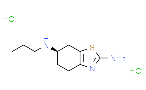
| SMILES |
Cl[H].Cl[H].S1C(N([H])[H])=NC2=C1C([H])([H])[C@@]([H])(C([H])([H])C2([H])[H])N([H])C([H])([H])C([H])([H])C([H])([H])[H]
|
| InChi Key |
QMNWXHSYPXQFSK-XCUBXKJBSA-N
|
| InChi Code |
InChI=1S/C10H17N3S.2ClH/c1-2-5-12-7-3-4-8-9(6-7)14-10(11)13-8;;/h7,12H,2-6H2,1H3,(H2,11,13);2*1H/t7-;;/m1../s1
|
| 化学名 |
(6R)-6-N-propyl-4,5,6,7-tetrahydro-1,3-benzothiazole-2,6-diamine;dihydrochloride
|
| 别名 |
Dexpramipexole dihydrochloride; KNS-760704; KNS760704; KNS 760704; R-Pramipexole; 104632-27-1; DEXPRAMIPEXOLE DIHYDROCHLORIDE; Dexpramipexole (dihydrochloride); SND 919CL2x; (R)-Pramipexole Dihydrochloride; KNS 760704; CHEMBL3216394; I9038PKO43;
|
| HS Tariff Code |
2934.99.9001
|
| 存储方式 |
Powder -20°C 3 years 4°C 2 years In solvent -80°C 6 months -20°C 1 month 注意: 请将本产品存放在密封且受保护的环境中,避免吸湿/受潮。 |
| 运输条件 |
Room temperature (This product is stable at ambient temperature for a few days during ordinary shipping and time spent in Customs)
|
| 溶解度 (体外实验) |
H2O : ~100 mg/mL (~351.80 mM)
DMSO : ≥ 100 mg/mL (~351.80 mM) |
|---|---|
| 溶解度 (体内实验) |
配方 1 中的溶解度: ≥ 2.08 mg/mL (7.32 mM) (饱和度未知) in 10% DMSO + 40% PEG300 + 5% Tween80 + 45% Saline (这些助溶剂从左到右依次添加,逐一添加), 澄清溶液。
例如,若需制备1 mL的工作液,可将100 μL 20.8 mg/mL澄清DMSO储备液加入400 μL PEG300中,混匀;然后向上述溶液中加入50 μL Tween-80,混匀;加入450 μL生理盐水定容至1 mL。 *生理盐水的制备:将 0.9 g 氯化钠溶解在 100 mL ddH₂O中,得到澄清溶液。 配方 2 中的溶解度: ≥ 2.08 mg/mL (7.32 mM) (饱和度未知) in 10% DMSO + 90% (20% SBE-β-CD in Saline) (这些助溶剂从左到右依次添加,逐一添加), 澄清溶液。 例如,若需制备1 mL的工作液,可将 100 μL 20.8 mg/mL澄清DMSO储备液加入900 μL 20% SBE-β-CD生理盐水溶液中,混匀。 *20% SBE-β-CD 生理盐水溶液的制备(4°C,1 周):将 2 g SBE-β-CD 溶解于 10 mL 生理盐水中,得到澄清溶液。 View More
配方 3 中的溶解度: ≥ 2.08 mg/mL (7.32 mM) (饱和度未知) in 10% DMSO + 90% Corn Oil (这些助溶剂从左到右依次添加,逐一添加), 澄清溶液。 配方 4 中的溶解度: 100 mg/mL (351.80 mM) in PBS (这些助溶剂从左到右依次添加,逐一添加), 澄清溶液; 超声助溶. 1、请先配制澄清的储备液(如:用DMSO配置50 或 100 mg/mL母液(储备液)); 2、取适量母液,按从左到右的顺序依次添加助溶剂,澄清后再加入下一助溶剂。以 下列配方为例说明 (注意此配方只用于说明,并不一定代表此产品 的实际溶解配方): 10% DMSO → 40% PEG300 → 5% Tween-80 → 45% ddH2O (或 saline); 假设最终工作液的体积为 1 mL, 浓度为5 mg/mL: 取 100 μL 50 mg/mL 的澄清 DMSO 储备液加到 400 μL PEG300 中,混合均匀/澄清;向上述体系中加入50 μL Tween-80,混合均匀/澄清;然后继续加入450 μL ddH2O (或 saline)定容至 1 mL; 3、溶剂前显示的百分比是指该溶剂在最终溶液/工作液中的体积所占比例; 4、 如产品在配制过程中出现沉淀/析出,可通过加热(≤50℃)或超声的方式助溶; 5、为保证最佳实验结果,工作液请现配现用! 6、如不确定怎么将母液配置成体内动物实验的工作液,请查看说明书或联系我们; 7、 以上所有助溶剂都可在 Invivochem.cn网站购买。 |
| 制备储备液 | 1 mg | 5 mg | 10 mg | |
| 1 mM | 3.5180 mL | 17.5901 mL | 35.1803 mL | |
| 5 mM | 0.7036 mL | 3.5180 mL | 7.0361 mL | |
| 10 mM | 0.3518 mL | 1.7590 mL | 3.5180 mL |
1、根据实验需要选择合适的溶剂配制储备液 (母液):对于大多数产品,InvivoChem推荐用DMSO配置母液 (比如:5、10、20mM或者10、20、50 mg/mL浓度),个别水溶性高的产品可直接溶于水。产品在DMSO 、水或其他溶剂中的具体溶解度详见上”溶解度 (体外)”部分;
2、如果您找不到您想要的溶解度信息,或者很难将产品溶解在溶液中,请联系我们;
3、建议使用下列计算器进行相关计算(摩尔浓度计算器、稀释计算器、分子量计算器、重组计算器等);
4、母液配好之后,将其分装到常规用量,并储存在-20°C或-80°C,尽量减少反复冻融循环。
计算结果:
工作液浓度: mg/mL;
DMSO母液配制方法: mg 药物溶于 μL DMSO溶液(母液浓度 mg/mL)。如该浓度超过该批次药物DMSO溶解度,请首先与我们联系。
体内配方配制方法:取 μL DMSO母液,加入 μL PEG300,混匀澄清后加入μL Tween 80,混匀澄清后加入 μL ddH2O,混匀澄清。
(1) 请确保溶液澄清之后,再加入下一种溶剂 (助溶剂) 。可利用涡旋、超声或水浴加热等方法助溶;
(2) 一定要按顺序加入溶剂 (助溶剂) 。
| NCT Number | Recruitment | interventions | Conditions | Sponsor/Collaborators | Start Date | Phases |
| NCT03430596 | Completed | Drug: Trihexyphenidyl hydrochloride Drug: Pramipexole |
Extrapyramidal Syndrome C | Shanghai Mental Health Center | May 1, 2018 | Early Phase 1 |
| NCT01525641 | Completed | Drug: Mirapex LA | Parkinson Disease | Boehringer Ingelheim | February 2012 | N/A |
InvivoChem的所有产品仅用于作科学研究,不面向患者销售
Copyright 2020 InvivoChem LLC | All Rights Reserved 粤ICP备20063088号-1


 COA
COA
 463611831
