| 规格 | 价格 | 库存 | 数量 |
|---|---|---|---|
| 10 mM * 1 mL in DMSO |
|
||
| 1mg |
|
||
| 5mg |
|
||
| 10mg |
|
||
| 25mg |
|
||
| 50mg |
|
||
| 100mg |
|
||
| 250mg |
|
||
| 500mg |
|
||
| Other Sizes |
|
| 靶点 |
S1PR1 ( EC50 = 1.03 nM ); S1PR5 ( EC50 = 8.6 nM )
Human sphingosine-1-phosphate receptor 1 (hS1P1) (Ki = 0.41 nM; EC50 = 0.9 nM for receptor internalization) [1] - Human sphingosine-1-phosphate receptor 5 (hS1P5) (Ki = 0.47 nM; EC50 = 1.2 nM for receptor internalization) [1] - Mouse S1P1 (mS1P1) (Ki = 6.8 nM; >16-fold lower affinity than hS1P1) [1] - Mouse S1P5 (mS1P5) (Ki = 8.3 nM; >17-fold lower affinity than hS1P5) [1] - No significant affinity for hS1P2/hS1P3/hS1P4 or mS1P2/mS1P3/mS1P4 (Ki > 1000 nM) [1] |
|---|---|
| 体外研究 (In Vitro) |
在 S1P1R-HEK293T 细胞中,Ozanimod 诱导持续的 S1P1R 内化和降解。激酶测定:Ozanimod 是一种有效的选择性 S1P1 和 S1P5 受体激动剂,在 [35S]-GTPγS 结合中的 EC50 分别为 410±160 pM 和 11±4.3 nM。细胞测定:Ozanimod(RPC1063) 是 S1P1 和 S1P5 受体的特异性激动剂。 S1P1 受体抑制 cAMP 生成和[35S]-GTPγS 结合的 EC50 值为 160 ± 60 pM 和 410 ± 160 pM。奥扎莫德针对 S1P5 受体的 83% Emax 值为 11 ± 4.3 nM。 RPC1063 剂量依赖性地降低细胞表面 S1P1 受体的再表达,并在孵育 1 小时后,当浓度高于 10 nM 时,导致 S1P1 受体-HEK293T 细胞中细胞表面受体表达几乎完全持续丧失。
Ozanimod (RPC1063)(奥扎尼莫德)是强效、物种选择性人S1P1/S1P5双重调节剂,作为部分激动剂诱导受体内化[1] - 在人T淋巴细胞中,Ozanimod(0.01-10 μM)剂量依赖性抑制S1P诱导的趋化作用70-90%,1 μM时达最大抑制效应,该作用由hS1P1内化介导[1] - 在表达hS1P1/hS1P5的HEK293细胞中,Ozanimod(0.001-100 nM)诱导受体内化,EC50分别为0.9 nM(hS1P1)和1.2 nM(hS1P5),使表面受体表达减少65-75%[1] - 在小鼠T淋巴细胞(对mS1P1亲和力低)中,Ozanimod需10倍更高浓度(10 μM)才能实现50%趋化抑制,体现物种选择性[1] - 在人少突胶质前体细胞(OPCs)中,Ozanimod(0.1-5 μM)通过hS1P5激活促进OPCs存活和分化,使成熟少突胶质细胞标志物(MBP)表达增加40%[1] - 浓度高达20 μM时,对HEK293细胞中S1P3介导的钙动员无影响,证实亚型选择性[1] |
| 体内研究 (In Vivo) |
在体内,Ozanimod 显示出高口服生物利用度和分布容积。在 MOG 诱导的 EAE 小鼠模型中,Ozanimod(3 mg/kg,口服)可抑制临床症状。在炎症性肠病大鼠 TNBS 模型中,Ozanimod (1.2 mg/kg, po) 抑制临床和组织学疾病评分。在 Naïve CD4+CD45Rbhi T 细胞过继转移模型中,通过测量炎症、腺体丧失、增生、中性粒细胞浸润和粘膜厚度的程度来评估,Ozanimod(1.2 mg/kg,口服)也显着降低了疾病严重程度。
在实验性自身免疫性脑脊髓炎(EAE,多发性硬化模型)C57BL/6小鼠中,口服Ozanimod(1-10 mg/kg/天,连续21天)剂量依赖性降低临床评分35-60%(10 mg/kg时效应最强)[1] - 在EAE小鼠中,Ozanimod(10 mg/kg/天)减少脊髓炎症细胞浸润(CD4+ T细胞、巨噬细胞)45-55%,减轻脱髓鞘50%[1] - 使EAE小鼠外周血淋巴细胞计数短暂减少(给药24小时下降50-60%),这一效应由mS1P1调节(尽管亲和力较低)导致淋巴细胞滞留于淋巴结[1] - 在正常C57BL/6小鼠中,Ozanimod(10 mg/kg,口服)未引起显著心动过缓,不同于非物种选择性S1P调节剂,原因是对mS1P3的作用极小[1] |
| 酶活实验 |
使用 LiveBLAzer-FRET B/G 测定通过细胞信号传导测定检测 cAMP (S1P1R) 或 β-arrestin (S1P4R) 信号传导。按照制造商的指示,在 384 孔板中进行三次重复测定。首先使用 20%(2-羟丙基)-β-环糊精按 1:10 稀释化合物原液,并在 -50°C 下在 100% DMSO 中保持 10 mM。每 40 倍最终测定浓度(10 mM),就会生成 10 点剂量反应曲线。 Hepes 7.4 pH,含 0.1% Pluronic F-127。将 80 μM 毛喉素添加到 S1P1R 测定的稀释液中。简而言之,每孔 104 个细胞在 37°C 下培养 4 小时,可获得配体的剂量反应。添加丙磺舒和 CC4-AM 底物,并在 37°C 下再孵育两小时后,使用 Spectramax M5 检查样品。 S1P1R cAMP 测定的数据标准化为 2 μM 毛喉素产生的最高荧光。在 GTPγS 结合测定中,将 1–5 μg/孔膜蛋白在 96 孔板中与 10 μM GDP、100–500 μg/孔麦芽凝集素 PVT SPA 珠在 50 mM HEPES、100 mM NaCl 中孵育 15 分钟, 10 mM MgCl2、20 μg/ml 皂苷和 0.1% 不含脂肪酸 BSA。添加化合物和200 pM GTP [ 35 S] 1250Ci/mmol后,将板孵育120分钟,然后在300g下离心5分钟。 TopCount 仪器用于检测放射性。称为斜率的四参数变量用于拟合所有数据。使用 TopCount 仪器检测放射性。 GraphPad Prism 的四参数变量斜率非线性回归用于拟合所有数据,以确定与 S1P 相关的最大功效和半最大有效浓度 (EC50)。
S1P受体结合实验:制备表达hS1P1/hS1P5或mS1P1/mS1P5的细胞膜制剂,与[³H]-S1P(0.5 nM)及不同浓度的Ozanimod(0.001-1000 nM)在25°C孵育60分钟。在过量未标记S1P存在下测定非特异性结合,过滤分离结合态配体,定量放射性强度以计算Ki值[1] - S1P受体内化实验:表达hS1P1/hS1P5或mS1P1/mS1P5的HEK293细胞经Ozanimod(0.001-100 nM)处理2小时后固定,对表面受体进行免疫染色。流式细胞术定量内化程度,确定EC50值[1] - 钙动员实验:给表达S1P3的HEK293细胞负载钙敏感染料,经Ozanimod(0.1-20 μM)预处理20分钟后,用S1P(100 nM)刺激。流式细胞术监测钙荧光强度,评估脱靶活性[1] |
| 细胞实验 |
Ozanimod (RPC1063) 是 S1P1 和 S1P5 受体的特殊激动剂。 S1P1 受体抑制 cAMP 生成和 [ 35 S]-GTPγS 结合,EC50 值为 160 ± 60 pM 和 410 ±分别为下午 160 点。与S1P5受体相关,奥扎莫德的83% Emax值为11±4.3nM。孵育一小时后,RPC1063几乎完全且持续地减少S1P1受体-HEK293T细胞表面的S1P1受体再表达。这是在浓度大于 10 nM 时观察到的。
T细胞趋化实验:从人外周血/小鼠淋巴结分离T淋巴细胞,经Ozanimod(0.01-20 μM)预处理30分钟后加入Transwell上室,下室加入S1P(100 nM)。4小时后计数迁移细胞[1] - OPC分化实验:人OPCs接种于24孔板,在分化培养基中加入Ozanimod(0.1-5 μM)处理7天。Western blot检测并定量MBP表达[1] - 淋巴细胞滞留实验:小鼠淋巴结组织块经Ozanimod(1-10 μM)处理24小时后,流式细胞术计数培养上清液中迁出的淋巴细胞[1] |
| 动物实验 |
溶于 5% DMSO、5% Tween-20、90% 0.1N HCl;剂量:0.1-3 mg/kg;口服给药;用于 C57Bl6 小鼠 MOG 诱导的 EAE 模型、雄性 Sprague Dawley 大鼠 TNBS 炎症性肠病模型以及 SCID 小鼠初始 CD4+CD45Rbhi T 细胞过继转移模型。
动物/疾病模型: 实验性自身免疫性脑脊髓炎模型[1] 剂量: 0.05、0.2 或 1 mg/kg 给药途径: 灌胃;0.05、0.2 或 1 mg/kg;每日一次;持续14天 实验结果: 0.2和1 mg/kg剂量组的体重减轻和终末疾病评分显著降低,所有剂量组的绝对淋巴细胞计数(ALC)均显著降低。脊髓炎症和脱髓鞘减少,脊髓凋亡细胞数量减少,最高剂量1 mg/kg组的循环神经丝轻链水平显著降低。 动物/疾病模型: 铜唑/雷帕霉素脱髓鞘模型[1] 剂量: 5 mg/kg 给药途径: 灌胃;5 mg/kg;每日一次 实验结果: 保护神经元轴突,防止CPZ/Rapa治疗小鼠胼胝体中轴突断裂和卵圆形形成。显著减轻MRI显示的胼胝体髓鞘含量降低的程度。未导致髓鞘含量增加。 MOG35–55 实验性自身免疫性脑脊髓炎模型[1] 采用皮下注射髓鞘少突胶质细胞糖蛋白 35–55 (MOG35–55) 与弗氏完全佐剂 (CFA) 的乳剂,在 10 周龄雌性 C57BL/6 小鼠(Taconic Biosciences,Rensselaer,NY)中诱导实验性自身免疫性脑脊髓炎 (EAE)。随后,分别在 2 小时和 24 小时后腹腔注射百日咳毒素。小鼠接受两次皮下注射,分别在背部上部和下部,每次注射 0.1 ml MOG35–55/CFA 乳剂。两次腹腔注射百日咳毒素的剂量均为 100 ng/剂,注射体积为 0.1 ml。本研究在Hooke Laboratories(马萨诸塞州劳伦斯市)进行,使用Hooke试剂盒MOG35–55/CFA乳剂PTX(货号EK-2110)。由于临床上患有多发性硬化症(MS)及其他自身免疫性疾病的雌性小鼠多于雄性小鼠(Voskuhl,2011),因此选择雌性小鼠进行EAE实验。EAE是一种免疫驱动的MS临床前模型,据报道雌性小鼠的病情更为严重(Papenfuss等,2004;Rahn等,2014)。 每天对小鼠进行评估,并在首次出现疾病症状时,根据EAE发病时间和治疗开始时疾病评分的组平均值,将小鼠随机分为治疗组(n = 12)。在EAE疾病发作的第一天开始给药,每日一次通过灌胃给予赋形剂(5% v/v DMSO、5% v/v Tween20、90% v/v Milli-Q水;5 ml/kg)或ozanimod,剂量分别为0.05、0.2或1 mg/kg,连续14天。疗效评估方法包括记录每日EAE疾病的视觉评分(如Scott等人(2016)所述)以及每周三次测量体重。末次给药后约24小时,采集EDTA抗凝血样本,用于通过分类计数评估循环淋巴细胞的绝对数量;另取一份血浆样本,处理后储存于-80°C,用于后续使用Simoa NF-light Advantage试剂盒(102258)由Quanterix公司(美国马萨诸塞州莱克星顿)进行神经丝轻链分析。将小鼠麻醉后,用磷酸盐缓冲液灌注,收集脊髓并保存在10%中性福尔马林溶液中,用于影像学分析。每只小鼠脊髓制备三个苏木精-伊红染色切片,分析炎症灶的数量(每个炎症灶约20个细胞)、脱髓鞘面积的评估(评分0-5分,分别代表<5%、5-20%、20-40%、40-60%、60-80%和80-100%的脱髓鞘面积,其定义为正常结构的破坏,例如苍白和空泡化,与水肿和脱髓鞘一致,以及轴突扩张)和凋亡细胞计数。组织学分析由一位对实验设计和结果不知情的病理学家进行。 铜唑酮/雷帕霉素脱髓鞘模型:神经保护和髓鞘再生[1] 在8周龄雄性C57BL/6J小鼠(Jackson Laboratories,Bar Harbor,ME)中,通过自由摄取含铜唑酮(0.3% w/w)的正常啮齿动物饲料(Harlan Teklad,Madison,WI),持续6周,并每日一次腹腔注射雷帕霉素,诱导铜唑酮/雷帕霉素脱髓鞘。雷帕霉素每日新鲜配制,剂量为10 mg/kg,体积为5 ml/kg,溶于5% v/v纯乙醇/5% v/v吐温80/5% PEG1000水溶液中。年龄匹配的对照组小鼠可自由摄取不含铜嗪的相同饲料,并每日腹腔注射赋形剂。小鼠以4至5只/笼饲养,每周更换三次新鲜饲料。所有小鼠均可自由饮用经反渗透过滤、酸化且适口性良好的饮用水,pH值为2.5至3.0。该研究由Renovo Neural公司(位于俄亥俄州克利夫兰市)完成。选择雄性小鼠作为脱髓鞘模型,是因为多项研究表明雌性小鼠对毒素的抵抗力更强,因此在雄性小鼠中观察到更严重的脱髓鞘(MacArthur 和 Papanikolaou,2014)。 查看更多小鼠适应环境两周后,随机分配到不同剂量组,按照图 1 所示的给药和样本采集/检测方案,受试者每日一次经口灌胃给予赋形剂(5% v/v DMSO、5% v/v Tween20、90% v/v Milli-Q 水;5 ml/kg)或 ozanimod 5 mg/kg。为评估 ozanimod 对神经保护和脱髓鞘的作用,于第 1 天与 cuprizone/rapamycin 同时开始给药,并持续每日一次,共 6 周。为评估 ozanimod 对髓鞘再生的作用,也于第 1 天开始每日给药,但给药时间在 6 周 cuprizone/rapamycin 治疗结束后继续,持续 12 周(研究的第 7-18 周)。在髓鞘再生评估组中,小鼠在6周的挑战期结束后停止喂食铜唑酮饮食和每日腹腔注射雷帕霉素,并恢复正常啮齿动物饮食。采用体内脑磁共振成像(MRI)监测6周铜唑酮/雷帕霉素治疗的效果,并在脱髓鞘挑战后12周(研究第6周和第18周)再次进行监测。使用7T/20 Bruker-Biospec系统对小鼠进行成像,以在同一动物体内纵向获取高质量的三维MRI图像。小鼠使用1%至3%的异氟烷进行镇静,并将呼吸频率调整至约50至80次/分钟。在MRI扫描过程中持续监测麻醉诱导水平。该系统的加热床将动物体温维持在35°C,直至实验结束。扫描结束后,停止异氟烷麻醉,并将小鼠放回笼中恢复。为了量化髓鞘丢失敏感的磁化转移比率的变化,采集了磁化转移加权磁共振成像(MRI)图像。根据图像质量和动物在MRI机器中的稳定性剔除异常值后,每组小鼠数量为6至9只。 在实验终止当天,小鼠未接受任何治疗。每组12只动物(年龄匹配的对照组为6只)在接受铜唑酮/雷帕霉素治疗6周后被安乐死,其余动物继续接受治疗直至第9、12和18周,此时处死这些动物并采集样本(第9和12周每组n=6,第18周每组n=12)。用磷酸盐缓冲液灌注动物,取出脑组织,并在4%多聚甲醛中于4℃固定过夜。使用定制的脑切片模具解剖脑组织,并进一步修剪以分离胼胝体,然后将其固定于2.5%戊二醛/4%多聚甲醛混合液中至少12小时。根据特定的形态学标志识别出一小块胼胝体,然后将其切割并包埋于Epon树脂中。将脑组织的头侧和尾侧部分(切片的两侧)置于4°C的冷冻保护液中过夜。使用切片机将头侧部分切成30 μm厚的游离切片;每只动物取两张切片,分别用SMI-32(非磷酸化神经丝蛋白H)或髓鞘蛋白脂蛋白(PLP)抗体染色,并用3,3′-二氨基联苯胺显色。对 SMI-32 染色切片进行评估,以评价白质(胼胝体)中的轴突卵圆形结构;对 PLP 染色切片进行评估,以评价海马和皮质的髓鞘再生程度。 药代动力学[1] 奥扎尼莫德及其主要活性啮齿动物代谢物 RP101075 在雄性和雌性 C57BL/6J 小鼠中的药代动力学特征相似,因此在连续 7 天每日口服奥扎尼莫德后,对 8 周龄雄性 C57BL/6J 小鼠(杰克逊实验室)的血浆和脑组织进行了评估。在MOG35-55 EAE和铜唑酮/雷帕霉素体内疗效研究中,奥扎尼莫德的给药剂量为1或5 mg/kg,溶剂与上述研究相同。在第七次每日给药后3、6和24小时采集终末血浆和脑组织样本。值得注意的是,在临床应用中,奥扎尼莫德的给药需要进行剂量滴定,以避免潜在的机制性心动过缓风险。但在临床前疗效评估研究中,则不采用剂量滴定,而是直接使用待评估剂量进行给药。使用Biospec Bead Beater-16(含1 mm玻璃珠)将脑组织在乙腈中以1:3 (w/v)的比例匀浆,然后用乙腈以1:10的比例进一步沉淀蛋白质,最终浓度为1:30 (w/v)。血浆蛋白也以1:3 (v/v)的比例用乙腈沉淀。样品经离心后,取上清液进行液相色谱-质谱联用(LC-MS/MS)分析。组织分析中,使用未经处理动物的脑组织匀浆制备标准曲线。每次生物分析均包含一条浓度范围为0.046 nM至500 nM的ozanimod或RP101075的10点标准曲线。色谱柱为Kinetex C18 2.6μm 30 × 3 mm(Phenomenex公司,美国加利福尼亚州托兰斯市),流动相A为0.1%甲酸去离子水,流动相B为0.1%甲酸乙腈。数据采集和分析使用Analyst软件1.5.1版本。 EAE(多发性硬化症)小鼠模型:雌性 C57BL/6 小鼠(8-10 周龄)用 MOG₃₅₋₅₅ 肽免疫以诱导 EAE。从免疫后第 7 天开始,连续 21 天,每天口服 1、3 和 10 mg/kg 的 Ozanimod(溶于 0.5% 羧甲基纤维素钠 (CMC-Na) 溶液中)。评估临床评分、脊髓组织病理学(炎症、脱髓鞘)和外周血淋巴细胞计数 [1] - 未免疫小鼠心动过缓试验:雄性 C57BL/6 小鼠(20-25 g)植入遥测心率监测装置。将奥扎尼莫德(10 mg/kg)悬浮于 0.5% CMC-Na 溶液中,经口给予,并连续记录 24 小时心率 [1] |
| 药代性质 (ADME/PK) |
吸收、分布和排泄
口服后,奥扎尼莫德经胃肠道吸收。奥扎尼莫德的血药浓度峰值 (Cmax) 为 0.244 ng/mL,给药后 6 至 8 小时达到,约 102 小时达到稳态。AUC 为 4.46 ng/mL。其吸收延迟可减少首次给药后可能出现的不良反应,例如心率变化。由于分布容积大,奥扎尼莫德的血浆峰浓度较低。 肾脏并非奥扎尼莫德的主要排泄途径。给予 0.92 mg 放射性标记的奥扎尼莫德后,约 26% 的标记药物经尿液排出,37% 经粪便排出,主要以非活性代谢物的形式存在。 奥扎尼莫德的平均分布容积为 5590 L。另一份参考文献提到,其分布容积范围为 73-101 L/kg。该药物可穿过血脑屏障。 根据处方信息,奥扎尼莫德的平均表观口服清除率为 192 L/h。另一份参考文献指出,其口服清除率为 233 L/h。 代谢/代谢物 奥扎尼莫德有两种主要活性代谢物 CC112273 和 CC1084037,以及一些次要活性代谢物,例如 RP101988、RP101075 和 RP101509,它们靶向 S1P1 和 S1P5 受体。参与奥扎尼莫德代谢的酶包括 ALDH/ADH、NAT-2、单胺氧化酶 B 和 AKR 1C1/1C2。代谢后,循环系统中主要含有奥扎尼莫德 (6%)、CC112273 (73%) 和 CC1084037 (15%)。 生物半衰期 奥扎尼莫德的半衰期为 17-21 小时。 口服生物利用度:小鼠口服 10 mg/kg 后约为 80% [1] - 消除半衰期:小鼠为 18-22 小时 [1] - 血浆蛋白结合率:人血浆中为 99.2%(浓度范围:0.1-10 μg/mL)[1] - 分布:小鼠分布容积 (Vd) = 1.5 L/kg,可有效穿过血脑屏障 (BBB) 并分布至脊髓 [1] |
| 毒性/毒理 (Toxicokinetics/TK) |
肝毒性
在针对多发性硬化症患者的大型奥扎尼莫德对照试验中,血清ALT升高较为常见(约5%的受试者),但通常程度较轻且无症状,停药后数月内即可恢复至基线水平,即使继续治疗也常常如此。奥扎尼莫德组4%的受试者出现转氨酶升高超过正常值上限(ULN)3倍,而安慰剂组不足1%;转氨酶升高超过ULN 5倍的受试者占1%。在这些上市前临床试验中,未出现急性肝炎或临床上明显的肝损伤病例,但约1%的受试者因肝功能检查异常而停药。虽然奥扎尼莫德与淋巴细胞减少症相关,且长期治疗与单纯疱疹和带状疱疹病毒感染复发的风险相关,但尚未发现其与乙型肝炎病毒复发病例相关,尽管芬戈莫德曾报道过一例乙型肝炎病毒复发病例。因此,治疗期间出现轻度至中度短暂的血清酶升高并不罕见,但尚未有因奥扎尼莫德引起临床上明显的肝损伤伴黄疸的报道,尽管其临床应用经验有限。 可能性评分:E(疑似但未证实是临床上明显的肝损伤的原因)。 妊娠和哺乳期影响 ◉ 哺乳期用药概述 尽管奥扎尼莫德及其活性代谢物在母体血浆中高度结合,不太可能大量进入母乳,但它对母乳喂养的婴儿仍有潜在毒性。由于尚无关于哺乳期使用奥扎尼莫德的已发表经验,专家意见通常建议在哺乳期避免使用与其密切相关的药物芬戈莫德,尤其是在哺乳新生儿或早产儿时。一些指南建议由于缺乏数据而在哺乳期避免使用奥扎尼莫德;然而,制造商的标签并未建议哺乳期妇女禁用奥扎尼莫德。 ◉ 对母乳喂养婴儿的影响 截至修订日期,未找到相关的已发表信息。 ◉ 对泌乳和母乳的影响 截至修订日期,未找到相关的已发表信息。 蛋白质结合 奥扎尼莫德及其代谢物的血浆蛋白结合率超过 98%。 急性毒性:小鼠口服 LD50 > 500 mg/kg [1] - 亚慢性毒性(EAE 小鼠 21 天口服给药):剂量高达 10 mg/kg/天时,未见明显的肝毒性或肾毒性;体重、血清肌酐、尿素氮或ALT/AST水平无变化[1] - 在治疗剂量(最高达10 mg/kg)下,未接受过治疗的小鼠未观察到明显的心动过缓或心律失常[1] |
| 参考文献 | |
| 其他信息 |
药效学
奥扎尼莫德可减少循环淋巴细胞,从而减轻与多发性硬化症相关的神经炎症,进而减轻衰弱症状,并可能延缓疾病进展。在临床试验中,奥扎尼莫德可减少多发性硬化症相关脑容量在多个区域的丢失。奥扎尼莫德可导致外周淋巴细胞滞留,从而减少胃肠道内的循环淋巴细胞。 奥扎尼莫德 (RPC1063) 是一种物种选择性的双重调节剂,可作用于人 S1P1 和 S1P5 受体,用于治疗多发性硬化症 (MS) [1] - 其核心机制涉及两个关键作用:hS1P1 介导的免疫调节(阻断 T 淋巴细胞从淋巴结迁移以减少中枢神经系统炎症)和 hS1P5 介导的神经保护(促进少突胶质前体细胞分化和髓鞘再生)[1] - 物种选择性(对人 S1P1/S1P5 的亲和力高于小鼠)最大限度地减少了临床前模型中的脱靶心脏效应(例如心动过缓),因为小鼠 S1P3(与心脏副作用相关)并非其靶点 [1] - 它能有效穿过血脑屏障,从而发挥外周免疫调节和中枢神经保护作用在多发性硬化症模型中的作用[1] - S1P1/S1P5的部分激动剂活性避免了受体完全脱敏,从而通过每日一次口服给药提供持续的治疗效果[1] |
| 分子式 |
C23H24N4O3
|
|
|---|---|---|
| 分子量 |
404.46
|
|
| 精确质量 |
404.184
|
|
| 元素分析 |
C, 68.30; H, 5.98; N, 13.85; O, 11.87
|
|
| CAS号 |
1306760-87-1
|
|
| 相关CAS号 |
Ozanimod hydrochloride; 1618636-37-5
|
|
| PubChem CID |
52938427
|
|
| 外观&性状 |
White to off-white solid powder
|
|
| 密度 |
1.3±0.1 g/cm3
|
|
| 沸点 |
648.3±65.0 °C at 760 mmHg
|
|
| 熔点 |
134-137
|
|
| 闪点 |
345.9±34.3 °C
|
|
| 蒸汽压 |
0.0±2.0 mmHg at 25°C
|
|
| 折射率 |
1.635
|
|
| LogP |
4.25
|
|
| tPSA |
104.2
|
|
| 氢键供体(HBD)数目 |
2
|
|
| 氢键受体(HBA)数目 |
7
|
|
| 可旋转键数目(RBC) |
7
|
|
| 重原子数目 |
30
|
|
| 分子复杂度/Complexity |
609
|
|
| 定义原子立体中心数目 |
1
|
|
| SMILES |
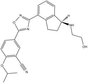
N#CC1=CC(C2=NC(C3=CC=CC4=C3CC[C@@H]4NCCO)=NO2)=CC=C1OC(C)C
|
|
| InChi Key |
XRVDGNKRPOAQTN-FQEVSTJZSA-N
|
|
| InChi Code |
InChI=1S/C23H24N4O3/c1-14(2)29-21-9-6-15(12-16(21)13-24)23-26-22(27-30-23)19-5-3-4-18-17(19)7-8-20(18)25-10-11-28/h3-6,9,12,14,20,25,28H,7-8,10-11H2,1-2H3/t20-/m0/s1
|
|
| 化学名 |
5-[3-[(1S)-1-(2-hydroxyethylamino)-2,3-dihydro-1H-inden-4-yl]-1,2,4-oxadiazol-5-yl]-2-propan-2-yloxybenzonitrile
|
|
| 别名 |
|
|
| HS Tariff Code |
2934.99.9001
|
|
| 存储方式 |
Powder -20°C 3 years 4°C 2 years In solvent -80°C 6 months -20°C 1 month |
|
| 运输条件 |
Room temperature (This product is stable at ambient temperature for a few days during ordinary shipping and time spent in Customs)
|
| 溶解度 (体外实验) |
|
|||
|---|---|---|---|---|
| 溶解度 (体内实验) |
配方 1 中的溶解度: ≥ 2.5 mg/mL (6.18 mM) (饱和度未知) in 10% DMSO + 40% PEG300 + 5% Tween80 + 45% Saline (这些助溶剂从左到右依次添加,逐一添加), 澄清溶液。
例如,若需制备1 mL的工作液,可将100 μL 25.0 mg/mL澄清DMSO储备液加入到400 μL PEG300中,混匀;然后向上述溶液中加入50 μL Tween-80,混匀;加入450 μL生理盐水定容至1 mL。 *生理盐水的制备:将 0.9 g 氯化钠溶解在 100 mL ddH₂O中,得到澄清溶液。 配方 2 中的溶解度: ≥ 2.5 mg/mL (6.18 mM) (饱和度未知) in 10% DMSO + 90% Corn Oil (这些助溶剂从左到右依次添加,逐一添加), 澄清溶液。 例如,若需制备1 mL的工作液,可将 100 μL 25.0 mg/mL 澄清 DMSO 储备液添加到 900 μL 玉米油中并混合均匀。 View More
配方 3 中的溶解度: 5%DMSO + Corn oil: 2.0mg/ml (4.94mM) 1、请先配制澄清的储备液(如:用DMSO配置50 或 100 mg/mL母液(储备液)); 2、取适量母液,按从左到右的顺序依次添加助溶剂,澄清后再加入下一助溶剂。以 下列配方为例说明 (注意此配方只用于说明,并不一定代表此产品 的实际溶解配方): 10% DMSO → 40% PEG300 → 5% Tween-80 → 45% ddH2O (或 saline); 假设最终工作液的体积为 1 mL, 浓度为5 mg/mL: 取 100 μL 50 mg/mL 的澄清 DMSO 储备液加到 400 μL PEG300 中,混合均匀/澄清;向上述体系中加入50 μL Tween-80,混合均匀/澄清;然后继续加入450 μL ddH2O (或 saline)定容至 1 mL; 3、溶剂前显示的百分比是指该溶剂在最终溶液/工作液中的体积所占比例; 4、 如产品在配制过程中出现沉淀/析出,可通过加热(≤50℃)或超声的方式助溶; 5、为保证最佳实验结果,工作液请现配现用! 6、如不确定怎么将母液配置成体内动物实验的工作液,请查看说明书或联系我们; 7、 以上所有助溶剂都可在 Invivochem.cn网站购买。 |
| 制备储备液 | 1 mg | 5 mg | 10 mg | |
| 1 mM | 2.4724 mL | 12.3622 mL | 24.7243 mL | |
| 5 mM | 0.4945 mL | 2.4724 mL | 4.9449 mL | |
| 10 mM | 0.2472 mL | 1.2362 mL | 2.4724 mL |
1、根据实验需要选择合适的溶剂配制储备液 (母液):对于大多数产品,InvivoChem推荐用DMSO配置母液 (比如:5、10、20mM或者10、20、50 mg/mL浓度),个别水溶性高的产品可直接溶于水。产品在DMSO 、水或其他溶剂中的具体溶解度详见上”溶解度 (体外)”部分;
2、如果您找不到您想要的溶解度信息,或者很难将产品溶解在溶液中,请联系我们;
3、建议使用下列计算器进行相关计算(摩尔浓度计算器、稀释计算器、分子量计算器、重组计算器等);
4、母液配好之后,将其分装到常规用量,并储存在-20°C或-80°C,尽量减少反复冻融循环。
计算结果:
工作液浓度: mg/mL;
DMSO母液配制方法: mg 药物溶于 μL DMSO溶液(母液浓度 mg/mL)。如该浓度超过该批次药物DMSO溶解度,请首先与我们联系。
体内配方配制方法:取 μL DMSO母液,加入 μL PEG300,混匀澄清后加入μL Tween 80,混匀澄清后加入 μL ddH2O,混匀澄清。
(1) 请确保溶液澄清之后,再加入下一种溶剂 (助溶剂) 。可利用涡旋、超声或水浴加热等方法助溶;
(2) 一定要按顺序加入溶剂 (助溶剂) 。
Prospective Evaluation of Sequencing From antiCD-20 Therapies to Ozanimod
CTID: NCT06529406
Phase: Phase 4 Status: Recruiting
Date: 2024-11-08
 SKI II
 JTE-013 HCl
 辛波莫德
辛波莫德
 辛波莫德富马酸盐
InvivoChem的所有产品仅用于作科学研究,不面向患者销售
Copyright 2020 InvivoChem LLC | All Rights Reserved 粤ICP备20063088号-1


 COA
COA
 463611831
