| 规格 | 价格 | 库存 | 数量 |
|---|---|---|---|
| 5mg |
|
||
| 10mg |
|
||
| 25mg |
|
||
| 50mg |
|
||
| 100mg |
|
||
| 250mg |
|
||
| Other Sizes |
|
| 靶点 |
Natural product; TMEM16A chloride channel; PKM2; NF-κB
|
|---|---|
| 体外研究 (In Vitro) |
Shikonin,TMEM16A 氯通道抑制剂,IC50 为 6.5 μM[1]。此外,Shikonin激肽特异性抑制 PKM2 [2]。此外,它还可以阻止核因子-κB (NF-κB) 通路的激活并抑制肿瘤坏死因子-α (TNF-α)。与对照相比,当暴露于浓度大于 50 μM 的紫草素时,正常人角质形成细胞 (NHK) 的活力显着降低 (P<0.05)。紫草素预处理两小时可抑制 TNF-α 诱导的 NF-κB p65 核转位 [3]。用 5 和 7.5 μM 紫草素处理 12 小时后,细胞活力显着降低。与 0 小时组相比,两种细胞系的抑制作用也显示出时间依赖性模式。结果发现,在24至48小时的时间段内,5μM紫草素比2.5μM紫草素具有更强的抑制作用。当U87和U251细胞用2.5、5和7.5 μM紫草素处理24和48小时(p<0.01)时,它们的侵袭性远低于对照组[4]。
在这项研究中,我们发现10μmol/L的Shikonin/紫草素刺激了正常人角质形成细胞的生长,1μmol/L的苦参素促进了人皮肤成纤维细胞的生长。然而,紫草素在体外不能直接诱导皮肤成纤维细胞中COL1 mRNA的表达和PIP的产生。此外,1μmol/L的紫草素抑制了肿瘤坏死因子-α刺激皮肤成纤维细胞诱导的NF-κB p65从细胞质向细胞核的易位。此外,紫草素抑制蛋白酶体的胰凝乳蛋白酶样活性,并与皮肤成纤维细胞中磷酸化抑制剂κB-α的积累有关。 结论:这些结果表明,Shikonin/紫草素可能通过其促进细胞生长的活性促进伤口愈合,并通过抑制蛋白酶体的活性抑制皮肤炎症。因此,紫草素可能是伤口愈合和炎症性皮肤病的潜在治疗试剂。[3] 紫草素/Shikonin是从紫草根中提取的蒽醌衍生物。紫草素传统上用于治疗炎症和感染性疾病,如肝炎。紫草素还抑制各种肿瘤的增殖并诱导其凋亡。然而,紫草素对胶质瘤的影响尚未完全阐明。本研究旨在探讨紫草素对人胶质母细胞瘤细胞迁移和侵袭的影响及其潜在机制。U87和U251人胶质母细胞瘤细胞用2.5、5和7.5μmol/L的紫草素处理,用CCK8、划痕愈合、体外Transwell迁移和侵袭试验评估细胞存活率、迁移和侵袭性。检测基质金属蛋白酶-2(MMP-2)和基质金属蛋白酶-9(MMP-9)的表达和活性,以及磷酸化β-catenin(p-β-catenmin)和磷酸化PI3K/Akt的表达。结果显示,紫草素显著抑制U87和U251细胞的增殖、迁移、侵袭和MMP-2和MMP-9的表达。β-catenin在两种细胞系中的表达趋势相反。它在U87细胞中受到显著抑制,在U251细胞中得到促进。本研究的结果表明,紫草素通过抑制MMP-2和-9的表达和活性,对胶质瘤细胞的迁移和侵袭具有抑制作用。此外,紫草素还抑制了p-PI3K和p-Akt的表达,以减弱两种细胞系中的细胞迁移和侵袭以及MMP-2和MMP-9的表达,这可以被PI3K/Akt通路激动剂胰岛素样生长因子-1(IGF-1)逆转。[4] 紫草素/Shikonin是一种高度亲脂性的萘醌,存在于紫草根中,在中药中具有多效性。基于其已报道的解热和抗炎特性,我们研究了紫草素是否抑制NLRP3炎性小体的激活。炎症小体是细胞质蛋白复合物,作为胱天蛋白酶-1募集和激活的支架,进而导致促炎细胞因子IL-1β和IL-18的切割和分泌。NLRP3炎性小体激活包括两个步骤:启动,即NF-κB通路的激活,以及炎性小体组装。虽然之前有报道称紫草素可以抑制启动步骤,但我们证明紫草素还可以抑制由可溶性和颗粒NLRP3激动剂在启动的永生化小鼠骨髓源性巨噬细胞中诱导的炎症小体激活的第二步。紫草宁比乙酰紫草宁更能有效地降低黑色素对NLRP3炎性体的激活。我们的研究结果表明,紫草素还可以通过双链DNA抑制AIM2炎性小体的激活。紫草素抑制小鼠巨噬细胞中ASC斑点的形成和caspase-1的激活,并抑制分离的caspase-1活性,表明它直接靶向caspase-1。将紫草素与β-乳球蛋白复合可以降低其毒性,同时保持对NLRP3炎性体激活的抑制作用,这表明具有提高生物利用度的紫草素可能对炎性体介导的疾病的治疗应用感兴趣[7]。 (-)-Alkannin 的 IC50 分别为 2.38 和 4.53 μM,抑制 HCT-116 和 SW-480 细胞的发育 [8]。 |
| 体内研究 (In Vivo) |
与骨关节炎组相比,紫草素显着阻止骨关节炎大鼠模型中IL-1β和TNF-α表达水平的升高(P<0.01)。在骨关节炎大鼠模型中,与骨关节炎组相比,紫草素显着降低了 NF-κB 蛋白表达量(P<0.01)。在用紫草素治疗的大鼠骨关节炎模型中,与骨关节炎组相比,诱导的iNOS水平降低(P<0.01)。与骨关节炎组相比,紫草素治疗显着降低了骨关节炎大鼠模型中COX-2蛋白表达的增加(P<0.01)。紫草素治疗的骨关节炎大鼠模型中caspase-3活性的增加明显低于骨关节炎组(P<0.01)。接受紫草素治疗后,骨关节炎组大鼠骨关节炎模型中Akt磷酸化显着恢复(P<0.01)[5]。
分泌性腹泻仍然是全球健康负担,并导致儿童死亡。抗腹泻疗法可能会减少腹泻疾病中的液体流失和肠道运动,这引起了人们的关注。在本研究中,我们使用基于细胞的荧光淬灭试验确定了紫草素是TMEM16A氯通道活性的抑制剂。紫草素的IC50值为6.5μM。短路电流测量表明,紫草素以剂量依赖的方式抑制Eact诱导的Cl(-)电流,IC50值为1.5μM。短路电流测量表明,紫草素对CCh诱导的小鼠结肠上皮细胞Cl(-)电流具有抑制作用,但不影响细胞质Ca(2+)浓度以及其他主要的肠细胞氯离子通道电导调节因子。特征研究发现,紫草素抑制基底外侧K(+)通道活性,而不影响Na(+)/K(+)-ATP酶活性。体内研究表明,紫草素显著延迟了小鼠的肠道运动,降低了轮状病毒腹泻新生小鼠模型的粪便含水量,而不影响体内病毒感染过程。综上所述,研究结果表明,紫草素抑制肠上皮细胞钙激活的氯通道,其抑制作用部分是通过抑制基底外侧K(+)通道活性实现的,紫草素可能是治疗轮状病毒分泌性腹泻的先导化合物。[1] 丙酮酸激酶M2(PKM2)的M2亚型已被证明在人类皮肤癌中上调。为了测试PKM2是否可能是化学预防的靶点,在一项化学诱导的小鼠皮肤癌变研究中使用了紫草素,紫草素是紫草根的天然产物,也是PKM2的特异性抑制剂。结果表明,紫草素治疗抑制了皮肤肿瘤的形成。皮肤表皮组织的形态学检查和免疫组织化学染色表明,紫草素抑制细胞增殖,但不诱导细胞凋亡。尽管紫草素单独抑制PKM2活性,但在皮肤癌变研究结束时,它并没有抑制肿瘤启动子诱导的皮肤表皮组织中PKM2的激活。为了揭示紫草素的潜在化学预防机制,进行了抗体微阵列分析,结果表明,化学致癌物处理上调了转录因子ATF2及其下游靶点Cdk4;而紫草素抑制了这些上调。在可促进的皮肤细胞模型中,ATF2的核水平在肿瘤促进过程中增加,而这种增加被紫草素抑制。此外,敲除ATF2降低了Cdk4和Fra-1(激活蛋白1的关键亚基)的表达水平。总之,这些结果表明,紫草素在皮肤癌变过程中抑制ATF2通路,而不是在体内抑制PKM2。[2] 紫草素先前已被证明具有抗肿瘤、抗炎、抗病毒和广泛的药理作用。本研究旨在探讨紫草素的保护作用是否通过抑制炎症和软骨细胞凋亡来介导,并阐明骨关节炎大鼠模型中的潜在分子机制。在健康雄性Sprague-Dawley大鼠中建立骨关节炎模型,每天腹腔注射10mg/kg紫草素4天。研究发现,紫草素治疗显著抑制了骨关节炎大鼠的炎症反应。与假手术组相比,骨关节炎显著增加了白细胞介素(IL)-1β、肿瘤坏死因子(TNF)-α和诱导型一氧化氮合酶(iNOS)水平。然而,紫草素治疗显著抑制了骨关节炎大鼠IL-1β、TNF-α和iNOS水平的升高。此外,与假手术组相比,骨关节炎大鼠的半胱氨酸天冬氨酸蛋白酶-3活性和环氧化酶(COX)-2蛋白表达显著增加,磷酸化Akt蛋白表达大大受到抑制。紫草素给药减轻了骨关节炎大鼠半胱氨酸天冬氨酸蛋白酶-3活性、COX-2表达和Akt磷酸化的变化。这些结果表明,紫草素通过调节骨关节炎大鼠模型中的磷酸肌醇3-激酶/Akt信号通路来抑制炎症和软骨细胞凋亡[5]。 |
| 酶活实验 |
碘化物流入荧光分析[1]
为了测量紫草素对TMEM16A的抑制作用,将表达TMEM16A的FRT细胞接种在黑壁透明底部96孔板中,直至融合。然后用PBS洗涤细胞三次,然后用不同浓度的紫草素孵育10分钟。用配备HQ 535/30M(535±15nm)发射和HQ500/20X(500±10nm)激发滤光片和注射泵的FLUOstar Galaxy微孔板读数器记录荧光数据。连续记录荧光14秒,以2秒的速度将ATP(200μM)与碘化物一起泵入系统。碘化物流入率(d[I-]/dt)如先前研究所述计算(Kristidis等人,1992)。 基于细胞的蛋白酶体活性测定[3] 在白壁96孔板中,约4000个HDF/孔在37°C下用0.1%DMSO、1μmol/L的Shikonin/紫草素或10μmol/L的内酰胺胱氨酸处理2小时,然后用50 ng/ml的TNF-α刺激30分钟。然后根据制造商的方案,将细胞与蛋白酶体Glo™细胞基检测试剂一起孵育15分钟。通过Centro LB 960微孔板光度计检测每个反应产生的发光。 Caspase-1活性测定[7] 使用BioVision的caspase-1抑制剂药物筛选试剂盒分析潜在的caspase-1抑制剂。紫草素和阳性抑制对照(Z-VAD-FMK)在PBS中制备,并应用于黑色96孔荧光板。加入活性胱天蛋白酶-1,然后加入胱天蛋白酶1底物YVAD-AFC。在37°C下孵育45分钟后,使用SinergyMx平板读数器测量样品的荧光。 |
| 细胞实验 |
免疫荧光研究[3]
将细胞接种到六孔板的盖玻片上,并在含有5%FBS的培养基中附着过夜。细胞在无血清培养基中饥饿24小时后,在用TNF-α(50 ng/ml)刺激之前,用1μmol/L的Shikonin/紫草素或0.1%的DMSO预处理细胞2小时。然后,在去除培养基后,用磷酸缓冲盐水冲洗细胞,并在4°C下用甲醇固定8分钟。在室温下用1%BSA的PBS溶液进行阻断步骤30分钟。细胞在室温下用1%BSA/PBS中的抗NF-κB p65(C-20)抗体(1:100稀释)免疫染色2小时,然后在室温下与FITC偶联的抗兔IgG-pAb(1:100稀释剂)孵育1小时。用BX 51TRF荧光显微镜观察载玻片。 免疫印迹分析[3] 人皮肤成纤维细胞用1μmol/L的Shikonin/紫草素或0.1%的DMSO预处理2小时,然后用50 ng/ml的TNF-α刺激30分钟。然后,根据制造商的说明,用核提取试剂盒提取细胞质蛋白。蛋白质在5-20%梯度凝胶上通过十二烷基硫酸钠聚丙烯酰胺凝胶电泳分离,并使用iBlot®系统通过半干转移法转移到硝化纤维膜上。 细胞增殖试验[4] 根据文献,用CCK8检测试剂盒测量细胞增殖。简而言之,将U87和U251细胞在标准DMEM中以每孔1×10⁴个细胞的密度接种到96孔板中,并在标准条件下(37°C和5%CO2)孵育24小时。我们之前的数据显示,24小时后,Shikonin/紫草素的IC50值对U251细胞为1.84±0.34μmol/L,对U87细胞为2.02±0.44μmol/L。因此,本研究中使用的浓度为2.5、5和7.5μmol/L。然后将培养基替换为空白、无血清DMEM或含紫草素的DMEM,浓度分别为2.5、6和7.5μol/L。每个孔的总体积为200μL。将胶质瘤细胞在这些溶液中孵育0、12、24、36、48或72小时,然后在37°C下用每个孔中的20μL CCK8处理1.5小时。最后,轻轻摇动平板,使用ELISA平板读数器在570nm(OD570)处记录光密度。至少进行了三次独立实验。抑制率按以下公式计算:(ControlOD570-实验组OD570)/ControlOD570×100%。 体外迁移试验[4] 根据文献,在具有8-μm孔径Transwell插入物的24孔板中评估了人胶质母细胞瘤细胞的迁移能力。将亲本U87或U251细胞胰蛋白酶化,以5×10⁵/mL的密度重新悬浮在无血清DMEM中,并将200μL细胞悬浮液加入上室。将500微升条件培养基(DMEM培养基,补充10%FBS)放入下腔室,作为细胞迁移的诱导剂。无血清DMEM作为阴性对照。将紫草素以2.5、5或7.5μmol/L的浓度添加到亲本U87细胞或U251细胞的悬浮液中。还添加了PIRES2-p-β-catenin、shRNA-p-β-cantin、LY294002(20μmol/L)或紫草素(5μmol/L)与PI3K/Akt激动剂胰岛素样生长因子-1(IGF-1)(20μg/mL,Proteintech)的组合。孵育24或48小时后,取出插入物,用棉签仔细去除过滤器上表面残留的细胞。迁移到下侧表面的细胞用PBS轻轻洗涤一次,在室温下用甲醇和冰醋酸(按3:1混合)固定30分钟,并在Giemsa染色中染色15分钟。在六个随机高倍视野(×400)中计数迁移细胞的平均数量。 划痕愈合试验[4] 如前所述,进行划痕愈合试验以评估胶质母细胞瘤细胞的迁移能力。简而言之,将细胞以1.0×10⁵/孔的密度接种到六孔板中,直到它们达到80%的融合。用移液管尖端在融合的U87或U251细胞单层中产生划痕。在实验开始时,伤口的宽度被评估为相同。用PBS冲洗孔三次,以去除漂浮的细胞和碎片。为了测试紫草素对人胶质母细胞瘤细胞迁移的影响,将亲本U87或U251细胞接种在有或没有紫草素(2.5、5或7.5μmol/L)的无血清DMEM中。然后将这些细胞孵育0-48小时。培养板在37°C和5%CO2中孵育。在0、24和48小时时,使用相差显微镜测量伤口愈合情况,并随着时间的推移用照片记录。 体外侵袭试验[4] 如前所述,使用具有8-μm孔径插入物的Transwell侵袭试验检查了紫草素对人胶质母细胞瘤细胞侵袭的影响。Transwell过滤器插入物的膜上涂有以1:7的比例用介质稀释的Matrigel。如上所述制备亲本U87或U251细胞。将500微升补充有10%FBS的DMEM放入下腔室。无血清DMEM作为阴性对照。将紫草素(2.5、5或7.5μmol/L)、pIRES2-p-β-catenin、shRNA-p-β-calenin、LY294002(20μmol/L)或紫草素(5μmol/L)与IGF-1(20μg/mL,Proteintech:Chicago,IL,USA)联合加入上腔细胞悬浮液中。孵育0-48小时后,取出插入物,如上所述在显微镜下进行观察。在6个随机高倍视野(×400)中计数侵袭细胞的平均数量。 蛋白质印迹分析[4] 为了测定p-β-catenin的表达,进行了Western blot分析。U87或U251细胞用浓度为2.5、5和7.5μmol/L的Shikonin/紫草素处理48小时。用冰冷的PBS洗涤细胞三次以停止刺激。然后,收集细胞并在含有50 mmol/L Tris(PH 7.4)、150 mmol/L NaCl、1%Triton X-100、1%脱氧胆酸钠、0.1%SDS、1 mmol/L原钒酸钠、50 mmol/L氟化钠和1 mmol/L EDTA的冰冷放射免疫沉淀分析裂解缓冲液中裂解30分钟。然后用超声波破碎机破碎沉淀物,在4°C下以17000 rpm离心样品60分钟。收集上清液作为可溶性部分,并转移到新的试管中。立即在沸水浴中加热样品管5分钟以使蛋白质变性。用BCA蛋白测定试剂盒测定可溶性物质的蛋白质浓度。 |
| 动物实验 |
肠道动力测定[1]
ICR小鼠禁食24小时后,口服给予紫草素(5.8 μg)。30分钟后,口服给予20 mg溶于5%阿拉伯胶的10%活性炭溶液。再过30分钟后,处死动物并取出小肠。蠕动指数的计算方法为活性炭在小肠内移动的长度与小肠总长度的比值。 轮状病毒腹泻小鼠模型[1] 将新生ICR小鼠(4-7日龄,体重2-3克)用聚乙烯管(外径0.6毫米,内径0.3毫米)和胰岛素注射器进行灌胃,接种30微升轮状病毒(病毒滴度1.2 × 10⁷ pfu/mL)。之后将小鼠放回母鼠身边哺乳。每日通过轻柔触诊腹部收集粪便样本。在一组实验中,紫草素处理组小鼠在病毒接种前一天口服紫草素(0.4 和 1.7 μg,溶于 30 μL PBS),之后每天三次,直至接种后第 3 天。对照组小鼠仅接受 30 μL PBS。阳性对照组(CaCCinh-A01)小鼠在病毒接种前一天腹腔注射 34 μg CaCCinh-A01(溶于 20 μL PBS),之后每天两次,直至接种后第 3 天。在另一组实验中,紫草素处理组小鼠在病毒接种后第二天口服紫草素(溶于 PBS),之后每天三次,直至接种后第 3 天。阴性对照组小鼠仅接受 30 μL PBS。阳性对照小鼠在病毒接种前一天腹腔注射CaCCinh-A01,之后每天两次,直至第3天。 化学诱导的小鼠皮肤癌变[2] 60只6-8周龄的雌性DBA/2小鼠(对皮肤癌变相对敏感)被分为4组:DMSO组、DMBA/TPA组、SKN组和SKN+DMBA/TPA组。DMSO组(5只小鼠)接受DMSO处理作为溶剂对照;DMBA/TPA组先单次局部涂抹200 nmol DMBA,持续2周,随后单次局部涂抹5 μg TPA(12-O-十四烷酰佛波醇-13-乙酸酯),每天一次,每周三次,持续14周。 SKN组按照与DMBA/TPA处理相同的方案,局部应用10 μg紫草素。SKN+DMBA/TPA组先接受紫草素(SKN)处理,30分钟后接受TPA处理。皮肤致癌性研究结束后,小鼠用戊巴比妥钠(150 mg/kg,腹腔注射)处死。收集实验部位的皮肤样本,并按以下所述进行生化和形态学分析。 实验组和处理[5] 大鼠随机分为三组:假手术组(n=10)、骨关节炎模型组(n=10)和紫草素处理组(n=10)。在假手术组中,仅在无菌条件下暴露麻醉大鼠的右膝关节,并腹腔注射0.1 ml/100 g生理盐水。在骨关节炎模型组中,骨关节炎模型大鼠腹腔注射0.1 ml/100 g生理盐水。在紫草素治疗组中,骨关节炎模型大鼠在建立骨关节炎模型后,每日一次腹腔注射10 mg/kg紫草素,连续4天(14,15)。 ELISA分析[5] 用10 mg/kg紫草素治疗后,从各组大鼠(n=10)的腹主动脉采集外周血。血液在4℃下以12,000 × g离心10分钟,取上清液,使用ELISA试剂盒(北京4A生物科技有限公司)按照制造商说明书分析IL-1β、TNF-α和iNOS的含量。 Western blot分析[5] 给予10 mg/kg紫草素治疗后,大鼠经腹腔注射50 mg/kg戊巴比妥钠麻醉,断头处死,收集关节组织样本(每组n=10)。将样本用RIPA裂解缓冲液匀浆。匀浆液在4℃下以12,000 × g离心10分钟,使用BCA蛋白定量试剂盒进行分析。将约 50 µg 蛋白质通过 12% SDS-聚丙烯酰胺凝胶电泳分离,然后转移至硝酸纤维素滤膜上。使用小鼠抗核因子 (NF)-κB p65 (sc-29311; 1:500)、抗环氧合酶 (COX)-2 (sc-23984; 1:300)、抗 Akt (sc-8312; 1:500)、抗磷酸化 Akt (抗 p-Akt; sc-135650; 1:1,000) 和抗 β-肌动蛋白 (BB-2101-1; 1:5,000) 抗体检测蛋白质,随后使用辣根过氧化物酶标记的山羊抗小鼠二抗 (sc-2777; 1:5,000) 进行检测。使用 AlphaEase FC 软件测定蛋白质表达的相对量。 Caspase-3 活性分析 [5] 用 10 mg/kg 紫草素治疗 4 天后,处死大鼠并收集骨关节炎样本。将样本用 RIPA 裂解缓冲液匀浆。匀浆液在 4°C 下以 12,000 × g 离心 10 分钟,并使用 BCA 蛋白定量试剂盒进行分析。将 20 µg 蛋白质与底物 Ac-DEVD-pNA 在反应缓冲液中混合,并在 37°C 下避光孵育 2 小时。然后在 405 nm 波长处检测吸光度。 |
| 药代性质 (ADME/PK) |
吸收、分布和排泄
紫草素和紫草苷是天然存在的羟基萘醌类化合物,具有广泛的伤口愈合、抗菌、抗炎和抗氧化活性。近年来,大量的科学研究集中于它们对多种肿瘤的疗效及其抗肿瘤机制。脂质体已被证明是一种有效的药物载体,与传统制剂相比具有显著优势,例如可控释放和靶向给药,因此市场上出现了多种脂质体制剂,其中一些用于抗癌药物。本研究旨在首次制备载有紫草苷的脂质体,以提高紫草苷的治疗指数。我们开发了一种基于薄膜水化法的优化技术,并从理化特性、药物包封率和释放曲线等方面对脂质体进行了表征。结果表明,使用1,2-二棕榈酰磷脂酰胆碱和卵磷脂酰胆碱两种脂质体均成功将紫草素包封于脂质体中。所得脂质体具有良好的理化性质、较高的包封率和令人满意的体外释放曲线。此外,还测试了脂质体对三种人类癌细胞系(乳腺癌、胶质瘤和非小细胞肺癌)的体外细胞毒性,结果显示其具有中等的生长抑制活性。实际应用:紫草素是一种天然存在的羟基萘醌类化合物,近年来,针对其对多种肿瘤的疗效及其抗肿瘤作用机制,开展了大量的科学研究(包括体外、体内和临床试验)。本研究旨在制备并表征载有紫草素的脂质体,以期将其作为一种新型的紫草素药物递送系统。与传统剂型相比,脂质体制剂具有显著优势,例如可控释放和靶向递送抗癌药物。因此,脂质体可以降低紫草素的副作用,提高其对癌细胞的选择性,并保护紫草素免受体内生物转化和不稳定性(氧化和聚合)的影响。此外,脂质体递送有助于克服紫草素水溶性低的问题,这是其口服和内服的主要障碍,因为紫草素无法溶解并从受体进一步吸收。药代动力学研究表明,紫草素通过灌胃和肌肉注射给药时吸收迅速,1分钟后几乎检测不到血浆中的紫草素,灌胃给药的生物利用度约为34%(Wang et al., 1988)。在本研究中,用于肠道运动(0.38 mg/kg)和轮状病毒感染小鼠模型(0.69 mg/kg)的剂量均低于被认为具有毒性的剂量。[1] |
| 毒性/毒理 (Toxicokinetics/TK) |
本研究观察到紫草素可降低轮状病毒感染新生小鼠的粪便含水量,这可能是通过紫草素的抗分泌作用实现的,该作用涉及抑制CaCCGI氯离子通道的活性。尽管TMEM16A存在于肠细胞中,但一些研究者提出,轮状病毒非结构蛋白NSP4引起的腹泻主要是通过激活肠道上皮细胞中的TMEM16A实现的。一项使用小分子TMEM16A抑制剂T16Ainh-A01的研究表明,TMEM16A仅是肠道上皮CaCC的次要成分(Namkung等,2011)。我们之前的研究表明,TMEM16A 和 CaCCGI 具有不同的特性,因为木脂素类化合物柯布辛和桉树素对 TMEM16A 和 CaCCGI 的影响不同,前者抑制 TMEM16A,后者激活 CaCCGI(Jiang 等,2015)。本研究表明,紫草素在细胞培养模型和离体小鼠结肠中均能抑制 CaCCGI 介导的短路电流。此外,体内研究表明,紫草素能降低新生小鼠腹泻模型中的水分含量,且不影响轮状病毒感染过程(图 6)。这些发现支持了以下观点:轮状病毒感染引起的水样腹泻的主要途径是通过 NSP4 激活 CaCCGI 而非 TMEM16A,从而加剧体液积聚。此外,紫草素对TMEM16A的抑制作用可延缓胃肠蠕动,延长液体吸收时间,从而进一步减少净液体分泌。
尽管紫草素具有诸多益处,但它并非没有毒性。腹腔注射紫草素已被证实会产生一定的毒性,其LD50为20 mg/kg(Sankawa等,1977)。药代动力学研究表明,紫草素经灌胃和肌肉注射后吸收迅速,1分钟后几乎检测不到血浆中的紫草素,灌胃给药的生物利用度约为34%(Wang等,1988)。在本研究中,用于肠道蠕动(0.38 mg/kg)和小鼠轮状病毒感染(0.69 mg/kg)的剂量均低于被认为具有毒性的剂量。[1] 72521trattLD50toralt>1 gm/kgtNahrung. Chemistry, Biochemistry, Microbiology, Technology, Nutrition., 15(505), 1971 [PMID:5172860] 72521tmousetLD50toralt3 gm/kgtNahrung. Chemistry, Biochemistry, Microbiology, Technology, Nutrition., 15(505), 1971 [PMID:5172860] 解毒剂和紧急处理 /SRP:/ 立即急救:确保已进行充分的去污处理。如果患者停止呼吸,应立即开始人工呼吸,最好使用按需呼吸机、球囊面罩或简易呼吸面罩,并按照培训内容进行操作。必要时进行心肺复苏。立即用流动清水冲洗受污染的眼睛。不要催吐。如果发生呕吐,应将患者身体前倾或置于左侧卧位(如果可能,头部向下),以保持呼吸道通畅并防止误吸。保持患者安静并维持正常体温。寻求医疗救助。/A类和B类中毒/ /SRP:/ 基本治疗:建立通畅的呼吸道(必要时使用口咽或鼻咽通气道)。必要时进行吸痰。观察呼吸功能不全的迹象,必要时辅助通气。使用无创呼吸面罩以10至15升/分钟的流量给予氧气。监测肺水肿,必要时进行治疗……。监测休克,必要时进行治疗……。预判癫痫发作并根据需要进行治疗……。如眼睛受到污染,立即用水冲洗眼睛。转运过程中,持续用0.9%生理盐水(NS)冲洗每只眼睛……。不要使用催吐剂。如误服,漱口后,如果患者能够吞咽、有强烈的咽反射且不流口水,则给予5 mL/kg至200 mL的水进行稀释……。皮肤烧伤经去污后,用干燥的无菌敷料覆盖……。/A类和B类毒物/ /SRP:/ 高级治疗:对于意识不清、严重肺水肿或严重呼吸窘迫的患者,考虑进行口咽或鼻咽气管插管以控制气道。使用球囊面罩进行正压通气可能有效。考虑药物治疗肺水肿……。考虑使用β受体激动剂(如沙丁胺醇)治疗严重支气管痉挛……。监测心律,必要时治疗心律失常……开始静脉输注5%葡萄糖溶液/SRP:“保持血管通畅”,最小流速/。如果出现低血容量的迹象,使用0.9%生理盐水(NS)或乳酸林格氏液。对于伴有低血容量迹象的低血压,谨慎输液。注意液体过量的迹象……用地西泮或劳拉西泮治疗癫痫发作……使用盐酸丙美卡因辅助眼部冲洗……/毒物A和B/Currance, PL Clements, B., Bronstein, AC (编).; 危险物质暴露的紧急护理。第3版,Elsevier Mosby,圣路易斯,密苏里州,2005年,第101页。 160-1 人体毒性摘录 /替代和体外试验/ 紫草素具有预防或用于治疗芳胺类药物诱导的膀胱移行细胞癌的潜力。/研究人员/通过测量乙酰化2-氨基芴 (AF) 的含量、AF-DNA 加合物的含量、/N-乙酰转移酶 (NAT)/ mRNA 的变化以及 NAT 酶的含量来评估其有效性。将 T24 人膀胱癌细胞与 30 μM AF 和不同浓度的紫草素孵育不同时间。然后收集用紫草素 (16 μM) 处理的 T24 细胞,并用于以下两项实验:1).将T24细胞与22.5 μM AF和紫草素(0、16 μM)(共处理)孵育6、12、18、24和48小时。将T24细胞与不同浓度的AF和紫草素(0、16 μM)孵育24小时,并通过高效液相色谱法(HPLC)测定AF和AAF的含量。然后,在制备的人T24细胞胞质溶胶中加入不同浓度的AF和紫草素,以测定NAT的动力学常数。接下来,检测并测定经紫草素处理或未处理的人T24细胞中AF-DNA加合物的含量。最后两步包括测定经紫草素处理或未处理后NAT抗原-抗体复合物的含量,以及评估紫草素对NAT基因的影响。较高浓度的紫草素会降低AF的乙酰化水平。研究发现,在相同紫草素浓度下,培养时间越长,AF乙酰化水平的差异越大。同时,AAF的增加与孵育时间呈正比。在16 μM紫草素存在下,AF的N-乙酰化水平降低了72-84%。紫草素在所有检测的AF剂量下均能降低人T24细胞中AAF的生成量。胞质NAT的Km和Vmax值在加入紫草素后均降低。此外,紫草素在所有检测的AF剂量下均能降低人724细胞中AAF的生成量和AF-DNA加合物的形成。紫草素处理后,抗体染色细胞的百分比发生了显著变化,尤其是在较高紫草素浓度下。随着紫草素浓度(16-24 μM)的增加,NAT1 mRNA 水平和 NAT1/β-肌动蛋白比值显著降低。紫草素影响人膀胱肿瘤 T24 细胞中的 NAT 活性、基因表达(NAT1 mRNA)、AF-DNA 加合物的形成以及 NAT 抗原-抗体的形成……PMID:15011747 /替代和体外试验/ 从中药紫草根中分离得到的紫草素具有抗炎特性。/研究人员/评估了紫草素的化疗潜力,并研究了其在组织培养中对人皮肤肿瘤的可能作用机制。与 SV-40 转染的角质形成细胞相比,紫草素以浓度和时间依赖的方式优先抑制人表皮样癌细胞的生长,表明其对该癌细胞系具有抗增殖作用。此外,紫草素降低了EGFR、ERK1/2和蛋白酪氨酸激酶的磷酸化水平,同时提高了JNK1/2的磷酸化水平。总体而言,紫草素治疗与人表皮样癌细胞中凋亡相关蛋白的细胞内磷酸化水平升高以及增殖相关蛋白水平降低有关。PMID:14568164 /替代和体外试验/ 本研究探讨了紫草素作为抗肝癌药物的潜力,并建立了体外人肝癌模型。本研究采用HepG2细胞系作为肝癌模型。通过MTT法检测紫草素对HepG2细胞生长的抑制作用。为了探究紫草素抑制细胞生长的潜在机制,本研究检测了HepG2细胞的细胞周期分布、DNA片段化、线粒体膜电位破坏以及Bax和Bcl-2的表达。通过流式细胞术和电镜分别检测Annexin V信号和CD95表达,研究了紫草素诱导细胞凋亡的活性。结果表明,紫草素以剂量依赖的方式抑制HepG2细胞的生长,其IC50值(抑制细胞生长50%的浓度)为4.30 mg/mL。紫草素以剂量依赖的方式抑制细胞生长,并阻滞HepG2细胞周期于S期。在不同浓度的紫草素处理组中观察到线粒体形态的改变,以及线粒体膜电位的剂量依赖性降低。Western blot分析显示,紫草素抑制Bcl-2的表达并诱导Bax的表达。紫草素可增强Annexin V信号和CD95(Fas/APO)表达,导致HepG2细胞凋亡。此外,电镜观察发现,紫草素处理48小时后,HepG2细胞中出现核内染色质团块形成、细胞核固缩、微绒毛消失、线粒体空泡变性、粗面内质网减少以及游离核糖体溶解等与细胞凋亡相关的病理改变。紫草素可能通过诱导早期细胞凋亡途径抑制HepG2细胞,并能通过CD95恢复HepG2细胞的凋亡敏感性,因此应被视为预防或治疗人类肝癌的候选药物。PMID:21164560 /替代和体外试验/ 紫草素(SK)是从草药紫草(紫草)中分离鉴定出的关键生物活性成分。/本研究/探讨了SK在乳腺癌细胞(MCF-7、T47D和MDA-MB-231细胞)中的抗雌激素活性。在人乳腺癌细胞中,我们观察到SK处理可抑制雌激素受体α(ERα)阳性乳腺癌细胞的生长,但对ERα阴性乳腺癌细胞无抑制作用。SK与雌激素联合处理可抑制雌激素依赖性细胞生长。本研究探讨了SK抑制雌激素作用的潜在分子机制……SK对ERα mRNA表达无影响,但会降低其蛋白水平。这种作用与ERα泛素化水平升高导致其降解有关。结果表明,SK通过蛋白酶体介导的途径下调ERα蛋白。……SK处理可抑制雌激素诱导的雌激素反应元件报告基因活性。此外,SK抑制ERα在雌激素依赖性基因启动子处的募集,进而抑制基因表达。最后,与SK联合治疗增强了乳腺癌细胞对内分泌治疗的敏感性……PMID:19760501 非人类毒性摘录 /实验动物:亚慢性或慢性前暴露/ /本研究的目的是/探讨紫草素对早期和已建立的小鼠胶原诱导性关节炎(CIA)的抗炎或免疫调节作用。/小鼠/在CIA发病期间、之前或之后,腹腔注射紫草素(5 mg/kg),持续10天。通过宏观评分对关节炎反应进行视觉监测。采用逆转录-聚合酶链式反应和蛋白质印迹法检测CIA小鼠髌骨及其邻近滑膜中细胞因子的mRNA和蛋白表达。膝关节组织学用于评估软骨破坏和骨侵蚀的发生情况。单独使用紫草素(5 mg/kg)治疗对早期胶原诱导性关节炎(CIA)的宏观评分和关节炎发生率没有影响。然而,在已建立CIA模型的小鼠中,紫草素治疗10天后,宏观评分和软骨破坏均显著改善。此外,与对照组相比,治疗组小鼠滑膜组织和关节软骨中Th1细胞因子(肿瘤坏死因子-α和白细胞介素(IL)-12)的mRNA水平显著降低,而Th2细胞因子(IL-10和IL-4)的mRNA和蛋白水平在整个治疗期间持续升高。此外,在已建立CIA模型的小鼠中,紫草素治疗后炎症细胞因子IL-6的mRNA和蛋白水平也下调。与对照组相比,紫草素组T细胞表达的T-box蛋白(T-bet)mRNA水平降低,而GATA-3 mRNA水平升高。紫草素治疗已建立的胶原诱导性关节炎(CIA)小鼠可抑制Th1细胞因子的表达,并诱导Th2细胞因子的表达。紫草素对Th1细胞因子表达的抑制作用可能不仅通过T-bet机制抑制Th1反应,还可能通过GATA-3依赖性机制诱导IL-10和IL-4等抗炎介质的产生。PMID:18781399 |
| 参考文献 | |
| 其他信息 |
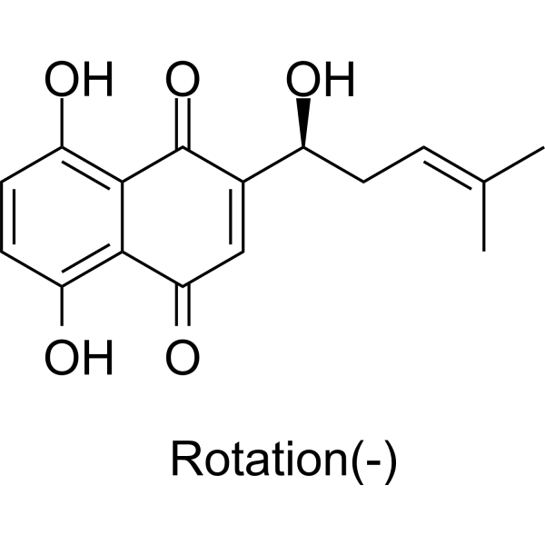
紫草素是一种羟基-1,4-萘醌。
据报道,紫草素存在于匍匐紫草(Arnebia decumbens)、紫草(Arnebia euchroma)以及其他有相关数据的生物体中。 紫草素是一种羟基-1,4-萘醌。 据报道,紫草素存在于匍匐紫草(Arnebia decumbens)、紫草(Arnebia euchroma)以及其他有相关数据的生物体中。 另见:水杨梅(Arnebia guttata)根(部分);紫草(Arnebia euchroma)根(部分);紫草(Lithospermum erythrorhizon)根(部分)。 作用机制 研究人员此前开发了一种基于基因枪的体内筛选系统,并将紫草素鉴定为肿瘤坏死因子-α (TNF-α) 基因表达的强效抑制剂。在此……紫草素选择性地在RNA剪接水平抑制TNF-α的表达。用紫草素处理脂多糖刺激的人原代单核细胞和THP-1细胞后,TNF-α的转录诱导正常,但未剪接的前体mRNA积累,导致功能性mRNA减少。这种效应在非细胞毒性剂量的紫草素下即可发生,且具有高度特异性,因为管家基因和另一种炎症细胞因子基因白细胞介素-8 (IL-8) 的mRNA生成均未受到影响。此外,LPS与紫草素联合处理可增加IL-8的最终蛋白生成,同时抑制双链RNA激活的蛋白激酶(PKR)通路。由于PKR失活已被证实会下调TNF-α RNA的剪接过程并干扰翻译,我们的研究结果表明,紫草素可能通过PKR通路失活实现对细胞因子蛋白表达的差异性调节,并揭示了TNF-α前体mRNA剪接的调控可能构成未来抗炎应用的一个有前景的靶点。紫草素是从中药紫草(Lithospermum erythrorhizon)的根中分离得到的,并被认为具有抗炎特性。研究人员评估了紫草素的化疗潜力,并在组织培养中研究了其在人皮肤肿瘤中的可能作用机制。与SV-40转染的角质形成细胞相比,紫草素以浓度和时间依赖的方式优先抑制人表皮样癌细胞的生长,表明其对该癌细胞系具有抗增殖作用。此外,紫草素降低了EGFR、ERK1/2和蛋白酪氨酸激酶的磷酸化水平,同时提高了JNK1/2的磷酸化水平。总体而言,紫草素处理与人表皮样癌细胞中凋亡相关蛋白的细胞内磷酸化水平升高以及增殖相关蛋白水平降低有关。 ……/先前的研究表明/紫草素,一种从紫草(Lithospermun erythrorhizon Sieb. Et Zucc)中分离得到的天然化合物,能够抑制脂肪生成和脂肪堆积。本研究旨在探讨紫草素抗脂肪生成作用的分子机制。我们利用小干扰RNA(siRNA)转染技术进行了基因敲低实验,以阐明β-catenin在紫草素抗脂肪生成作用中的关键作用。在源自小鼠胚胎的前脂肪细胞——3T3-L1细胞的脂肪生成过程中,紫草素能够阻止β-catenin的下调,并提高其转录产物cyclin D1的水平。siRNA介导的基因敲低实验表明,β-catenin是紫草素抗脂肪生成作用的关键介质。紫草素诱导的脂肪生成主要转录因子(包括过氧化物酶体增殖物激活受体γ和CCAAT/增强子结合蛋白α)以及脂质代谢酶(包括脂肪酸结合蛋白4和脂蛋白脂肪酶)的表达降低,以及细胞内脂肪的积累,均可通过siRNA介导的β-catenin基因敲低而显著恢复。在WNT/β-catenin信号通路中的基因中,WNT10B和DVL2的表达水平显著上调,而AXIN的表达水平则被紫草素下调。本研究表明,紫草素可通过体外调节WNT/β-catenin信号通路抑制脂肪生成,并提示WNT/β-catenin信号通路可作为治疗肥胖及相关疾病的潜在靶点,例如利用紫草素等天然化合物进行治疗。紫草素是紫草(紫草属植物紫草的干燥根)的主要成分,因其抗炎活性而被广泛应用(Chen et al., 2002)。据报道,紫草素具有抗氧化、抗菌、抗寄生虫、抗病毒和促进伤口愈合等活性(Andujar et al., 2013)。紫草素可能用于治疗哮喘。Takano-Ohmuro等(2008)着重研究了紫草素的抗炎活性,并探讨了其在哮喘治疗中的应用(Takano-Ohmuro et al., 2008)。其他研究者利用小鼠哮喘模型证实,紫草素在体外可抑制骨髓来源树突状细胞的成熟,在体内可抑制过敏反应和气管高反应性(Lee et al., 2010)。由于TMEM16A在气道平滑肌细胞中表达并参与平滑肌收缩(Huang et al., 2012),我们推测TMEM16A可能参与紫草素介导的哮喘抑制作用。在本研究中,我们发现紫草素可抑制TMEM16A氯离子通道活性,表明紫草素可能通过抑制气管平滑肌收缩来缓解哮喘。[1] 综上所述,本研究结果表明,紫草素能有效抑制化学诱导的皮肤癌变,其主要机制是通过抑制皮肤癌变过程中的细胞增殖。本研究中发现的潜在靶点ATF2将在未来的实验中进行验证。[2] 总之,我们的研究结果表明,紫草素通过一种目前未知的机制促进皮肤细胞增殖,但紫草素不会诱导HDFs中COL1的表达。此外,紫草素抑制HDFs中的NF-κB信号通路和蛋白酶体活性,提示紫草素具有抗炎作用。因此,紫草素可能是一种潜在的治疗药物,可用于伤口愈合和炎症性皮肤病的治疗。因此,紫草素可能最适合用于治疗难治性炎症性皮肤溃疡。[3] 紫草素通过抑制MMP-2和MMP-9来减弱人胶质母细胞瘤细胞的增殖、迁移和侵袭能力。在p53野生型胶质瘤细胞中,该机制与磷酸化β-catenin Y333和p-PI3K/p-Akt表达的下调有关。在p53突变型胶质瘤细胞中,它与PI3K/Akt通路抑制相关。[4] 总之,本研究结果证实,紫草素通过调节PI3K/Akt信号通路抑制骨关节炎大鼠模型中的炎症和软骨细胞凋亡。这些发现表明,紫草素具有治疗骨关节炎的潜力。[5] |
| 分子式 |
C16H16O5
|
|---|---|
| 分子量 |
288.2952
|
| 精确质量 |
288.099
|
| 元素分析 |
C, 66.66; H, 5.59; O, 27.75
|
| CAS号 |
517-88-4
|
| 相关CAS号 |
Shikonin;517-89-5;Alkannin;23444-65-7
|
| PubChem CID |
72521
|
| 外观&性状 |
Brown to red solid powder
|
| 密度 |
1.4±0.1 g/cm3
|
| 沸点 |
567.4±50.0 °C at 760 mmHg
|
| 熔点 |
149°
|
| 闪点 |
311.0±26.6 °C
|
| 蒸汽压 |
0.0±1.6 mmHg at 25°C
|
| 折射率 |
1.642
|
| LogP |
4.35
|
| tPSA |
94.83
|
| 氢键供体(HBD)数目 |
3
|
| 氢键受体(HBA)数目 |
5
|
| 可旋转键数目(RBC) |
3
|
| 重原子数目 |
21
|
| 分子复杂度/Complexity |
501
|
| 定义原子立体中心数目 |
1
|
| SMILES |
CC(=CC[C@@H](C1=CC(=O)C2=C(C=CC(=C2C1=O)O)O)O)C
|
| InChi Key |
NEZONWMXZKDMKF-JTQLQIEISA-N
|
| InChi Code |
InChI=1S/C16H16O5/c1-8(2)3-4-10(17)9-7-13(20)14-11(18)5-6-12(19)15(14)16(9)21/h3,5-7,10,17-19H,4H2,1-2H3/t10-/m0/s1
|
| 化学名 |
5,8-dihydroxy-2-[(1S)-1-hydroxy-4-methylpent-3-enyl]naphthalene-1,4-dione
|
| 别名 |
(-)-Alkannin; 517-88-4; Anchusin; Alkanna Red; Anchusa acid; Alkannin (VAN); Anchusin (VAN); Alkanna red (VAN);
|
| HS Tariff Code |
2934.99.9001
|
| 存储方式 |
Powder -20°C 3 years 4°C 2 years In solvent -80°C 6 months -20°C 1 month |
| 运输条件 |
Room temperature (This product is stable at ambient temperature for a few days during ordinary shipping and time spent in Customs)
|
| 溶解度 (体外实验) |
DMSO : ~33.33 mg/mL (~115.61 mM)
|
|---|---|
| 溶解度 (体内实验) |
配方 1 中的溶解度: ≥ 2.5 mg/mL (8.67 mM) (饱和度未知) in 10% DMSO + 40% PEG300 + 5% Tween80 + 45% Saline (这些助溶剂从左到右依次添加,逐一添加), 澄清溶液。
例如,若需制备1 mL的工作液,可将100 μL 25.0 mg/mL澄清DMSO储备液加入到400 μL PEG300中,混匀;然后向上述溶液中加入50 μL Tween-80,混匀;加入450 μL生理盐水定容至1 mL。 *生理盐水的制备:将 0.9 g 氯化钠溶解在 100 mL ddH₂O中,得到澄清溶液。 配方 2 中的溶解度: ≥ 2.5 mg/mL (8.67 mM) (饱和度未知) in 10% DMSO + 90% (20% SBE-β-CD in Saline) (这些助溶剂从左到右依次添加,逐一添加), 澄清溶液。 例如,若需制备1 mL的工作液,可将 100 μL 25.0 mg/mL澄清DMSO储备液加入900 μL 20% SBE-β-CD生理盐水溶液中,混匀。 *20% SBE-β-CD 生理盐水溶液的制备(4°C,1 周):将 2 g SBE-β-CD 溶解于 10 mL 生理盐水中,得到澄清溶液。 请根据您的实验动物和给药方式选择适当的溶解配方/方案: 1、请先配制澄清的储备液(如:用DMSO配置50 或 100 mg/mL母液(储备液)); 2、取适量母液,按从左到右的顺序依次添加助溶剂,澄清后再加入下一助溶剂。以 下列配方为例说明 (注意此配方只用于说明,并不一定代表此产品 的实际溶解配方): 10% DMSO → 40% PEG300 → 5% Tween-80 → 45% ddH2O (或 saline); 假设最终工作液的体积为 1 mL, 浓度为5 mg/mL: 取 100 μL 50 mg/mL 的澄清 DMSO 储备液加到 400 μL PEG300 中,混合均匀/澄清;向上述体系中加入50 μL Tween-80,混合均匀/澄清;然后继续加入450 μL ddH2O (或 saline)定容至 1 mL; 3、溶剂前显示的百分比是指该溶剂在最终溶液/工作液中的体积所占比例; 4、 如产品在配制过程中出现沉淀/析出,可通过加热(≤50℃)或超声的方式助溶; 5、为保证最佳实验结果,工作液请现配现用! 6、如不确定怎么将母液配置成体内动物实验的工作液,请查看说明书或联系我们; 7、 以上所有助溶剂都可在 Invivochem.cn网站购买。 |
| 制备储备液 | 1 mg | 5 mg | 10 mg | |
| 1 mM | 3.4686 mL | 17.3430 mL | 34.6861 mL | |
| 5 mM | 0.6937 mL | 3.4686 mL | 6.9372 mL | |
| 10 mM | 0.3469 mL | 1.7343 mL | 3.4686 mL |
1、根据实验需要选择合适的溶剂配制储备液 (母液):对于大多数产品,InvivoChem推荐用DMSO配置母液 (比如:5、10、20mM或者10、20、50 mg/mL浓度),个别水溶性高的产品可直接溶于水。产品在DMSO 、水或其他溶剂中的具体溶解度详见上”溶解度 (体外)”部分;
2、如果您找不到您想要的溶解度信息,或者很难将产品溶解在溶液中,请联系我们;
3、建议使用下列计算器进行相关计算(摩尔浓度计算器、稀释计算器、分子量计算器、重组计算器等);
4、母液配好之后,将其分装到常规用量,并储存在-20°C或-80°C,尽量减少反复冻融循环。
计算结果:
工作液浓度: mg/mL;
DMSO母液配制方法: mg 药物溶于 μL DMSO溶液(母液浓度 mg/mL)。如该浓度超过该批次药物DMSO溶解度,请首先与我们联系。
体内配方配制方法:取 μL DMSO母液,加入 μL PEG300,混匀澄清后加入μL Tween 80,混匀澄清后加入 μL ddH2O,混匀澄清。
(1) 请确保溶液澄清之后,再加入下一种溶剂 (助溶剂) 。可利用涡旋、超声或水浴加热等方法助溶;
(2) 一定要按顺序加入溶剂 (助溶剂) 。
|
|
|
InvivoChem的所有产品仅用于作科学研究,不面向患者销售
Copyright 2020 InvivoChem LLC | All Rights Reserved 粤ICP备20063088号-1


 463611831
