| 规格 | 价格 | 库存 | 数量 |
|---|---|---|---|
| 5mg |
|
||
| Other Sizes |
|

| 靶点 |
Natural product; anti-tumor agent
|
|---|---|
| 体外研究 (In Vitro) |
Arnebin-1对HUVECs增殖的影响[1]
PCNA是核细胞增殖的标志物。为了确定arnebin-1是否促进HUVEC的增殖,通过蛋白质印迹分析测量PCNA水平。在1×10⁻³µM至10−1µM的浓度范围内,单独使用arnebin-1对PCNA水平没有显著影响(图1B)。然而,在VEGF(1 ng/ml)存在的情况下,arnebin-1以浓度依赖的方式显著增加了PCNA的表达(图1C)。与我们之前的研究结果一致,我们发现arnebin-1对细胞存活率和增殖没有明显影响(因为在测试范围内通过MTT法评估PCNA表达没有变化),但与VEGF具有协同作用,因为它促进了HUVEC的增殖(图5A)。 Arnebin-1激活VEGFR2信号通路[1] 据报道,VEGFR2磷酸化激活了广泛的下游信号底物,这些底物与内皮细胞增殖、迁移和管形成密切相关。为了研究arnebin-1是否激活VEGFR2及其下游信号分子,我们筛选了一些与VEGFR2信号通路相关的基本激酶。如图2所示,arnebin-1以浓度依赖的方式显著增加了VEGF(1 ng/ml)诱导的VEGFR2、FAK、ERK和Src的磷酸化,这表明arnebin-1通过直接靶向VEGFR2并随后激活VEGFR2诱导的下游信号级联来发挥其促血管生成作用。这些结果与我们之前的研究一致,该研究表明,在VEGF存在的情况下,arnebin-1以浓度依赖的方式促进HUVEC的增殖、迁移和管形成。 Arnebin-1以PI3K依赖的方式上调HUVECs中eNOS、VEGF和HIF-1α的表达水平[1] 随后,我们研究了arnebin-1对HUVEC中eNOS和VEGF表达水平的影响。与载体处理的(对照)细胞相比,在1×10⁻³µM至10−1µM的浓度范围内,arnebin-1以浓度依赖的方式显著增加了HUVEC中eNOS的蛋白质表达(图3A)。此外,与对照组相比,10−2和10−1µM的arnebin-1也显著增加了VEGF蛋白的表达和分泌(图3B和C)。同样,arnebin-1也显著上调了HIF-1α的表达(图3D)。我们进一步研究了arnebin-1在HUVEC中上调eNOS、VEGF和HIF-1α是否是通过其对PI3K通路的影响介导的。在用10-1µM arnebin-1刺激前1小时用2µM LY294002处理,HIF-1α的蛋白表达显著降低(图3E)。同样,用2µM LY294002预处理后,eNOS的蛋白表达以及VEGF蛋白的表达和分泌也显著降低(图3F–H)。 Arnebin-1通过PI3K依赖途径促进HUVEC的增殖、迁移和管形成[1] 在之前的一项研究中,我们证实arnebin-1在VEGF(1 ng/ml)存在下以浓度依赖的方式显著促进HUVEC的增殖、迁移和管形成,在10-1µM时效果最大。在本研究中,我们研究了arnebin-1产生这些作用的机制。如图5所示,与对照组相比,低浓度VEGF(1ng/ml)刺激可增强HUVEC的增殖、迁移和管形成。此外,10−1µM的arnebin-1和VEGF具有协同作用,与单独用VEGF处理的细胞相比,显著增加了这些过程。然而,当HUVEC用PI3K抑制剂LY294002预处理时,arnebin-1和VEGF对细胞增殖、迁移和管形成的协同作用被消除(图5)。如图所示,LY294002预处理减弱了arnebin-1诱导的eNOS、VEGF和HIF-1α表达水平的增加(图3)。总的来说,这些结果表明,arnebin-1通过以PI3K依赖的方式上调eNOS、VEGF和HIF-1α,促进与血管生成相关的内皮细胞增殖、迁移和管形成过程。 |
| 体内研究 (In Vivo) |
Arnebin-1对糖尿病创面HIF-1α、eNOS和VEGF表达的影响[1]
为了研究促进血管新生的机制,在用arnebin-1治疗后,我们测量了HIF-1α及其靶基因、VEGF和eNOS的体内表达水平。Western blot分析显示,与非糖尿病伤口相比,糖尿病伤口中HIF-1α、eNOS和VEGF的蛋白表达水平显著降低(图6)。糖尿病组和赋形剂治疗组的HIF-1α、eNOS和VEGF水平没有显著差异。然而,用arnebin-1治疗后,糖尿病伤口中HIF-1α的表达显著增加(图6A)。与糖尿病组和赋形剂治疗组相比,arnebin-1治疗组的eNOS表达水平更高(图6B)。同样,arnebin-1在第7天显著增加了VEGF的蛋白表达(图6C)。综上所述,这些结果表明,arnebin-1通过上调HIF-1α、eNOS和VEGF的表达水平,促进糖尿病大鼠伤口的新生血管形成。 Arnebin-1对血管新生和糖尿病伤口愈合的影响[1] 在体外实验中,我们证明arnebin-1和低浓度VEGF显著增加了PCNA的表达,而不含VEGF的arnebin-1给药没有达到相同的结果。在体内,糖尿病伤口组织中的VEGF水平仍然较低,与糖尿病组和载体治疗组相比,局部应用arnebin-1软膏可上调PCNA的表达(图7A),这与我们的体外结果一致。为了确定arnebin-1在糖尿病伤口新生血管中的作用,检测了血管生成的生化标志物CD31的表达,以分析arnebin-1的作用。在我们之前的研究中,通过组织学分析,我们证明了用arnebin-1治疗的糖尿病伤口在受伤后第4天和第7天毛细血管密度增加。在本研究中,用抗CD31抗体对内皮细胞进行免疫荧光染色后,非糖尿病大鼠的伤口出现了阳性染色(图7B)。在糖尿病对照动物和载体治疗的糖尿病动物的伤口中,这种染色似乎显著减少。我们发现,在用arnebin-1治疗后的第7天,肉芽形成区域周围的CD31阳性血管数量增加。定量分析结果显示,arnebin-1治疗组的毛细血管密度明显大于糖尿病组(图7C)。此外,蛋白质印迹分析的结果表明,与未用arnebin-1治疗的其他糖尿病组相比,用arnebin1治疗后CD31的蛋白水平显著升高(图7D)。 |
| 细胞实验 |
细胞增殖试验[1]
通过线粒体MTT四氮唑试验检测细胞增殖。将HUVEC以3×10³个细胞/孔的速度铺在96孔板上。一夜之间,用或不用LY294002(2µM)预处理HUVEC,然后用补充有载体[二甲亚砜(DMSO)]和Arnebin-1(10-1µM)的测试培养基替换培养基,添加或不添加1ng/ml VEGF。孵育24小时后,根据制造商的说明使用MTT试剂检测活细胞的数量。简而言之,将10µl MTT(5mg/ml)加入100µl培养基中,在37°C下培养4小时。去除上清液后,通过加入DMSO溶解甲赞晶体。使用Biotek Elx-800平板读数器测定培养基的吸光度(570nm)。 细胞迁移试验[1] 如前所述,使用Transwell室进行细胞迁移测定。该装置的底部腔室装有600µl的测试介质。将HUVEC(5×10⁴个细胞/孔)加入上腔,在含有2%FBS的M199培养基中培养。孵育24小时后,去除膜表面上方的未迁移细胞。将迁移细胞用甲醇固定15分钟,然后用0.1%结晶紫染色20分钟。然后用30%冰醋酸冲洗膜。最后,在540nm下检查洗涤溶液以计数HUVEC的数量。 试管形成试验[1] 为了检查促血管生成作用Arnebin-1,我们使用了如前所述的实验性体外Matrigel系统。生长因子降低的Matrigel基底膜基质在4°C的冰上解冻过夜,所有移液管和96孔平底板在使用前均已预冷。在37°C下,每孔用50µl Matrigel涂覆96孔板30分钟。将HUVEC以每孔4×10⁴个细胞的速度接种在100µl的检测培养基中。孵育16小时后,使用倒置显微镜拍摄管状结构。使用ImageJ软件量化总管长。 |
| 动物实验 |
动物及糖尿病诱导[1]
所有动物实验程序均经中山大学实验动物中心批准。如前所述,雄性Sprague-Dawley (SD)大鼠(体重250-300 g)饲养于无特定病原体(SPF)级不锈钢笼中。大鼠饲养于温度恒定在18-22℃、光暗周期为12小时的受控环境中,并可自由摄取食物和水。实验开始前,大鼠适应环境4周。大鼠禁食12小时后,每隔一天腹腔注射溶于生理盐水的阿洛克生单水合物(剂量为100 mg/kg),以诱导糖尿病。连续注射阿洛克生3天后,使用血糖仪测定大鼠的空腹血糖(FBG)水平。本研究将空腹血糖(FBG)水平 >16.7 mmol/L 的大鼠确诊为糖尿病大鼠。实验前后均监测了 FBG 水平。动物随机分为 4 组(1 组非糖尿病组和 3 组糖尿病组;n=6),具体如下:i) 非糖尿病组:大鼠连续 7 天给予蒸馏水(非糖尿病组);ii) 第一糖尿病组:糖尿病大鼠给予蒸馏水(糖尿病组);iii) 第二糖尿病组:糖尿病大鼠给予赋形剂(不含 Arnebin-1 的软膏;DM-赋形剂;D+V 组);iv) 第三糖尿病组:糖尿病大鼠接受 Arnebin-1 软膏治疗(DM-Arnebin-1;D+A 组),持续 7 天。 软膏的制备 [1] 如我们之前的研究所述,将含有西瑞奇(1.5 g)、蜂蜡(5 g)和猪油(0.15 g)的软膏在 70–75°C 下加热至溶解,然后加入 6.65 mg Arnebin-1(0.1%)并混合均匀。最后,将混合物搅拌至冷却至室温。该软膏用作测试化合物。 给药 [1] 如上所述,每只糖尿病大鼠背部有 3 个伤口,非糖尿病大鼠有 1 个伤口。在 D + V 组中,仅背部上方的伤口用赋形剂(不含测试化合物)处理。在糖尿病组中,仅尾部附近的伤口用蒸馏水处理。在D+A组中,仅对中间的伤口使用Arnebin-1(0.1%软膏)进行治疗。因此,每组大鼠治疗的伤口区域各不相同。顶部的伤口作为治疗伤口的载体对照。试验化合物软膏和载体每隔一天涂抹一次,用量足以薄薄地覆盖伤口。所有治疗持续到处死当天。大鼠通过腹腔注射过量巴比妥类药物处死。 组织采集[1] 在受伤后第7天,用过量戊巴比妥钠(200 mg/kg,腹腔注射)麻醉大鼠。切除伤口及其周围约5 mm的未受伤皮肤。将这些伤口组织快速冷冻于液氮中,直至进行蛋白质分离处理。 蛋白质印迹分析[1] 为了检测组织中PCNA、CD31、HIF-1α、VEGF和eNOS的水平,在伤后第7天收集用Arnebin-1或载体处理的伤口。切除后,将组织在裂解缓冲液中匀浆。如上所述,通过蛋白质印迹分析测定VEGF、eNOS和HIF-1α的表达水平。 |
| 参考文献 | |
| 其他信息 |
萘醌衍生物阿涅宾-1在紫草(一种传统伤口愈合中药)的伤口愈合特性中发挥着关键作用。研究表明,阿涅宾-1与血管内皮生长因子(VEGF)协同作用,对人脐静脉内皮细胞(HUVECs)具有促血管生成作用,并能加速糖尿病伤口的愈合。然而,阿涅宾-1对HUVECs的促血管生成作用及其对糖尿病伤口愈合作用的具体机制尚未完全阐明。本研究旨在阐明阿涅宾-1的作用机制,我们采用蛋白质印迹法分析了阿涅宾-1对VEGF处理的HUVECs中VEGF受体2(VEGFR2)和磷脂酰肌醇3-激酶(PI3K)依赖性信号通路的影响。本研究采用MTT法、Transwell小室迁移实验和体外成管实验,评估了arnebin-1对人脐静脉内皮细胞(HUVECs)的促血管生成作用,包括其对细胞增殖和迁移的影响。通过Western blot分析检测了HUVECs以及非糖尿病和糖尿病大鼠伤口组织中缺氧诱导因子(HIF)-1α、内皮型一氧化氮合酶(eNOS)和血管内皮生长因子(VEGF)的表达水平。采用免疫荧光染色法评估了大鼠伤口组织中CD31的表达。结果发现,arnebin-1增强了VEGF诱导的VEGFR2信号通路的激活。Arnebin-1通过PI3K依赖性通路促进内皮细胞的增殖、迁移和成管。此外,Arnebin-1显著提高了HUVECs中eNOS、VEGF和HIF-1α的表达水平,并通过PI3K依赖性信号通路加速了糖尿病伤口的愈合。与未治疗的糖尿病大鼠相比,经阿尼宾-1治疗的糖尿病大鼠伤口中CD31表达显著增强。因此,本研究结果表明,阿尼宾-1通过诱导促血管生成反应促进糖尿病大鼠的伤口愈合。[1] 综上所述,基于本研究结果并结合我们之前的研究数据,我们证实阿尼宾-1在体外显著促进人脐静脉内皮细胞(HUVECs)的血管生成,并且局部应用阿尼宾-1软膏可通过PI3K依赖性信号通路诱导eNOS、VEGF和HIF-1α的表达,从而加速I型糖尿病大鼠的伤口愈合。因此,局部应用阿尼宾-1软膏可能是一种治疗糖尿病足溃疡的新型策略。需要进行临床试验来确定阿尼宾-1治疗是否能促进糖尿病患者的伤口愈合。阿尼宾-1对成纤维细胞和角质形成细胞的确切作用仍有待研究。[1]
|
| 分子式 |
C21H22O6
|
|---|---|
| 分子量 |
370.40
|
| 精确质量 |
370.142
|
| CAS号 |
5162-01-6
|
| 相关CAS号 |
β,β-Dimethylacrylalkannin;34539-65-6
|
| PubChem CID |
32465
|
| 外观&性状 |
Light brown to brown solid powder
|
| 密度 |
1.269g/cm3
|
| 沸点 |
587.345 °C
|
| 闪点 |
206.409 °C
|
| 来源 |
Arnebia hispidissima
|
| LogP |
3.637
|
| tPSA |
100.9
|
| 氢键供体(HBD)数目 |
2
|
| 氢键受体(HBA)数目 |
6
|
| 可旋转键数目(RBC) |
6
|
| 重原子数目 |
27
|
| 分子复杂度/Complexity |
706
|
| 定义原子立体中心数目 |
0
|
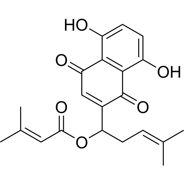
| SMILES |
CC(=CCC(C1=CC(=O)C2=C(C=CC(=C2C1=O)O)O)OC(=O)C=C(C)C)C
|
| InChi Key |
BATBOVZTQBLKIL-UHFFFAOYSA-N
|
| InChi Code |
InChI=1S/C21H22O6/c1-11(2)5-8-17(27-18(25)9-12(3)4)13-10-16(24)19-14(22)6-7-15(23)20(19)21(13)26/h5-7,9-10,17,22-23H,8H2,1-4H3
|
| 化学名 |
[1-(5,8-dihydroxy-1,4-dioxonaphthalen-2-yl)-4-methylpent-3-enyl] 3-methylbut-2-enoate
|
| 别名 |
Dmask; Arnebin I; [1-(5,8-dihydroxy-1,4-dioxonaphthalen-2-yl)-4-methylpent-3-enyl] 3-methylbut-2-enoate; 2-Butenoic acid, 3-methyl-, 1-(1,4-dihydro-5,8-dihydroxy-1,4-dioxo-2-naphthalenyl)-4-methyl-3-pentenyl ester, (+)-; 1-(5,8-DIHYDROXY-1,4-DIOXONAPHTHALEN-2-YL)-4-METHYLPENT-3-EN-1-YL 3-METHYLBUT-2-ENOATE; 2-Butenoic acid, 3-methyl-, 1-(1,4-dihydro-5,8-dihydroxy-1,4-dioxo-2-naphthalenyl)-4-methyl-3-pentenyl ester; Crotonic acid, 3-methyl-, ester with 5,8-dihydroxy-2-(1-hydroxy-4-methyl-3-pentenyl)-1,4-naphthoquinone; ...; 5162-01-6;
|
| HS Tariff Code |
2934.99.9001
|
| 存储方式 |
Powder -20°C 3 years 4°C 2 years In solvent -80°C 6 months -20°C 1 month 注意: 请将本产品存放在密封且受保护的环境中(例如氮气保护),避免吸湿/受潮和光照。 |
| 运输条件 |
Room temperature (This product is stable at ambient temperature for a few days during ordinary shipping and time spent in Customs)
|
| 溶解度 (体外实验) |
DMSO: 25 mg/mL (67.49 mM)
|
|---|---|
| 溶解度 (体内实验) |
注意: 如下所列的是一些常用的体内动物实验溶解配方,主要用于溶解难溶或不溶于水的产品(水溶度<1 mg/mL)。 建议您先取少量样品进行尝试,如该配方可行,再根据实验需求增加样品量。
注射用配方
注射用配方1: DMSO : Tween 80: Saline = 10 : 5 : 85 (如: 100 μL DMSO → 50 μL Tween 80 → 850 μL Saline)(IP/IV/IM/SC等) *生理盐水/Saline的制备:将0.9g氯化钠/NaCl溶解在100 mL ddH ₂ O中,得到澄清溶液。 注射用配方 2: DMSO : PEG300 :Tween 80 : Saline = 10 : 40 : 5 : 45 (如: 100 μL DMSO → 400 μL PEG300 → 50 μL Tween 80 → 450 μL Saline) 注射用配方 3: DMSO : Corn oil = 10 : 90 (如: 100 μL DMSO → 900 μL Corn oil) 示例: 以注射用配方 3 (DMSO : Corn oil = 10 : 90) 为例说明, 如果要配制 1 mL 2.5 mg/mL的工作液, 您可以取 100 μL 25 mg/mL 澄清的 DMSO 储备液,加到 900 μL Corn oil/玉米油中, 混合均匀。 View More
注射用配方 4: DMSO : 20% SBE-β-CD in Saline = 10 : 90 [如:100 μL DMSO → 900 μL (20% SBE-β-CD in Saline)] 口服配方
口服配方 1: 悬浮于0.5% CMC Na (羧甲基纤维素钠) 口服配方 2: 悬浮于0.5% Carboxymethyl cellulose (羧甲基纤维素) 示例: 以口服配方 1 (悬浮于 0.5% CMC Na)为例说明, 如果要配制 100 mL 2.5 mg/mL 的工作液, 您可以先取0.5g CMC Na并将其溶解于100mL ddH2O中,得到0.5%CMC-Na澄清溶液;然后将250 mg待测化合物加到100 mL前述 0.5%CMC Na溶液中,得到悬浮液。 View More
口服配方 3: 溶解于 PEG400 (聚乙二醇400) 请根据您的实验动物和给药方式选择适当的溶解配方/方案: 1、请先配制澄清的储备液(如:用DMSO配置50 或 100 mg/mL母液(储备液)); 2、取适量母液,按从左到右的顺序依次添加助溶剂,澄清后再加入下一助溶剂。以 下列配方为例说明 (注意此配方只用于说明,并不一定代表此产品 的实际溶解配方): 10% DMSO → 40% PEG300 → 5% Tween-80 → 45% ddH2O (或 saline); 假设最终工作液的体积为 1 mL, 浓度为5 mg/mL: 取 100 μL 50 mg/mL 的澄清 DMSO 储备液加到 400 μL PEG300 中,混合均匀/澄清;向上述体系中加入50 μL Tween-80,混合均匀/澄清;然后继续加入450 μL ddH2O (或 saline)定容至 1 mL; 3、溶剂前显示的百分比是指该溶剂在最终溶液/工作液中的体积所占比例; 4、 如产品在配制过程中出现沉淀/析出,可通过加热(≤50℃)或超声的方式助溶; 5、为保证最佳实验结果,工作液请现配现用! 6、如不确定怎么将母液配置成体内动物实验的工作液,请查看说明书或联系我们; 7、 以上所有助溶剂都可在 Invivochem.cn网站购买。 |
| 制备储备液 | 1 mg | 5 mg | 10 mg | |
| 1 mM | 2.6998 mL | 13.4989 mL | 26.9978 mL | |
| 5 mM | 0.5400 mL | 2.6998 mL | 5.3996 mL | |
| 10 mM | 0.2700 mL | 1.3499 mL | 2.6998 mL |
1、根据实验需要选择合适的溶剂配制储备液 (母液):对于大多数产品,InvivoChem推荐用DMSO配置母液 (比如:5、10、20mM或者10、20、50 mg/mL浓度),个别水溶性高的产品可直接溶于水。产品在DMSO 、水或其他溶剂中的具体溶解度详见上”溶解度 (体外)”部分;
2、如果您找不到您想要的溶解度信息,或者很难将产品溶解在溶液中,请联系我们;
3、建议使用下列计算器进行相关计算(摩尔浓度计算器、稀释计算器、分子量计算器、重组计算器等);
4、母液配好之后,将其分装到常规用量,并储存在-20°C或-80°C,尽量减少反复冻融循环。
计算结果:
工作液浓度: mg/mL;
DMSO母液配制方法: mg 药物溶于 μL DMSO溶液(母液浓度 mg/mL)。如该浓度超过该批次药物DMSO溶解度,请首先与我们联系。
体内配方配制方法:取 μL DMSO母液,加入 μL PEG300,混匀澄清后加入μL Tween 80,混匀澄清后加入 μL ddH2O,混匀澄清。
(1) 请确保溶液澄清之后,再加入下一种溶剂 (助溶剂) 。可利用涡旋、超声或水浴加热等方法助溶;
(2) 一定要按顺序加入溶剂 (助溶剂) 。
InvivoChem的所有产品仅用于作科学研究,不面向患者销售
Copyright 2020 InvivoChem LLC | All Rights Reserved 粤ICP备20063088号-1


 463611831
