| 规格 | 价格 | 库存 | 数量 |
|---|---|---|---|
| 1g |
|
||
| 2g |
|
||
| 5g |
|
||
| 10g |
|
||
| 50g |
|
||
| Other Sizes |
|

| 靶点 |
VDR/vitamin D receptor; PXR; FXR; Microbial Metabolite; Endogenous Metabolite
|
|---|---|
| 体外研究 (In Vitro) |
Lithocholic Acid 阻断 GW4064 和 CDCA 产生的 FXR 激活的 IC50 分别为 0.7 μM 和 1.4 μM [5]。在 HepG2 细胞中,10-30 μM 石胆酸可在 24 小时内抑制 100 nM GW4064 诱导的 BSEP 表达 [5]。 Lithocholic Acid (0-500 μM) 以剂量依赖性方式抑制神经母细胞瘤细胞生长(BE(2)-m17、SK-n-SH、SK-n-MCIXC 和 Lan-1)[3]。
|
| 体内研究 (In Vivo) |
Lithocholic acid (LCA) 是一种有效的胆汁淤积诱导剂,其作用机制和造模方法如下:
致病原理 • LCA 作为有毒次级胆汁酸,可通过改变肝细胞膜组分和胆汁分泌功能导致肝内胆汁淤积 • 其毒性作用表现为促进胆管上皮细胞损伤、炎症细胞浸润及胆汁酸代谢紊乱 • 通过激活FXR/PXR等核受体通路加剧胆汁淤积病理过程 动物模型构建方法 大鼠模型(急性) • 品系:Wistar雄性(250-300g) • 给药方案: o 剂量:0.2 μmol/100g o 途径:静脉注射(溶于7.5%牛血清白蛋白+0.45%生理盐水) o 处理时间:1小时后处死 小鼠模型(亚急性) • 品系:ICR雄性(5-7周龄) • 给药方案: o 剂量:150 mg/kg o 途径:口服灌胃(溶于玉米油) o 频次:每日2次,连续5次 o 处死时间:末次给药后12小时 检测指标 1. 血清生化: o 总胆红素(TBIL)、直接胆红素(DBIL)显著升高 o ALT/AST/ALP等肝酶活性增加 2. 组织病理: o 肝细胞空泡变性、胆管增生 o 汇管区炎症细胞浸润 o 毛细血管胆汁淤积 3. 功能变化: o 早期胆汁流量短暂增加,后期显著减少 o 胆汁酸(TBA)代谢紊乱 注意事项 • 静脉给药需严格控制浓度避免溶血 • 灌胃给药建议在光照周期固定时段进行以减少昼夜节律影响 需设置溶剂对照组(牛血清白蛋白/玉米油)排除载体干扰 当石胆酸以 0.6% 的比例添加到食物中连续 7 天时,会提高雄性 C57BL/6 小鼠肝脏中 TGFB1、TGFBR1 和 TGFBR2 mRNA 的水平,激活 SMAD3,并引起胆道损伤 [4]。腹腔注射石胆酸(125 mg/kg,每天两次,持续四天)的雄性 C57BL/6 小鼠会出现肝损伤,并且 AST、ALT 和 ALP 水平升高 [2]。 胆汁淤积性肝损伤是由α-异硫氰酸萘酯(ANIT)、3,5-二乙氧羰基-1,4-二氢吡啶(DDC)和Lithocholic acid(LCA)引起的。结果表明,三种小鼠胆汁淤积模型血浆中胆汁酸水平普遍升高,精氨酸水平降低。ANIT和LCA诱导的肝内PBC和PSC的血浆谷胱甘肽水平分别降低。但是,DDC诱导的肝外PSC中肝脏谷胱甘肽含量降低。ANIT和DDC模型中的血浆磷脂水平升高,而LCA模型中的磷脂水平降低。DDC模型肝脏原卟啉IX显著升高。这些代谢组学数据可能有助于区分三种胆汁淤积的代谢差异,从而有助于理解胆汁淤积性肝损伤的潜在机制。[2] 石胆酸/Lithocholic acid(LCA)诱导肝脏中TGFB1和受体TGFBR1和TGFBR2的表达。此外,免疫组织化学显示,在石胆酸/LCA暴露后,门静脉周围的TGFβ表达增加,SMAD3缺失小鼠肝细胞中SMAD3磷酸化减少。血清代谢组学表明,LCA暴露后胆汁酸增加,溶血磷脂酰胆碱(LPC)减少。有趣的是,在Smad3缺失的小鼠中,代谢改变减弱了。在Smad3缺失的小鼠中,LCA诱导的溶血磷脂酰胆碱酰基转移酶4(LPCAT4)和有机溶质转运蛋白β(OSTβ)表达显著降低,而TGFβ诱导的原代小鼠肝细胞中LPCAT4和OSTβ表达显著降低。此外,SMAD3的引入增强了TGFβ诱导的人肝细胞癌细胞系HepG2中LPCAT4和OSTβ的表达。总之,考虑到Smad3缺失小鼠的血清ALP活性减弱,这是胆管细胞损伤的诊断指标,这些结果强烈支持TGFβ-Smad3信号传导介导肝脏炎症和胆管损伤后磷脂和胆汁酸代谢改变的观点[4] 暴露于石胆酸可提高肝脏中TGFβ和受体mRNA水平[4] 使用用合成AIN93G饮食(Cont)和添加0.6%LCA的AIN93G膳食(LCA)处理7天的C57BL/6小鼠,研究了石胆酸(LCA)暴露对TGFβ信号传导的影响。肝脏TGFB1、TGFBR1和TGFBR2 mRNA水平在LCA暴露后升高,尽管肝脏中TGFBR3 mRNA水平没有变化(图1)。这些结果表明,LCA暴露刺激了肝脏中的TGFβ信号传导。 Smad3缺失小鼠Lithocholic acid/LCA诱导的肝损伤减轻[4] TGFβ通过TGFβ受体激活SMAD3。因此,为了研究SMAD3是否参与了石胆酸/Lithocholic acid/LCA诱导的肝损伤,用对照饮食和LCA饮食治疗SMAD3缺失小鼠6天。LCA暴露后,Smad3缺失小鼠的肝脏质量小于LCA治疗的野生型小鼠(图2A)。此外,在Smad3缺失的小鼠中,LCA增加的血清ALP活性显著减弱,尽管血清ALT活性没有变化(图2B,C)。此外,在Smad3缺失小鼠中,肝脏组织学显示门静脉周围有轻度炎性细胞浸润,而在类似治疗的野生型小鼠中没有观察到这种情况(图2D)。免疫组织化学显示,与野生型小鼠相比,Smad3缺失小鼠门静脉周围的TGFβ蛋白表达较低(图2E),表明TGFβ对肝脏Smad3激活的刺激较低(补充图I)。此外,在SMAD3缺失小鼠的肝细胞核中观察到SMAD3磷酸化信号的显著衰减(图2E)。这些结果表明,TGFβ-SMAD3信号传导与LCA诱导的胆管损伤有关,并增加了TGFβ-SMARD3信号传导改变肝脏代谢的可能性。 暴露于石胆酸/Lithocholic acid/LCA后,野生型和Smad3缺失小鼠血清代谢组的差异[4] 为了检测血清代谢物,使用来自喂食石胆酸/Lithocholic acid/LCA或对照饮食的小鼠血清的UPLC-ESI-QTOFMS阴性模式数据进行PLS和贡献分析。PLS分析显示,LCA处理的野生型(图3A)和LCA治疗的Smad3缺失组之间存在分离,并用负荷图进行了进一步检查(图3B)。贡献分析显示,10个增强离子和10个衰减离子是引起分离的顶级离子。与野生型小鼠相比,Smad3缺失小鼠的溶血磷脂酰胆碱(LPC)和脂肪酸片段被确定为升高的离子(表1)。最低的离子来自胆汁盐(表2)。LCA喂养后,Smad3缺失小鼠的血清代谢组在LPC和胆汁盐方面与野生型小鼠有很大不同。 |
| 细胞实验 |
用Lithocholic acid/石胆酸/LCA、过氧化氢或z-DEVD-fmk[3]处理细胞
首先在100%二甲基亚砜(DMSO)中制备不同浓度的Lithocholic acid/LCA储备溶液。为了治疗培养的人NB、人BC和大鼠GL细胞,然后将这些储备溶液稀释至指定的LCA终浓度(DMSO的终浓度始终保持在1%),稀释液为DMEM和Ham的F-12营养混合物的1:1混合物,补充了10%FBS(适用于SK-n-MCIXC和BE(2)-m17细胞),EMEM补充了10%的FBS(适用于SK-n-SH细胞),DMEM补充了10%BCS(适用于Lan-1细胞)。为了用LCA治疗人类原代神经元,将在100%DMSO中制备的不同浓度的LCA储备溶液在含有0.225%碳酸氢钠、1 mM丙酮酸钠、2 mM L-谷氨酰胺、0.1%葡萄糖和5%FBS的EMEM中稀释至LCA的指定终浓度(DMSO的终浓度始终保持在1%)。为了用LCA治疗培养的癌症细胞或原代神经元,将它们在所示最终浓度的LCA存在下孵育48小时;对照细胞仅用空的DMSO载体处理。对于用过氧化氢处理的细胞,其30%的储备溶液在无菌H2O中稀释,并在用LCA或空DMSO载体预处理24小时后直接加入细胞培养物中;然后将细胞孵育24小时。在涉及用z-DEVD-fmk进行细胞处理的实验中,这种半胱氨酸天冬氨酸蛋白酶-3抑制剂与LCA或空的DMSO载体同时添加至终浓度为5μM。 细胞活力测定[3] 使用基于MTT(3-(4,5-二甲基噻唑-2-基)-2,5-二苯基溴化四唑)的CellTiter 96非放射性细胞增殖测定法测量暴露于Lithocholic acid/LCA的培养物中活细胞的数量。在该试验中,只有活的、代谢活跃的细胞能够还原MTT的黄色四唑盐,形成紫色甲赞产物。通过加入洗涤剂使这种不溶性产物溶解。然后使用96孔板读数器在570nm波长下用分光光度法检测所得的细胞内紫色甲赞。使用630nm的波长校正信号以考虑细胞碎片。用荧光染料Hoechst在培养基中以4μM的终浓度显示暴露于LCA和/或过氧化氢和/或z-DEVD-fmk的细胞中的染色质,并使用荧光显微镜进行观察。对于每种细胞培养物,计算携带完整、非碎片化核(含非浓缩染色质)的活非凋亡细胞的百分比。死亡的凋亡细胞携带含有浓缩染色质的碎裂核,这是凋亡死亡的标志性事件。 荧光显微镜观察线粒体和测量线粒体膜电位[3] 使用培养基中浓度为125 nM的MitoTracker Red CMXRos对用Lithocholic acid/LCA处理的细胞的线粒体形态进行可视化。使用荧光显微镜观察细胞,并计算显示线粒体碎片的细胞百分比。使用四甲基罗丹明乙酯(TMRE)(一种细胞渗透性阳离子荧光染料)测量线粒体膜电位(∆Ψ)。线粒体对TMRE的可逆隔离程度与∆Ψ的值成正比。将细胞与50 nM TMRE孵育20分钟,并使用荧光显微镜直接观察。计算了显示可检测水平∆Ψ的TMRE阳性细胞的百分比。 瞬时转染试验[5] 转染前24小时,将HepG2细胞以3.2×10⁴个细胞/孔的密度接种在含有10%FBS的DMEM中。根据制造商的说明,使用FuGENE6转染试剂在无血清Opti-MEM I培养基中用转染混合物转染细胞。通常,每个孔的转染混合物含有0.405μl FuGENE6、3 ng pcDNA3.1-GAL4-hFXR(LBD)表达载体、3 ng pc DNA3.1-hRXRα表达构建体、60 ng pUAS(5X)-tk LUC报告载体和60 ng pCMV-lacZ作为转染效率的内部对照。细胞在37°C、10%CO2的气氛中在转染混合物中孵育4小时。然后将细胞在含有5%木炭剥离FBS的新鲜DMEM中孵育约40-48小时,可添加或不添加不同浓度的配体。根据制造商的指示,使用报告裂解缓冲液生产细胞裂解物。在ML3000光度计中使用萤光素酶测定缓冲液测定细胞提取物中的萤光素酶活性。如前所述,使用β-d-吡喃半乳糖苷测定β-半乳糖苷酶活性。每个孔的萤光素酶活性分别归一化为β-半乳糖苷酶活性。当检测Lithocholic acid/LCA的拮抗活性时,在100 nm GW4064的存在下,将细胞与越来越高浓度的LCA一起孵育。 |
| 动物实验 |
动物/疾病模型:雄性小鼠 (C57BL/6)[4]。
剂量:0.6% LCA 补充剂饮食,以 AIN93G 饮食作为对照。 给药途径:饲料中添加,持续 6 天。 实验结果:诱导肝损伤。激活 TGFβ-SMAD3 信号通路。血清 ALP 活性升高。 动物/疾病模型:雄性小鼠 (C57BL/6)[2]。 剂量: 125 mg/kg,溶于玉米油 给药途径: 腹腔注射,每日两次,连续四天 实验结果: 诱导肝损伤,产生坏死和中性粒细胞浸润(HE染色)。AST、ALT和ALP水平升高。 动物实验[2] 雄性C57BL/6小鼠(6-8周龄)饲养于温度和湿度可控的环境中,光照/黑暗周期为12小时/12小时。30只小鼠随机分为六组:(1)ANIT对照组(ANIT-C);(2)ANIT组;(3)DDC对照组(DDC-C);(4)DDC组;(5)石胆酸/LCA对照组(LCA-C);(6)LCA组。将溶于玉米油的ANIT以75 mg/kg的剂量单次灌胃给予小鼠,并在给药后48小时处死小鼠(Fang et al., 2017; Tang et al., 2016)。DDC的给药方案在前人报道的基础上略作修改(Dai et al., 2017; Fickert et al., 2007)。将溶于玉米油的DDC以100 mg/kg的剂量连续7天口服,并在末次给药后24小时处死小鼠。将溶于玉米油的LCA以125 mg/kg的剂量连续4天腹腔注射,每日两次(Beilke et al., 2009; Owen et al., 2010),并在末次给药后24小时处死小鼠。以玉米油处理的小鼠作为对照组。所有小鼠均采用二氧化碳吸入法处死。随后,收集小鼠血浆和肝脏样本进行生化分析、组织病理学分析和代谢组学分析。生化分析包括ALT、AST和ALP的测定。部分肝组织采用苏木精-伊红(H&E)染色进行组织病理学分析,方法如前所述(Hu et al., 2018),其余组织保存于−80 °C。 动物和饲料[4] 雄性小鼠(C57BL/6)、MAD同源物3(Smad3)基因敲除小鼠和背景匹配的野生型小鼠饲养于温度和光照可控的房间内,并自由摄取水和NIH-31颗粒饲料。在石胆酸/LCA研究中,小鼠饲喂添加0.6% LCA的饲料,并以AIN93G饲料作为对照。喂食对照饮食的三只野生型小鼠和三只 Smad3 基因敲除小鼠,喂食 LCA 饮食的五只野生型小鼠和四只 Smad3 基因敲除小鼠。 |
| 药代性质 (ADME/PK) |
代谢/代谢产物
石胆酸(24)C(14)在大鼠肝匀浆中转化为3α-6β-二羟基-5β-胆酸,发生7σ羟基化,可证实其与牛磺酸羟基化结合并形成3-硫酸酯。 将标记的石胆酸盐注射到胆结石患者和健康志愿者体内,胆汁中大部分放射性(50-60%)以硫酸酯结合物的形式存在。甘氨酸结合物的硫酸化程度高于牛磺酸结合物,这表明甘氨酸结合物更容易发生硫酸化。 石胆酸已知的代谢产物包括6α-羟基石胆酸。 |
| 毒性/毒理 (Toxicokinetics/TK) |
相互作用
皮肤肿瘤抑制作用按以下酸的顺序递减:鹅脱氧胆酸、石胆酸、脱氧胆酸和胆酸。 16α-氰基孕烯醇酮(5 mg,腹腔注射,每日两次,持续2天)在体外可使大鼠肝微粒体中石胆酸的6β-和7α-羟基化分别增加2倍和3-4倍。这可能是其预防石胆酸诱发胆结石的原因。 石胆酸(24)C(14)在大鼠肝匀浆中转化为3α-6β-二羟基-5β-胆酸。向酶系统中添加乙醇可抑制 3α,6β-二羟基-5β-胆酸的生成。 锂胆酸钠可增加 MNNG(N-甲基-N'-硝基-N-亚硝基胍)诱导的无菌和常规大鼠(F344)结肠肿瘤的发生率。/锂胆酸钠/ LCA 也被测试作为 N-硝基双(2-羟丙基)胺 (BHP) 诱导致癌作用的促进剂。两组 5 至 6 周龄的仓鼠(数量未说明)每周皮下注射一次 500 mg/kg BHP,持续 5 周,第三组未接受进一步治疗;第4组在饲料中添加0.5%的LCA,持续30周,所有动物均在第35周进行尸检。两组动物的采食量和体重均无差异。肝脏病变数量也无差异(第3组:15/15个增生性结节,2/15个肝细胞癌,1/15个胆管癌;第4组:22/22个增生性结节,3/22个肝细胞癌,3/22个胆管癌)。然而,胰腺肿瘤的数量存在显著差异:第3组有4/15个肉眼可见的肿瘤,5/15个癌,4/15个腺瘤;而第4组有13/22个肉眼可见的肿瘤,15/22个癌(P<0.04),2/22个腺瘤。在本实验条件下,LCA单独给药时不具有致癌性,但能有效促进BHP胰腺癌的发生。 小鼠口服LD50 3900 mg/kg NIH-NCI-EC-72-3252合同进展报告,由Litton Bionetics公司提交给美国国家癌症研究所,NCI-EC-72-3252(1973) |
| 参考文献 |
|
| 其他信息 |
六边形片状晶体(由醇类析出)或棱柱状晶体(由乙酸析出)或白色粉末。(NTP, 1992)
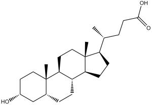
石胆酸是一种单羟基-5β-胆烷酸,其3位带有α-羟基取代基。它是一种胆汁酸,由鹅脱氧胆酸经细菌作用转化而来。它既是人体代谢产物,也是小鼠代谢产物,并具有抗衰老作用。它是一种胆汁酸、单羟基-5β-胆烷酸和C24甾体。它是石胆酸盐的结合酸。 已有关于石胆酸在智人体内被报道的数据。 一种由鹅脱氧胆酸盐经细菌作用形成的胆汁酸,通常与甘氨酸或牛磺酸结合。它作为一种表面活性剂,能够溶解脂肪以促进吸收,自身也能被吸收。它被用作利胆剂和利胆药。如上所述,胆管阻塞是原发性胆汁性胆管炎(PBC)和原发性硬化性胆管炎(PSC)的病理特征,在三种化学诱导的胆汁淤积模型中,包括TCDCA、T-α/β-MCA和TH/UDCA,多种胆汁酸水平普遍升高。除了这三种胆汁淤积模型中胆汁酸水平升高外,精氨酸水平也降低。ANIT和DDC模型破坏了脂质代谢,导致血浆和肝脏中溶血磷脂酰胆碱(LPC)和肉碱水平升高。与ANIT和LCA模型不同,DCC诱导的肝损伤导致肝脏谷胱甘肽(GSH)水平降低(图S3)。DDC模型诱导肝脏中原卟啉IX(PPIX)积累,而ANIT和LCA模型中PPIX水平降低。考虑到PPIX在体内转化为血红素进行氧气转移,ANIT和LCA模型中PPIX水平降低可能是细胞损伤改善的一种适应性反应。综上所述,这些变化可能部分揭示了三种胆汁淤积性肝损伤的差异,有助于理解不同类型胆汁淤积的潜在机制。本文数据证实了代谢组学方法在胆汁淤积性肝损伤生物标志物发现方面的优势。[2]衰老是癌症的主要危险因素之一。药物和饮食抗衰老干预可以延缓癌症的发生。我们最近在酵母细胞衰老模型中发现,石胆酸 (LCA) 可以延长寿命。本文表明,在对人神经元原代培养物无细胞毒性的浓度下,LCA 可以杀死神经母细胞瘤 (NB) 细胞系 BE(2)-m17、SK-n-SH、SK-n-MCIXC 和 Lan-1。在 BE(2)-m17、SK-n-SH 和 SK-n-MCIXC 细胞中,LCA 的抗肿瘤作用是由于细胞凋亡所致。相反,LCA诱导的Lan-1细胞死亡并非由细胞凋亡引起。低浓度LCA可使BE(2)-m17和SK-n-MCIXC细胞对过氧化氢诱导的线粒体介导的细胞凋亡更加敏感,但这些浓度的LCA却使原代培养的人类神经元对这种细胞死亡方式产生抵抗力。LCA杀死BE(2)-m17和SK-n-MCIXC细胞系,不仅通过激活线粒体外膜通透性和起始caspase-9激活所驱动的内在(线粒体)细胞凋亡途径,还通过激活起始caspase-8所介导的外在(死亡受体)细胞凋亡途径。基于这些数据,我们提出了LCA在培养的人类神经母细胞瘤(NB)细胞中发挥强效且选择性抗肿瘤作用的机制。此外,我们发现LCA能够杀死培养的人乳腺癌细胞和鼠神经胶质瘤细胞,这表明它对来源于不同组织和生物体的癌细胞具有广泛的抗肿瘤作用。[3]胆汁酸输出泵(BSEP)是肝脏中主要的胆汁酸转运蛋白。BSEP基因突变会导致进行性肝内胆汁淤积,这是一种严重的肝脏疾病,会损害胆汁流动并造成不可逆的肝损伤。BSEP是药物和异常胆汁酸代谢产物抑制和下调的靶点,这种抑制和下调可能导致胆汁酸潴留和肝内胆汁淤积。在本研究中,我们定量分析了FXR配体对原代人肝细胞和HepG2细胞中BSEP表达的调控作用。我们证实,核受体法尼醇X受体(FXR)的配体能够显著调控BSEP的表达。内源性FXR激动剂鹅脱氧胆酸(CDCA)和合成FXR配体GW4064均能有效增加两种细胞类型中BSEP mRNA的表达。这种上调最早可在3小时内检测到,且配体对BSEP的调控效力与其对FXR的内在活性相关。这些结果表明BSEP是FXR的直接靶点,并支持近期关于BSEP启动子可被FXR反式激活的报道。与CDCA和GW4064相反,疏水性胆汁酸石胆酸(LCA)是一种强效的胆汁淤积诱导剂,它能显著降低BSEP的表达。以往的研究并未发现LCA是细胞中的FXR拮抗剂配体,但我们在此证明LCA是细胞中的FXR拮抗剂,并具有部分激动剂活性。在体外共激活因子结合实验中,LCA 以 1 μM 的 IC50 值降低了 CDCA 和 GW4064 诱导的 FXR 激活。在 HepG2 细胞中,LCA 也有效拮抗了 GW4064 增强的 FXR 转录激活。这些数据表明,LCA 在动物体内的毒性和胆汁淤积作用可能是由于其通过 FXR 下调 BSEP 所致。综上所述,这些观察结果表明 FXR 在 BSEP 基因表达中发挥重要作用,FXR 配体可能成为治疗肝内胆汁淤积的潜在药物。[5] LCA 最近被鉴定为 PXR/SXR 激动剂配体。PXR 被认为是第二种胆汁酸受体,在肝脏解毒中起着关键作用 (27, 28)。然而,目前尚无证据表明 PXR 参与 BSEP 基因调控。事实上,PXR特异性配体利福平并不调节BSEP的表达,这表明PXR不参与BSEP的调控,并进一步支持了LCA通过拮抗FXR活性来下调BSEP表达的结论。BSEP的表达对于肝脏保护至关重要。FXR作为BSEP的重要调节因子,不仅为FXR介导的BSEP基因调控提供了分子机制,也提示FXR配体可能成为治疗肝内胆汁淤积和脂质代谢紊乱的潜在药物。[5] |
| 分子式 |
C24H40O3
|
|
|---|---|---|
| 分子量 |
376.57
|
|
| 精确质量 |
376.297
|
|
| 元素分析 |
C, 76.55; H, 10.71; O, 12.75
|
|
| CAS号 |
434-13-9
|
|
| 相关CAS号 |
Allolithocholic acid;2276-94-0;Isoallolithocholic acid;2276-93-9;Isolithocholic acid;1534-35-6;Lithocholic acid-d4;83701-16-0;Lithocholic acid-d5;52840-06-9
|
|
| PubChem CID |
9903
|
|
| 外观&性状 |
White to off-white solid powder
|
|
| 密度 |
1.1±0.1 g/cm3
|
|
| 沸点 |
511.0±23.0 °C at 760 mmHg
|
|
| 熔点 |
183-188 °C(lit.)
|
|
| 闪点 |
276.9±19.1 °C
|
|
| 蒸汽压 |
0.0±3.0 mmHg at 25°C
|
|
| 折射率 |
1.528
|
|
| LogP |
6.7
|
|
| tPSA |
57.53
|
|
| 氢键供体(HBD)数目 |
2
|
|
| 氢键受体(HBA)数目 |
3
|
|
| 可旋转键数目(RBC) |
4
|
|
| 重原子数目 |
27
|
|
| 分子复杂度/Complexity |
574
|
|
| 定义原子立体中心数目 |
9
|
|
| SMILES |
C[C@H](CCC(=O)O)[C@H]1CC[C@@H]2[C@@]1(CC[C@H]3[C@H]2CC[C@H]4[C@@]3(CC[C@H](C4)O)C)C
|
|
| InChi Key |
SMEROWZSTRWXGI-HVATVPOCSA-N
|
|
| InChi Code |
InChI=1S/C24H40O3/c1-15(4-9-22(26)27)19-7-8-20-18-6-5-16-14-17(25)10-12-23(16,2)21(18)11-13-24(19,20)3/h15-21,25H,4-14H2,1-3H3,(H,26,27)/t15-,16-,17-,18+,19-,20+,21+,23+,24-/m1/s1
|
|
| 化学名 |
3alpha-Hydroxy-5beta-cholan-24-oic acid
|
|
| 别名 |
|
|
| HS Tariff Code |
2934.99.9001
|
|
| 存储方式 |
Powder -20°C 3 years 4°C 2 years In solvent -80°C 6 months -20°C 1 month |
|
| 运输条件 |
Room temperature (This product is stable at ambient temperature for a few days during ordinary shipping and time spent in Customs)
|
| 溶解度 (体外实验) |
|
|||
|---|---|---|---|---|
| 溶解度 (体内实验) |
配方 1 中的溶解度: ≥ 2.08 mg/mL (5.52 mM) in 10% DMSO + 90% (20% SBE-β-CD in Saline) (这些助溶剂从左到右依次添加,逐一添加), 澄清溶液。
例如,若需制备1 mL的工作液,可将100 μL 20.8 mg/mL澄清DMSO储备液加入900 μL 20% SBE-β-CD生理盐水溶液中,混匀。 *20% SBE-β-CD 生理盐水溶液的制备(4°C,1 周):将 2 g SBE-β-CD 溶解于 10 mL 生理盐水中,得到澄清溶液。 配方 2 中的溶解度: ≥ 2.08 mg/mL (5.52 mM) (饱和度未知) in 10% DMSO + 90% Corn Oil (这些助溶剂从左到右依次添加,逐一添加), 澄清溶液。 例如,若需制备1 mL的工作液,可将 100 μL 20.8 mg/mL 澄清 DMSO 储备液加入到 900 μL 玉米油中并混合均匀。 View More
配方 3 中的溶解度: ≥ 1 mg/mL (2.66 mM) (饱和度未知) in 10% EtOH + 40% PEG300 + 5% Tween80 + 45% Saline (这些助溶剂从左到右依次添加,逐一添加), 澄清溶液。 配方 4 中的溶解度: ≥ 1 mg/mL (2.66 mM) (饱和度未知) in 10% EtOH + 90% (20% SBE-β-CD in Saline) (这些助溶剂从左到右依次添加,逐一添加), 澄清溶液。 例如,若需制备1 mL的工作液,可将100μL 10.0mg/mL澄清EtOH储备液加入到900μL 20%SBE-β-CD生理盐水中,混匀。 *20% SBE-β-CD 生理盐水溶液的制备(4°C,1 周):将 2 g SBE-β-CD 溶解于 10 mL 生理盐水中,得到澄清溶液。 配方 5 中的溶解度: ≥ 1 mg/mL (2.66 mM) (饱和度未知) in 10% EtOH + 90% Corn Oil (这些助溶剂从左到右依次添加,逐一添加),澄清溶液。 例如,若需制备1 mL的工作液,可将100 μL 10.0 mg/mL 澄清 EtOH 储备液添加到 900 μL 玉米油中并充分混合。 1、请先配制澄清的储备液(如:用DMSO配置50 或 100 mg/mL母液(储备液)); 2、取适量母液,按从左到右的顺序依次添加助溶剂,澄清后再加入下一助溶剂。以 下列配方为例说明 (注意此配方只用于说明,并不一定代表此产品 的实际溶解配方): 10% DMSO → 40% PEG300 → 5% Tween-80 → 45% ddH2O (或 saline); 假设最终工作液的体积为 1 mL, 浓度为5 mg/mL: 取 100 μL 50 mg/mL 的澄清 DMSO 储备液加到 400 μL PEG300 中,混合均匀/澄清;向上述体系中加入50 μL Tween-80,混合均匀/澄清;然后继续加入450 μL ddH2O (或 saline)定容至 1 mL; 3、溶剂前显示的百分比是指该溶剂在最终溶液/工作液中的体积所占比例; 4、 如产品在配制过程中出现沉淀/析出,可通过加热(≤50℃)或超声的方式助溶; 5、为保证最佳实验结果,工作液请现配现用! 6、如不确定怎么将母液配置成体内动物实验的工作液,请查看说明书或联系我们; 7、 以上所有助溶剂都可在 Invivochem.cn网站购买。 |
| 制备储备液 | 1 mg | 5 mg | 10 mg | |
| 1 mM | 2.6555 mL | 13.2777 mL | 26.5555 mL | |
| 5 mM | 0.5311 mL | 2.6555 mL | 5.3111 mL | |
| 10 mM | 0.2656 mL | 1.3278 mL | 2.6555 mL |
1、根据实验需要选择合适的溶剂配制储备液 (母液):对于大多数产品,InvivoChem推荐用DMSO配置母液 (比如:5、10、20mM或者10、20、50 mg/mL浓度),个别水溶性高的产品可直接溶于水。产品在DMSO 、水或其他溶剂中的具体溶解度详见上”溶解度 (体外)”部分;
2、如果您找不到您想要的溶解度信息,或者很难将产品溶解在溶液中,请联系我们;
3、建议使用下列计算器进行相关计算(摩尔浓度计算器、稀释计算器、分子量计算器、重组计算器等);
4、母液配好之后,将其分装到常规用量,并储存在-20°C或-80°C,尽量减少反复冻融循环。
计算结果:
工作液浓度: mg/mL;
DMSO母液配制方法: mg 药物溶于 μL DMSO溶液(母液浓度 mg/mL)。如该浓度超过该批次药物DMSO溶解度,请首先与我们联系。
体内配方配制方法:取 μL DMSO母液,加入 μL PEG300,混匀澄清后加入μL Tween 80,混匀澄清后加入 μL ddH2O,混匀澄清。
(1) 请确保溶液澄清之后,再加入下一种溶剂 (助溶剂) 。可利用涡旋、超声或水浴加热等方法助溶;
(2) 一定要按顺序加入溶剂 (助溶剂) 。
InvivoChem的所有产品仅用于作科学研究,不面向患者销售
Copyright 2020 InvivoChem LLC | All Rights Reserved 粤ICP备20063088号-1


 COA
COA
 463611831
