| 规格 | 价格 | 库存 | 数量 |
|---|---|---|---|
| 10 mM * 1 mL in DMSO |
|
||
| 1mg |
|
||
| 5mg |
|
||
| 10mg |
|
||
| 25mg |
|
||
| 50mg |
|
||
| 100mg |
|
||
| Other Sizes |
|

描述:1M7 是一种用于 RNA SHAPE-MaP 实验的试剂,可在 70 秒内以单核苷酸分辨率分析 RNA 二级结构。使用 1M7 进行的 SHAPE 化学反应能够准确报告天然条件下 RNase P 特异性结构域的已知结构。1M7 的反应活性可以检测受碱基配对和特异性非经典三级相互作用约束的核苷酸。SHAPE 化学反应能够非常精确地分析两种结构之间的差异,例如 Mg2+ 依赖性三级相互作用。1M7 易于在实验室操作,并可在 70 秒内以单核苷酸分辨率分析大型 RNA 结构。
| 体外研究 (In Vitro) |
选择性2'-羟基酰化引物延伸分析(SHAPE)化学方法能够以单核苷酸分辨率定量评估任何RNA中局部核苷酸的柔性。SHAPE化学利用核糖2'-羟基亲核反应性的结构门控,该门控取决于核苷酸的约束或柔性程度。SHAPE化学最初使用N-甲基异靛酸酐(NMIA)开发,但NMIA的亲电性较弱,且需要数十分钟才能形成核糖2'-O-加合物。本文设计并评估了一种效率更高、反应更快的SHAPE化学试剂。在反应性羰基的对位引入硝基,形成1-甲基-7-硝基异靛酸酐(1M7),得到的试剂不仅能更快地与RNA反应生成2'-O-加合物,而且更容易发生有利的自限性水解。使用 1M7,可在 70 秒内完成 RNA 结构的单核苷酸分辨率分析。利用 1M7 进行的 SHAPE 分析能够准确报告 RNase P 特异性结构域的二级和三级结构,并能以高达 91% 的准确率预测该 RNA 的二级结构 [1]。
1M7 是一种快速的 SHAPE(选择性 2'-羟基酰化引物延伸分析)试剂。它与构象灵活的 RNA 核苷酸的 2'-羟基反应,形成共价 2'-O-加合物。该反应具有结构选择性,柔性核苷酸表现出高反应活性,而受限核苷酸(例如,碱基配对的核苷酸)则表现出低反应活性。该修饰可在天然和变性条件下进行。 [2]在活细胞中,1M7(终浓度为10 mM)可在短时间内迅速修饰RNA。该试剂在约2分钟内即可通过水解完全终止反应。它能有效检测细胞质和细胞核中的核糖核蛋白(RNP)。与反应较慢的试剂NAI-N3(icSHAPE)相比,1M7无需后续富集步骤。[3] |
|---|---|
| 酶活实验 |
许多生物过程都由RNA介导,但大多数RNA的高级结构尚不清楚,这使得理解RNA结构如何调控其功能变得困难。本文介绍了一种基于引物延伸和突变谱分析的选择性2'-羟基酰化方法(SHAPE-MaP),该方法能够从头大规模地鉴定RNA功能基序。通过大规模平行测序,我们发现SHAPE介导的2'-羟基酰化位点在cDNA合成过程中编码为非互补核苷酸。SHAPE-MaP指导的建模方法在已知结构的复杂RNA中识别出超过90%的已知碱基对,我们利用该方法构建了HIV-1 RNA基因组的新模型。该HIV-1模型包含了所有已知的结构基序和先前未知的元件,包括经实验验证的假结。 SHAPE-MaP 可生成精确且高分辨率的二级结构模型,能够分析低丰度 RNA,在单次实验中解析序列多态性,并最终实现 RNA 结构分析的普及化 [2]。
在 SHAPE-MaP 中,逆转录是在一种能使逆转录酶错误读取 1M7 修饰核苷酸的条件下进行的。该反应使用莫洛尼鼠白血病病毒逆转录酶(SuperScript II),缓冲液包含 50 mM Tris-HCl(pH 8.0)、75 mM KCl、6 mM MnCl2 和 14 mM DTT。Mn2+ 的存在促进了酶在大体积 2'-O-加合物位点的通读。逆转录酶在加合物位点掺入与原始序列不互补的核苷酸,从而在 cDNA 中产生突变。[2] |
| 细胞实验 |
SHAPE-MaP 在 RNA 结构探测策略中独树一帜,它既能以单核苷酸分辨率测量 RNA 的柔性,又能量化这些测量的不确定性。我们提出了一种简便的分析框架,该框架纳入了这些不确定性,从而能够检测任意两种状态之间的 RNA 结构差异。我们利用该框架检测健康小鼠滋养层干细胞中的 RNA-蛋白质相互作用。我们通过分析三种模型胞质和核核糖核蛋白复合物,并在 2 分钟的细胞内探测实验中验证了该方法的有效性。相比之下,其他细胞内 SHAPE 探测方法产生的数据与 SHAPE-MaP 产生的数据相关性较差(r = 0.2),且无法提供准确的 RNA-蛋白质相互作用信号。随后,我们研究了 RNase MRP 复合物中的 RNA-蛋白质和 RNA-底物相互作用,并通过比较细胞内相互作用位点与疾病相关突变,从分子表型的角度表征了这些非编码突变。这些结果共同表明,SHAPE-MaP 能够在对所涉及的蛋白质或 RNA 了解有限的情况下,在天然细胞条件下确定真实的相互作用位点并推断 RNA 功能。[3]
1M7 用于小鼠滋养层干细胞 (TSC) 的细胞内 RNA 结构探测。用 PBS 洗涤活的 TSC,并加入 900 µL 新鲜培养基。然后,向细胞中加入 100 µL 100 mM 的 1M7 DMSO 溶液(最终浓度为 10 mM),并通过快速旋转培养皿混匀。将细胞在 37°C 下孵育 5 分钟。孵育后,移除培养基,用 PBS 洗涤细胞一次,并分离总 RNA。使用纯 DMSO 代替 1M7,以类似方式制备无试剂阴性对照。 [3] 对于离体分析,从 TSC 中温和提取 RNA,并在缓冲液(100 mM HEPES,pH 8.0,100 mM NaCl,10 mM MgCl2)中折叠。然后,将约 3 µg RNA 与终浓度为 10 mM 的 1M7 处理,并在 37°C 下孵育 5 分钟。[3] |
| 药代性质 (ADME/PK) |
1M7 在水溶液中的半衰期 (t1/2) 约为 17 秒,之后会因水解而完全猝灭。这种快速水解是其相对于 NAI-N3(t1/2 约为 30 分钟)等较慢试剂的关键优势。[3]
|
| 参考文献 |
[1]. A fast-acting reagent for accurate analysis of RNA secondary and tertiary structure by SHAPE chemistry. J Am Chem Soc . 2007 Apr 11;129(14):4144-5. doi: 10.1021/ja0704028.
[2]. RNA motif discovery by SHAPE and mutational profiling (SHAPE-MaP). Nat Methods . 2014 Sep;11(9):959-65. doi: 10.1038/nmeth.3029. [3]. Detection of RNA-Protein interactions in living cells with SHAPE. Biochemistry . 2015 Nov 24;54(46):6867-75. doi: 10.1021/acs.biochem.5b00977. |
| 其他信息 |
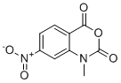
1-甲基-7-硝基异吲哚菁酐是一种3,1-苯并噁嗪-1,4-二酮,其N位带有甲基取代基,7位带有硝基。它既是苯并噁嗪化合物,也是C-硝基化合物。
1M7 是一种经过充分验证的快速SHAPE试剂,用于精确分析RNA的二级和三级结构。其半衰期短,特别适用于细胞内探测实验,因为它反应迅速且淬灭快,因此能够反映RNA的稳态结构,而不是RNP的组装、解离或细胞周转。相比之下,像NAI-N3这样的反应较慢的试剂对特定的离子和缓冲液的选择非常敏感,其较长的反应时间可能反映与RNA稳态结构无关的事件。 [3] 在 SHAPE-MaP 实验中,1M7 通常以 10 mM 的最终浓度使用,溶于纯 DMSO 中。体外折叠 RNA 的修饰反应在 37°C 下进行 3 分钟。[2] |
| 分子式 |
C9H6N2O5
|
|---|---|
| 分子量 |
222.15434
|
| 精确质量 |
222.027
|
| 元素分析 |
C, 48.66; H, 2.72; N, 12.61; O, 36.01
|
| CAS号 |
73043-80-8
|
| 相关CAS号 |
73043-80-8;
|
| PubChem CID |
12535373
|
| 外观&性状 |
Light yellow to yellow solid powder
|
| 密度 |
1.6±0.1 g/cm3
|
| 沸点 |
395.4±44.0 °C at 760 mmHg
|
| 闪点 |
192.9±28.4 °C
|
| 蒸汽压 |
0.0±0.9 mmHg at 25°C
|
| 折射率 |
1.623
|
| LogP |
0.4
|
| tPSA |
98.03
|
| 氢键供体(HBD)数目 |
0
|
| 氢键受体(HBA)数目 |
5
|
| 可旋转键数目(RBC) |
0
|
| 重原子数目 |
16
|
| 分子复杂度/Complexity |
350
|
| 定义原子立体中心数目 |
0
|
| SMILES |
O=C(O1)N(C)C2=CC([N+]([O-])=O)=CC=C2C1=O
|
| InChi Key |
MULNCJWAVSDEKJ-UHFFFAOYSA-N
|
| InChi Code |
InChI=1S/C9H6N2O5/c1-10-7-4-5(11(14)15)2-3-6(7)8(12)16-9(10)13/h2-4H,1H3
|
| 化学名 |
1-Methyl-7-nitro-2H-3,1-benzoxazine-2,4(1H)-dione
|
| 别名 |
1M7; 1-methyl-7-nitro-3,1-benzoxazine-2,4-dione
|
| HS Tariff Code |
2934.99.9001
|
| 存储方式 |
Powder -20°C 3 years 4°C 2 years In solvent -80°C 6 months -20°C 1 month 注意: 本产品在运输和储存过程中需避光。 |
| 运输条件 |
Room temperature (This product is stable at ambient temperature for a few days during ordinary shipping and time spent in Customs)
|
| 溶解度 (体外实验) |
DMSO : ~50 mg/mL (~225.07 mM)
|
|---|---|
| 溶解度 (体内实验) |
注意: 如下所列的是一些常用的体内动物实验溶解配方,主要用于溶解难溶或不溶于水的产品(水溶度<1 mg/mL)。 建议您先取少量样品进行尝试,如该配方可行,再根据实验需求增加样品量。
注射用配方
注射用配方1: DMSO : Tween 80: Saline = 10 : 5 : 85 (如: 100 μL DMSO → 50 μL Tween 80 → 850 μL Saline)(IP/IV/IM/SC等) *生理盐水/Saline的制备:将0.9g氯化钠/NaCl溶解在100 mL ddH ₂ O中,得到澄清溶液。 注射用配方 2: DMSO : PEG300 :Tween 80 : Saline = 10 : 40 : 5 : 45 (如: 100 μL DMSO → 400 μL PEG300 → 50 μL Tween 80 → 450 μL Saline) 注射用配方 3: DMSO : Corn oil = 10 : 90 (如: 100 μL DMSO → 900 μL Corn oil) 示例: 以注射用配方 3 (DMSO : Corn oil = 10 : 90) 为例说明, 如果要配制 1 mL 2.5 mg/mL的工作液, 您可以取 100 μL 25 mg/mL 澄清的 DMSO 储备液,加到 900 μL Corn oil/玉米油中, 混合均匀。 View More
注射用配方 4: DMSO : 20% SBE-β-CD in Saline = 10 : 90 [如:100 μL DMSO → 900 μL (20% SBE-β-CD in Saline)] 口服配方
口服配方 1: 悬浮于0.5% CMC Na (羧甲基纤维素钠) 口服配方 2: 悬浮于0.5% Carboxymethyl cellulose (羧甲基纤维素) 示例: 以口服配方 1 (悬浮于 0.5% CMC Na)为例说明, 如果要配制 100 mL 2.5 mg/mL 的工作液, 您可以先取0.5g CMC Na并将其溶解于100mL ddH2O中,得到0.5%CMC-Na澄清溶液;然后将250 mg待测化合物加到100 mL前述 0.5%CMC Na溶液中,得到悬浮液。 View More
口服配方 3: 溶解于 PEG400 (聚乙二醇400) 请根据您的实验动物和给药方式选择适当的溶解配方/方案: 1、请先配制澄清的储备液(如:用DMSO配置50 或 100 mg/mL母液(储备液)); 2、取适量母液,按从左到右的顺序依次添加助溶剂,澄清后再加入下一助溶剂。以 下列配方为例说明 (注意此配方只用于说明,并不一定代表此产品 的实际溶解配方): 10% DMSO → 40% PEG300 → 5% Tween-80 → 45% ddH2O (或 saline); 假设最终工作液的体积为 1 mL, 浓度为5 mg/mL: 取 100 μL 50 mg/mL 的澄清 DMSO 储备液加到 400 μL PEG300 中,混合均匀/澄清;向上述体系中加入50 μL Tween-80,混合均匀/澄清;然后继续加入450 μL ddH2O (或 saline)定容至 1 mL; 3、溶剂前显示的百分比是指该溶剂在最终溶液/工作液中的体积所占比例; 4、 如产品在配制过程中出现沉淀/析出,可通过加热(≤50℃)或超声的方式助溶; 5、为保证最佳实验结果,工作液请现配现用! 6、如不确定怎么将母液配置成体内动物实验的工作液,请查看说明书或联系我们; 7、 以上所有助溶剂都可在 Invivochem.cn网站购买。 |
| 制备储备液 | 1 mg | 5 mg | 10 mg | |
| 1 mM | 4.5015 mL | 22.5073 mL | 45.0146 mL | |
| 5 mM | 0.9003 mL | 4.5015 mL | 9.0029 mL | |
| 10 mM | 0.4501 mL | 2.2507 mL | 4.5015 mL |
1、根据实验需要选择合适的溶剂配制储备液 (母液):对于大多数产品,InvivoChem推荐用DMSO配置母液 (比如:5、10、20mM或者10、20、50 mg/mL浓度),个别水溶性高的产品可直接溶于水。产品在DMSO 、水或其他溶剂中的具体溶解度详见上”溶解度 (体外)”部分;
2、如果您找不到您想要的溶解度信息,或者很难将产品溶解在溶液中,请联系我们;
3、建议使用下列计算器进行相关计算(摩尔浓度计算器、稀释计算器、分子量计算器、重组计算器等);
4、母液配好之后,将其分装到常规用量,并储存在-20°C或-80°C,尽量减少反复冻融循环。
计算结果:
工作液浓度: mg/mL;
DMSO母液配制方法: mg 药物溶于 μL DMSO溶液(母液浓度 mg/mL)。如该浓度超过该批次药物DMSO溶解度,请首先与我们联系。
体内配方配制方法:取 μL DMSO母液,加入 μL PEG300,混匀澄清后加入μL Tween 80,混匀澄清后加入 μL ddH2O,混匀澄清。
(1) 请确保溶液澄清之后,再加入下一种溶剂 (助溶剂) 。可利用涡旋、超声或水浴加热等方法助溶;
(2) 一定要按顺序加入溶剂 (助溶剂) 。
| NCT Number | Recruitment | interventions | Conditions | Sponsor/Collaborators | Start Date | Phases |
| NCT05462145 | RECRUITING | Device: Globe Pulsed Field System | Paroxysmal Atrial Fibrillation Persistent Atrial Fibrillation |
Kardium Inc. | 2023-03-09 | Not Applicable |
|
|
 Apotracker Red
 18:1-18:1-C11 BODIPY 505/515 TG
 18:1-6:0 DNP-C11 BODIPY 505/515 TG
 Anisoin
InvivoChem的所有产品仅用于作科学研究,不面向患者销售
Copyright 2020 InvivoChem LLC | All Rights Reserved 粤ICP备20063088号-1


 COA
COA


 463611831
