| 规格 | 价格 | 库存 | 数量 |
|---|---|---|---|
| 5mg |
|
||
| 10mg |
|
||
| 25mg |
|
||
| 50mg |
|
||
| 100mg |
|
||
| 250mg |
|
||
| 500mg |
|
||
| Other Sizes |
|

| 靶点 |
mRNA N6-methyladenosine (m6A) demethylase FTO
|
|---|---|
| 体外研究 (In Vitro) |
当用 FB23 处理 72 小时时,急性髓系白血病 (AML) 细胞的 IC50 值为 44.8 μM,而 NB4 和 MONOMAC6 AML 细胞的 IC50 值为 23.6 μM [1]。 FB23 处理显着抑制 MYC 靶标、E2F 靶标和 G2M 检查点信号级联反应,这可能有助于解释 FTO 整流和 FTO KD 如何减少细胞周期和增殖。 FB23 给药可激活细胞展示和 p53 染料 [1]。
开发一种选择性和强效的FTO抑制剂(FB23)[1] 此前,我们确定MA是ALKBH5上FTO去甲基化的抑制剂(Huang等人,2015)。FTO/MA的结构复合体清楚地阐明了MA选择性的基本原理,促进了强效FTO抑制剂的结构指导设计。为此,我们应用了以下两个原则:i)保持苄基羧酸作为MA的关键元素,使其对FTO的选择性高于ALKBH5,ii)将二氯取代的苯扩展到一个更深的口袋,这个口袋可以被一个庞大的配体完全占据(图1A和S1A)。抑制剂的合成涉及通过交叉偶联化学将5元杂环引入MA(图S1B)。其中,FB23在抑制FTO介导的去甲基化方面比MA更有效,IC50为0.06μM(图1B),因此比MA增加了140倍(Huang等人,2015)。 为了验证FB23与FTO的直接结合,我们建立了与FTO蛋白结合的FB23的共晶结构。通过分子置换解决了晶体结构,并将其精炼至2.20Å的分辨率(表S1)。与dm3T配体或抑制剂结合的FTO结构复合物的叠加显示,整体蛋白质折叠没有明显差异(图S1C)。2Fo-Fc密度图的轮廓为1.0σ(图1C),模拟退火Fo-Fc-OMIT密度图的曲线为3.0σ(图S1C),表明FB23显示出与底物结合位点互补的非凡形状,占据了整个结合袋。与FTO/MA复合物中观察到的相互作用类似,带有羧酸取代基的FB23中的苯环与核苷酸识别盖形成疏水相互作用,从而排除了与RNA脱甲基酶ALKBH5或DNA修复酶ALKBH2和ALKBH3的非特异性结合。FB23中的羧基与FTO Ser229残基的侧链直接发生氢键。在FB23中,一个氯原子直接与FTO的Arg96中的胍基团接触。此外,在FB23的延伸杂环中的氮或氧与FTO的Glu234的酰胺骨架之间观察到额外的氢键,这可能使抑制剂FB23对FTO的抑制活性比MA更强。总的来说,FTO/FB23结构表明FB23对FTCO具有特异性和更好的抑制作用。 我们进一步研究了FTO和FB23之间的相互作用。在Carr-Purcell Meiboom-Gill(CPMG)核磁共振(NMR)滴定中观察到信号的剂量依赖性衰减(图1D和S1D),还检测到正饱和转移差(STD)信号(图1D),这表明FTO干扰了FB23的状态。我们还进行了细胞热转移分析(CETSA),以进一步验证它们在细胞条件下的相互作用(Martinez-Molina等人,2013)。正如预期的那样,FB23的存在诱导了NB4和MONOMAC6 AML细胞中FTO蛋白的明显热转移(图1E)。因此,NMR滴定和CETSA分析进一步证明FB23是一种直接的FTO抑制剂。 FB23显示出适度的抗增殖作用,其衍生物(FB23-2)显示出显著提高的活性[1] 接下来,我们试图研究FB23对AML细胞的抗增殖作用。然而,FB23仅适度抑制NB4和MONOMAC6细胞的增殖,IC50分别为44.8μM和23.6μM(图1F)。通过LC-MS/MS分析检测,我们发现NB4细胞中FB23的细胞内浓度仅为0.02 nmol/百万,MONOMAC6细胞中为0.015 nmol/万(图1G)。因此,FB23对AML细胞增殖的有限抑制作用可能是由于FB23的低细胞摄取。 FTO/FB23复合物的结构表明,对FB23羧酸的优化不会干扰FTO的亲和力和特异性。为了提高FB23的渗透性,我们根据生物异构原理合成了苄基羧酸衍生物。苯甲羟肟酸,称为FB23-2(图1H和S1B),对NB4和MONOMAC6细胞的抗增殖活性显著提高,IC50为0.8-1.5μM(图1I),并在体外保持对FTO去甲基化的抑制活性(图1J)。为了确定绝对构型,我们确定了FB23-2的X射线晶体结构,该结构明确地显示了羟肟酸的氨基氢和羰基之间的分子内氢键(图1H,右图)。此外,我们使用核奥弗豪瑟效应(NOE)分析了FB23-2在溶液中的相对构型,NOE是核自旋极化通过空间而不是化学键的转移。NOESY光谱中H-1和H-10之间强烈的NOE相关性也支持分子内氢键(图S1E)。有了这些证据,FB23-2与FTO的对接导致FB23-2在与结合到FTO的抑制剂FB23的晶体学确定的结合模式完美重叠的位置上非常吻合(图S1F)。接下来,我们通过LC-MS/MS定量检测了FB23-2的细胞摄取(图1K)。值得注意的是,在MONOMAC6和NB4细胞中检测到FB23-2约为0.05-0.2 nmol/百万细胞,这比FB23的细胞摄取高出几倍(见图1G)。同时,在FB23-2处理的AML细胞中也检测到少量的FB23,这可能是FB23-2的水解产物。FB23-2细胞内浓度的增加可能有助于其改善AML细胞的抗增殖作用。 FB23和FB23-2在AML细胞中靶向与FTO KD相似的信号通路[1] 为了研究哪些基因和信号通路负责FTO抑制剂的抗白血病功能,我们对FTO KD、FB23处理或FB23-2处理的NB4 AML细胞以及对照细胞进行了转录组全RNA测序(RNA-seq)分析。通过对三种不同比较的独立分析,我们发现FTO KD、FB23治疗和FB23-2治疗都显著抑制了MYC靶点、E2F靶点和G2M检查点信号级联,这可能有助于FTO抑制剂和FTO KD对细胞周期和增殖的抑制作用(图5A和S3A-S3D)。此外,所有三种治疗均持续激活细胞凋亡和p53通路(图5A)。全球基因集富集分析(GSEA)表明,FTO-KD和FB23或FB23-2治疗在调节一组功能重要的信号通路方面显示出相似的效果(图S3E-S3G)。值得注意的是,FTO-KD增加的绝大多数途径(43个中的41条,95.3%)也可以被FB23-2富集(图5B和S3H);同样,FB23-2抑制的大多数信号通路也受到FTO KD的抑制(图5C和S3H)。这些结果强烈表明,FTO抑制剂,尤其是FB23-2,对AML细胞中控制细胞周期、细胞增殖和细胞存活的关键信号通路具有与FTO KD相同的作用。 此外,我们将shFTO、FB23和FB23-2视为一个单独的组,并重新分析了该组和对照组(包括shNS组和DMSO组)之间的测序数据。FTO-KD和抑制持续刺激细胞凋亡和p53通路(图5D和S3I,以及表S4);同时,MYC靶点、G2M检查点和E2F靶点被抑制(图5E和S3I以及表S4)。FB23-2治疗显著下调了富含MYC靶V1、MYC靶V2、E2F靶和G2M检查点特征的基因;同时,AML细胞中富含凋亡和p53通路特征的基因下调(图5F)。 |
| 体内研究 (In Vivo) |
Sprague Dawley (SD) 腹腔内单次剂量 3 mg/kg FB23 的药代动力学参数。 FB23 的 Cmax 和 Tmax 值分别为 142.5 ng/mL 和 0.4 小时[1]。
|
| 酶活实验 |
FTO抑制剂对COX-1和COX-2酶的影响[1]
按照制造商的方案,使用COX荧光抑制剂筛查试剂盒评估FTO抑制剂FB23和FB23-2对COX1和COX2酶的抑制作用。简而言之,COX-1和COX-2分别与受试化合物在室温下孵育5分钟,然后将10μl ADHP(10-乙酰基-3,7-二羟基吩恶嗪)加入样品和背景孔中(不含COX酶)。通过快速加入10μl花生四烯酸引发反应,并在室温下孵育2分钟。使用535nm的激发波长和595nm的发射波长来获得信号。 基于HPLC的RNA中m6A去甲基化抑制作用的测定[1] 体外ssRNA去甲基化是在对报告的测定进行了一些修改后进行的(Huang等人,2015)。将含有0.25μM FTOΔN31或3μM ALKBH5ΔN66、5μM 15-mer ssRNA(5′-AUGUCA(m6A)CAGCAGC-3′)、300μg、280μg(NH4)2Fe(SO4)2、2 mM L-抗坏血酸和所需浓度的抑制剂的反应在50 mM Tris-HCl(pH 7.5-8.0)中在25°C下孵育30分钟。在90°C下加热5分钟终止反应,然后用核酸酶P1和碱性磷酸酶消化混合物。使用非线性回归,在GraphPad Prism 5.0™上进行剂量反应拟合,根据指定浓度抑制剂存在下m6A去甲基化的抑制百分比对IC50值进行定量。所有反应均进行三次。 FTO/FB23配合物的结晶和结构测定[1] 在18°C下用悬滴蒸汽扩散法进行结晶。将8mg/ml的FTOΔN31蛋白与5倍FB23一起孵育,并与含有100mM柠檬酸钠(pH 5.4)、11.5%(w/v)聚乙二醇3350和8%异丙醇的储液混合。使用额外的20%(v/v)甘油对晶体进行冷冻保护。在上海同步辐射研究所(SSRF)的BL18U1和BL17U1光束线上收集了衍射数据。所有X射线数据均使用HKL2000程序进行处理(Otwinowski和Minor,1997),并在CCP4程序中转换为结构因子(协作计算项目,1994)。以FTO/MA复合物(PDB代码4QKN)的结构为搜索模型,在Phaser中通过分子置换求解结构。利用REFMAC5程序对结构复杂FTO/FB23模型进行了计算改进。 核磁共振(NMR)滴定[1] 磷酸盐缓冲液(20 mM磷酸钠(pH 7.4)、100 mM NaCl、5%DMSO)用于在布鲁克Avance III-600 MHz光谱仪上采集NMR数据,该光谱仪配备有25°C的低温冷却探头。实验样品分别含有200μM的FB23和0μM、1μM、2μM和3μM的FTO蛋白。 细胞热位移测定(CETSA)[1] CETSA按照之前描述的方案进行(Martinez-Molina等人,2013)。收集NB4和MONOMAC6细胞,并在50 mM Tris-HCl(pH 7.5)、150 mM NaCl和2 mM DTT中裂解。将50μMFB23或DMSO加入上清液中,在25°C下孵育25分钟。在不同温度下变性5分钟后,对样品进行离心,并通过蛋白质印迹分析上清液。所有实验均一式三份。 |
| 细胞实验 |
细胞增殖试验将5000个细胞/孔NB4、FTO KO NB4和MONOMAC6 AML细胞接种并用DMSO或FTO抑制剂处理72小时。根据制造商的说明,用CellTiter 96®AQueous非放射性细胞增殖试验测定细胞增殖。接种10000个细胞/孔的人AML细胞(MA9.3ITD、MA9.3RAS、U937、ML2和MV4-11)和来自AML患者的四个原代细胞,并按指示进行FTO抑制剂处理96小时。将分离自AML小鼠的10000个细胞/孔MA9和FLT3/NPM1原代细胞以及5000个细胞/孔shNS和shFTO NB4细胞接种,并用FTO抑制剂处理24小时、48小时、72小时和96小时,以进行增殖测定。[1]
分别用10μM的FB23或FB23-2处理AML细胞NB4和MONOMAC6细胞24小时,以定量FB23FB23和FB23-2的细胞浓度。分析物在XSELECT™HSS T3柱(100 mm×3.0 mm,2.5μm;Waters,USA)上分离。用于洗脱的流动相为(A)0.1%(v/v)甲酸/水和(B)0.1%(v/v)甲酸-乙腈。质谱仪在负MRM模式下运行。对于FB23,母体到产物的转变分别为m/z 375.1→339.1、375.1→298.1,对于FB23-2,母体到产品的转变分别是m/z 390.3→318.0、390.3→289.9[1]。 |
| 动物实验 |
药代动力学[1]
抑制剂FB23-2配制成DMSO溶液,浓度为3 mg/ml。SD大鼠(雄性,7-8周龄,n=3)腹腔注射1 ml/kg配制好的化合物。分别于腹腔注射后5分钟、0.25小时、0.5小时、1小时、2小时、4小时、6小时、8小时和24小时,通过眼眶后静脉丛取血。血液收集于含EDTA的试管中,并以2000 g离心5分钟获得血浆。采用LC-MS/MS法定量血浆中FB23-2及其水解代谢物FB23的浓度。所有分析均使用Phoenix 1.4(Pharsight,美国)软件进行非房室模型分析。浓度-时间曲线下面积(AUC)采用梯形法计算。 AUC0−∞ = AUC0-t + Ct/ke,其中 ke 为消除速率常数。消除半衰期 (T1/2) = 0.693/ke,平均停留时间 (MRT) = AUMC/AUC。 血浆中 FB23-2 的定量分析 [1] FB23-2 和 FB23 的校准曲线浓度范围为 1.00 至 500 ng/ml。每次取样时,立即向 50 μl 大鼠血浆中加入 150 μl 乙腈沉淀,并涡旋振荡以稳定 FB23-2。将50 μl研究样品上清液、25 μl内标溶液(丙磺舒和雌酮-3-硫酸盐:400/100 nmol/l)和50.0 μl 5 mM乙酸铵溶液(含0.1%甲酸)加入1.5 ml聚丙烯管中,涡旋混匀后,以11,000 × g离心10 min,取上清液进行LC-MS/MS分析。采用LC-30AD液相色谱系统联用Triple Quad 5500质谱仪采集LC-MS/MS数据。分析物在Eclipse Plus C18色谱柱(100 mm × 4.6 mm内径,3.5 μm)上分离。等度洗脱所用的流动相为25% (A) 5 mM乙酸铵-甲酸 (100/0.1, v/v) 和75% (B) 乙腈。流速为0.6 mL/min。质谱仪采用负离子多反应监测 (MRM) 模式。母离子到子离子的离子跃迁分别为:FB23-2 的 m/z 390.2→318.0,丙磺舒(FB23-2 的内标)的 m/z 283.9→239.9,FB23 的 m/z 375.2→298.2,以及雌酮-3-硫酸酯(FB23 的内标)的 m/z 349.2→269.2。碰撞能量分别设置为 -16、-30、-28 和 -43 eV。每次转换的停留时间设置为 100 毫秒。 |
| 药代性质 (ADME/PK) |
微粒体稳定性测定[1]
该测定按照先前报道的方法进行(Di et al., 2006)。简而言之,将3 μM FB23-2与0.5 mg/ml大鼠肝微粒体蛋白在37 °C下于1 mM NADPH辅因子存在下孵育。分别孵育0、5、15、30和60分钟后,加入冷乙腈终止反应。离心后,采用与AML细胞中FB23和FB23-2定量类似的LC-MS/MS方法分析上清液。丙磺舒(FB23-2的内标)的母离子到子离子的转变是m/z 283.9→239.9。计算公式为 T1/2 = −0.693/k,固有清除率 CLint = (0.693/体外 T1/2) × (孵育体积/mg 微粒体蛋白) × (mg 微粒体蛋白/g 肝脏) × (g 肝脏/kg 体重)。每个时间点组均包含两次重复实验。 |
| 参考文献 | |
| 其他信息 |
FTO是一种mRNA N6-甲基腺苷(m6A)去甲基化酶,据报道可促进白血病发生。我们利用基于结构的合理设计,开发了两种有前景的FTO抑制剂,即FB23和FB23-2,它们可直接与FTO结合并选择性抑制FTO的m6A去甲基化酶活性。FB23-2模拟FTO缺失,在体外显著抑制人急性髓系白血病(AML)细胞系和原代AML原始细胞的增殖,并促进其分化/凋亡。此外,FB23-2在异种移植小鼠模型中显著抑制人AML细胞系和原代细胞的进展。综上所述,我们的数据表明FTO是一个可成药的靶点,使用小分子抑制剂靶向FTO有望用于治疗AML。[1] 与DNA和组蛋白表观遗传学的研究相比,目前已知的RNA甲基化调控抑制剂很少。本文报道,我们通过基于结构的理性设计,成功开发出更有效的FTO小分子抑制剂。MA衍生的抑制剂FB23在体外对m6A-RNA的FTO去甲基化表现出显著增强的抑制活性。接下来,我们优化了FB23的理化性质,从而鉴定出化合物FB23-2。FB23-2显著提高了抑制多种AML细胞系增殖的能力,并且还能抑制PDX小鼠中的原代AML LSC,这表明FTO可能作为LSC中抑制白血病发生的潜在分子靶点。FB23-2的发现及其对AML的抗增殖作用将进一步激发人们对RNA甲基化,特别是其药理学方面的浓厚兴趣。[1] 重要的是,我们发现我们的抑制剂靶向FTO并抑制其去甲基化,并且通过靶向FTO,我们的抑制剂产生了显著的生物学效应。我们证实了FTO抑制剂对急性髓系白血病(AML)的影响与某些下游靶点相关,例如MYC、CEBPA、RARA和ASB2 RNA转录本。然而,FB23-2是否会损害FTO与细胞内靶转录本的结合仍不清楚。现有抑制剂的靶点结合机制需要更深入的研究,这或许能够揭示这些抑制剂在推动表观转录组学领域发展方面的潜力。
总之,我们在此提供了一个概念验证,即靶向致癌FTO去甲基化酶的小分子药物可能是一种治疗AML的有效策略。我们的研究表明,减弱FTO去甲基化可以诱导AML细胞分化。这种效应可能是通过特异性地调控关键基因和信号通路的表达来实现的,而这种调控是由于FTO抑制剂诱导这些基因的mRNA转录本中m6A水平升高所致。由于 FTO 介导的去甲基化也与多种癌症类型有关,我们的发现可能通过靶向表观转录组 RNA 甲基化对癌症治疗产生广泛的影响。 |
| 分子式 |
C18H14CL2N2O3
|
|---|---|
| 分子量 |
377.22136259079
|
| 精确质量 |
376.04
|
| 元素分析 |
C, 57.31; H, 3.74; Cl, 18.80; N, 7.43; O, 12.72
|
| CAS号 |
2243736-35-6
|
| PubChem CID |
138393314
|
| 外观&性状 |
White to off-white solid powder
|
| LogP |
5.7
|
| tPSA |
75.4
|
| 氢键供体(HBD)数目 |
2
|
| 氢键受体(HBA)数目 |
5
|
| 可旋转键数目(RBC) |
4
|
| 重原子数目 |
25
|
| 分子复杂度/Complexity |
469
|
| 定义原子立体中心数目 |
0
|
| SMILES |
ClC1C=C(C=C(C=1NC1C=CC=CC=1C(=O)O)Cl)C1C(C)=NOC=1C
|
| InChi Key |
VUXZATVQMFSUCM-UHFFFAOYSA-N
|
| InChi Code |
InChI=1S/C18H14Cl2N2O3/c1-9-16(10(2)25-22-9)11-7-13(19)17(14(20)8-11)21-15-6-4-3-5-12(15)18(23)24/h3-8,21H,1-2H3,(H,23,24)
|
| 化学名 |
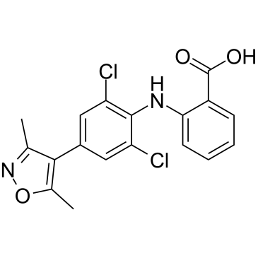
2-[2,6-dichloro-4-(3,5-dimethyl-1,2-oxazol-4-yl)anilino]benzoic acid
|
| 别名 |
FB23; FB 23; FB23; 2243736-35-6; 2-((2,6-Dichloro-4-(3,5-dimethylisoxazol-4-yl)phenyl)amino)benzoic acid; 2-[[2,6-bis(chloranyl)-4-(3,5-dimethyl-1,2-oxazol-4-yl)phenyl]amino]benzoic acid; Fb23 inhibitor; CHEMBL4572939; SCHEMBL23261118; BDBM589235;FB-23
|
| HS Tariff Code |
2934.99.9001
|
| 存储方式 |
Powder -20°C 3 years 4°C 2 years In solvent -80°C 6 months -20°C 1 month |
| 运输条件 |
Room temperature (This product is stable at ambient temperature for a few days during ordinary shipping and time spent in Customs)
|
| 溶解度 (体外实验) |
DMSO : ~125 mg/mL (~331.37 mM)
|
|---|---|
| 溶解度 (体内实验) |
注意: 如下所列的是一些常用的体内动物实验溶解配方,主要用于溶解难溶或不溶于水的产品(水溶度<1 mg/mL)。 建议您先取少量样品进行尝试,如该配方可行,再根据实验需求增加样品量。
注射用配方
注射用配方1: DMSO : Tween 80: Saline = 10 : 5 : 85 (如: 100 μL DMSO → 50 μL Tween 80 → 850 μL Saline)(IP/IV/IM/SC等) *生理盐水/Saline的制备:将0.9g氯化钠/NaCl溶解在100 mL ddH ₂ O中,得到澄清溶液。 注射用配方 2: DMSO : PEG300 :Tween 80 : Saline = 10 : 40 : 5 : 45 (如: 100 μL DMSO → 400 μL PEG300 → 50 μL Tween 80 → 450 μL Saline) 注射用配方 3: DMSO : Corn oil = 10 : 90 (如: 100 μL DMSO → 900 μL Corn oil) 示例: 以注射用配方 3 (DMSO : Corn oil = 10 : 90) 为例说明, 如果要配制 1 mL 2.5 mg/mL的工作液, 您可以取 100 μL 25 mg/mL 澄清的 DMSO 储备液,加到 900 μL Corn oil/玉米油中, 混合均匀。 View More
注射用配方 4: DMSO : 20% SBE-β-CD in Saline = 10 : 90 [如:100 μL DMSO → 900 μL (20% SBE-β-CD in Saline)] 口服配方
口服配方 1: 悬浮于0.5% CMC Na (羧甲基纤维素钠) 口服配方 2: 悬浮于0.5% Carboxymethyl cellulose (羧甲基纤维素) 示例: 以口服配方 1 (悬浮于 0.5% CMC Na)为例说明, 如果要配制 100 mL 2.5 mg/mL 的工作液, 您可以先取0.5g CMC Na并将其溶解于100mL ddH2O中,得到0.5%CMC-Na澄清溶液;然后将250 mg待测化合物加到100 mL前述 0.5%CMC Na溶液中,得到悬浮液。 View More
口服配方 3: 溶解于 PEG400 (聚乙二醇400) 请根据您的实验动物和给药方式选择适当的溶解配方/方案: 1、请先配制澄清的储备液(如:用DMSO配置50 或 100 mg/mL母液(储备液)); 2、取适量母液,按从左到右的顺序依次添加助溶剂,澄清后再加入下一助溶剂。以 下列配方为例说明 (注意此配方只用于说明,并不一定代表此产品 的实际溶解配方): 10% DMSO → 40% PEG300 → 5% Tween-80 → 45% ddH2O (或 saline); 假设最终工作液的体积为 1 mL, 浓度为5 mg/mL: 取 100 μL 50 mg/mL 的澄清 DMSO 储备液加到 400 μL PEG300 中,混合均匀/澄清;向上述体系中加入50 μL Tween-80,混合均匀/澄清;然后继续加入450 μL ddH2O (或 saline)定容至 1 mL; 3、溶剂前显示的百分比是指该溶剂在最终溶液/工作液中的体积所占比例; 4、 如产品在配制过程中出现沉淀/析出,可通过加热(≤50℃)或超声的方式助溶; 5、为保证最佳实验结果,工作液请现配现用! 6、如不确定怎么将母液配置成体内动物实验的工作液,请查看说明书或联系我们; 7、 以上所有助溶剂都可在 Invivochem.cn网站购买。 |
| 制备储备液 | 1 mg | 5 mg | 10 mg | |
| 1 mM | 2.6510 mL | 13.2549 mL | 26.5097 mL | |
| 5 mM | 0.5302 mL | 2.6510 mL | 5.3019 mL | |
| 10 mM | 0.2651 mL | 1.3255 mL | 2.6510 mL |
1、根据实验需要选择合适的溶剂配制储备液 (母液):对于大多数产品,InvivoChem推荐用DMSO配置母液 (比如:5、10、20mM或者10、20、50 mg/mL浓度),个别水溶性高的产品可直接溶于水。产品在DMSO 、水或其他溶剂中的具体溶解度详见上”溶解度 (体外)”部分;
2、如果您找不到您想要的溶解度信息,或者很难将产品溶解在溶液中,请联系我们;
3、建议使用下列计算器进行相关计算(摩尔浓度计算器、稀释计算器、分子量计算器、重组计算器等);
4、母液配好之后,将其分装到常规用量,并储存在-20°C或-80°C,尽量减少反复冻融循环。
计算结果:
工作液浓度: mg/mL;
DMSO母液配制方法: mg 药物溶于 μL DMSO溶液(母液浓度 mg/mL)。如该浓度超过该批次药物DMSO溶解度,请首先与我们联系。
体内配方配制方法:取 μL DMSO母液,加入 μL PEG300,混匀澄清后加入μL Tween 80,混匀澄清后加入 μL ddH2O,混匀澄清。
(1) 请确保溶液澄清之后,再加入下一种溶剂 (助溶剂) 。可利用涡旋、超声或水浴加热等方法助溶;
(2) 一定要按顺序加入溶剂 (助溶剂) 。
InvivoChem的所有产品仅用于作科学研究,不面向患者销售
Copyright 2020 InvivoChem LLC | All Rights Reserved 粤ICP备20063088号-1


 463611831
