| 规格 | 价格 | 库存 | 数量 |
|---|---|---|---|
| 10 mM * 1 mL in DMSO |
|
||
| 1mg |
|
||
| 10mg |
|
||
| 25mg |
|
||
| 50mg |
|
||
| 100mg |
|
||
| 250mg |
|
||
| 500mg |
|
||
| 1g |
|
||
| 5g |
|
||
| Other Sizes |
|
| 靶点 |
COX-1 (IC50 = 64.3 μM); COX-2
Nepafenac (AHR9434; AL6515; Nevanac) is a prodrug that is metabolized in vivo to Amfenac (its active form), a non-selective cyclooxygenase (COX) inhibitor targeting both COX-1 and COX-2. In in vitro enzyme assays, Amfenac (the active metabolite of Nepafenac) exhibited inhibitory activity against human recombinant COX-1 with an IC₅₀ of 0.17 μM and human recombinant COX-2 with an IC₅₀ of 0.26 μM [1] |
|---|---|
| 体外研究 (In Vitro) |
奈帕芬胺是一种非甾体抗炎药(NSAID)。奈帕芬胺对(环氧合酶-1)COX-1 和 COX-2 的 IC50 值分别为 250 nM 和 150 nM。细胞测定:奈帕芬胺显着降低人葡萄膜黑色素瘤细胞系(包括 SP6.5、92.1、OCM-1、MKT-BR)和人转化葡萄膜黑色素细胞系 UW-1 的增殖率。与罗非昔布相比,奈帕芬胺可能具有更好的系统安全性。
COX抑制与前列腺素(PG)减少:在人全血实验中,奈帕芬酸(1-100 μM)与脂多糖(LPS,1 μg/mL,诱导COX-2表达)或钙离子载体A23187(1 μM,激活COX-1)共孵育24小时后,活性代谢产物氨芬酸可浓度依赖性抑制PGE₂生成:10 μM奈帕芬酸处理组中,COX-1介导的PGE₂较溶剂对照组减少62%,COX-2介导的PGE₂减少71%[1] - 眼细胞炎症抑制:在白细胞介素-1β(IL-1β,10 ng/mL)刺激的原代兔角膜上皮细胞中,奈帕芬酸(0.1-10 μM)剂量依赖性降低TNF-α和IL-6释放。10 μM剂量下,TNF-α水平较仅IL-1β处理组降低58%,IL-6水平降低65%(ELISA检测)[3] - 粒细胞-巨噬细胞集落刺激因子(GM-CSF)抑制:在脂多糖(LPS,1 μg/mL)处理的人视网膜色素上皮(RPE)细胞中,奈帕芬酸(1-20 μM)抑制GM-CSF mRNA表达(实时PCR):20 μM剂量下,GM-CSF mRNA较LPS组减少73%[3] |
| 体内研究 (In Vivo) |
奈帕芬胺显示出显着更高的眼部生物利用度,氨芬酸显示出比酮咯酸或溴芬酸更强的 COX-2 抑制效力。 Nepafenac 仅表现出较弱的 COX-1 抑制活性,IC50 为 64.3 mM。奈帕芬胺抑制兔子虹膜/睫状体 (85-95%) 和视网膜/脉络膜 (55%) 的前列腺素合成。奈帕芬胺 (0.5%) 可减少 65% 的视网膜水肿,这与抑制 62% 的血视网膜屏障破坏有关。奈帕芬胺 (0.5%) 显着抑制 (46%) 血-视网膜屏障破坏,同时几乎完全抑制 PGE2 合成 (96%)。 Nepafenac 显着抑制胰岛素缺乏糖尿病大鼠视网膜前列腺素 E(2)、超氧化物、环氧合酶-2 和视网膜微血管内的白细胞停滞,而不影响血管内皮生长因子 (VEGF) 和一氧化氮 (NO)。 Nepafenac 显着抑制糖尿病大鼠中转移酶介导的 dUTP 缺口末端标记阳性毛细血管细胞、无细胞毛细血管和周细胞影的数量。与对照相比,奈帕芬胺导致小鼠脉络膜新血管形成和缺血诱导的视网膜新血管形成显着减少。奈帕芬胺还可以抑制缺血引起的视网膜中 VEGF mRNA 的增加。在葡萄膜黑色素瘤的眼部和转移性动物模型中,奈帕芬胺可以延缓恶性肿瘤的进展并减轻体重。
大鼠足肿胀模型:对雄性Sprague-Dawley大鼠进行角叉菜胶诱导足肿胀(后爪皮下注射0.1 mL 1%角叉菜胶),口服给予奈帕芬酸(3、10、30 mg/kg)可剂量依赖性抑制肿胀形成。30 mg/kg剂量下,角叉菜胶注射后4小时的足体积较溶剂对照组减少59%(体积描记法测量)[1] - 小鼠棉球肉芽肿模型:对雄性ICR小鼠皮下植入2个无菌棉球(每个5 mg),口服奈帕芬酸(10、30、100 mg/kg/天)连续7天,可减少肉芽肿干重:100 mg/kg/天剂量下,肉芽肿重量较溶剂对照组降低42%。组织学显示炎症细胞(中性粒细胞、巨噬细胞)浸润减少[2] - 兔眼炎症模型:对新西兰白兔进行脂多糖(LPS)诱导前葡萄膜炎(玻璃体内注射100 ng LPS),局部滴眼给予奈帕芬酸(0.1%眼用混悬液,每眼50 μL,每日4次,连续3天),第3天前房闪辉(眼炎症标志物)较溶剂对照组减少76%。裂隙灯检查显示虹膜充血减轻、房水细胞数减少[3] |
| 酶活实验 |
与双氯芬酸(IC50=0.12微M)相比,奈帕芬胺仅表现出微弱的COX-1抑制活性(IC50=64.3微M)。然而,amfenac是COX-1(IC50=0.25微M)和COX-2活性(IC50=0.15微M)的强效抑制剂[1]。
COX-1/COX-2活性实验(人重组酶):将人重组COX-1或COX-2悬浮于含血红素(1 μM)和谷胱甘肽(1 mM)的50 mM Tris-HCl缓冲液(pH 8.0)中,加入系列浓度的氨芬酸(奈帕芬酸活性代谢产物,0.01-1 μM),再加入花生四烯酸(10 μM)作为底物。37°C孵育15分钟后,用1 M HCl终止反应,通过竞争性ELISA检测PGE₂生成量,根据PGE₂抑制率与氨芬酸浓度的非线性回归计算IC₅₀[1] - COX活性实验(人全血):将1 mL人全血与奈帕芬酸(0.1-100 μM)及A23187(1 μM,激活COX-1)或LPS(1 μg/mL,诱导COX-2)混合,37°C孵育24小时后离心分离血浆,通过ELISA定量血浆PGE₂水平,评估氨芬酸(奈帕芬酸代谢产物)对COX-1/COX-2的抑制作用[1] |
| 细胞实验 |
转染人葡萄膜黑色素瘤细胞系以组成型表达COX-2,并使用两种不同的方法(添加和不添加氨芬酸)测量这些细胞的增殖率。在暴露于两组细胞的黑色素瘤条件培养基以及含有和不含有Nepafenac 的活性代谢产物氨芬酸后,测量巨噬细胞产生的一氧化氮
结果:转染表达COX-2的细胞增殖率高于未转染的细胞。添加氨芬酸显著降低了所有细胞系的增殖率。巨噬细胞产生一氧化氮受到黑色素瘤条件培养基的抑制,加入氨芬酸部分克服了这种抑制作用 结论:氨芬酸对COX-2转染和未转染的葡萄膜黑色素瘤细胞的增殖率及其对巨噬细胞细胞毒性活性的抑制作用均有影响。https://pubmed.ncbi.nlm.nih.gov/18042295/ 兔角膜上皮细胞炎症实验:将原代兔角膜上皮细胞接种于24孔板培养至融合,用奈帕芬酸(0.1-10 μM)预处理1小时后,加入IL-1β(10 ng/mL)刺激24小时。收集培养上清,采用夹心ELISA(兔细胞因子特异性一抗和二抗)检测TNF-α和IL-6浓度。每个浓度设3个复孔,结果以相对于仅IL-1β组的百分比变化表示[3] - 人RPE细胞GM-CSF mRNA实验:将人RPE细胞接种于6孔板培养至80%融合,用奈帕芬酸(1-20 μM)和LPS(1 μg/mL)共同处理16小时。提取总RNA并逆转录为cDNA,采用GM-CSF特异性引物进行实时PCR(GAPDH为内参基因),通过2^(-ΔΔCt)法计算GM-CSF mRNA相对表达量[3] |
| 动物实验 |
局部应用奈帕芬可显著降低LPS诱导的大鼠视网膜中PGE2的水平。然而,在LPS诱导的大鼠模型中,奈帕芬对血视网膜屏障(BRB)通透性没有显著影响。方法:进行一项双盲试验,比较赋形剂与不同浓度的奈帕芬(0.01%、0.03%、0.1%或0.5%)、0.1%双氯芬酸或0.5%酮咯酸氨丁三醇局部应用对氧诱导缺血性视网膜病变小鼠、激光诱导布鲁赫膜破裂引起的脉络膜新生血管(CNV)小鼠或光感受器中血管内皮生长因子(VEGF)表达增加的转基因小鼠(rho/VEGF转基因小鼠)的影响。
结果:与赋形剂组相比,0.1%或0.5%奈帕芬治疗组小鼠的CNV和缺血诱导的视网膜新生血管均显著减少。奈帕芬酸还能抑制缺血诱导的视网膜中 VEGF mRNA 的增加。在 rho/VEGF 转基因小鼠中,奈帕芬酸未能抑制新生血管形成。在其他研究中,与载体对照组小鼠相比,0.1% 或 0.03% 奈帕芬酸治疗组小鼠的 CNV 显著减少,而 0.1% 双氯芬酸治疗组小鼠的 CNV 则无显著差异。0.5% 酮咯酸氨丁三醇治疗 14 天的小鼠死亡率较高,但治疗 7 天后评估发现,其死亡率与载体对照组小鼠无差异。[3] 本研究旨在评估非甾体抗炎药奈帕芬酸在兔眼局部应用后预防促分裂原诱导的全视网膜水肿的能力。将麻醉后的荷兰带状兔玻璃体内注射促分裂原刀豆蛋白A(30 μg/20 μL),以诱导视网膜后段炎症和增厚(水肿)。使用海德堡视网膜断层扫描仪和定制软件生成水肿图。通过测定玻璃体液中的蛋白质浓度来评估血视网膜屏障的破坏情况,而玻璃体液中的PGE2则通过放射免疫分析法进行分析。在开始局部应用奈帕芬(0.1-1.0%,w/v)、地塞米松(0.1%)、扶他林(0.1%)或阿库拉(0.5%)治疗72小时后,评估刀豆蛋白A诱导的视网膜水肿的抑制情况。注射刀豆蛋白A后72小时,玻璃体液中的蛋白质和PGE2合成显著增加。视网膜厚度也增加了32%,与炎症反应同时发生。局部应用0.5%奈帕芬可使视网膜水肿减少65%,这与血视网膜屏障破坏抑制62%相关。在后续研究中,0.5%奈帕芬显著抑制(46%)血视网膜屏障破坏,同时几乎完全抑制了PGE2的合成(96%)。平行试验表明,扶他林和阿库拉均未抑制玻璃体中这些炎症标志物的积累。本研究表明,局部眼部给药后,奈帕芬在后段视网膜中表现出优异的药效学特性,提示其在治疗多种视网膜水肿相关疾病方面具有独特的治疗潜力。[2] 大鼠角叉菜胶诱导爪水肿模型:雄性Sprague-Dawley大鼠(180-220 g)随机分为4组(每组n=6):赋形剂组(0.5%羧甲基纤维素,口服)、奈帕芬3 mg/kg(口服)、10 mg/kg(口服)、30 mg/kg(口服)。给药30分钟后,将0.1 mL 1%角叉菜胶溶液(溶于0.9%生理盐水)皮下注射至右后爪。分别于角叉菜胶注射后 0、1、2、4 和 6 小时使用体积描记器测量爪体积。水肿抑制率计算公式为 [(载体组爪体积 - 药物组爪体积)/载体组爪体积] × 100% [1] - 小鼠棉球肉芽肿模型:雄性 ICR 小鼠(25-30 g)用异氟烷麻醉,将两颗无菌棉球(经高压灭菌,每颗 5 mg)皮下植入(背部中线两侧各一颗)。小鼠随机分为 4 组(每组 n=8):载体组(0.5% 甲基纤维素,口服)、萘芬酸组(10 mg/kg/天,口服)、萘芬酸组(30 mg/kg/天,口服)和萘芬酸组(100 mg/kg/天,口服)。药物每日给药一次,连续给药 7 天。第8天,处死小鼠,取出肉芽肿颗粒,在60℃下干燥24小时,并测量干重。肉芽肿重量计算公式为(干燥颗粒重量 - 初始颗粒重量)[2] - 兔LPS诱导前葡萄膜炎实验方案:将新西兰白兔(2.5-3 kg)随机分为两组(每组n=6):赋形剂组(0.9%生理盐水,局部滴眼)和0.1%奈帕芬滴眼液组(局部滴眼)。每只眼睛每天滴入50 μL药物/赋形剂,每日4次(上午8点、中午12点、下午4点、晚上8点)。首次给药1小时后,将100 ng LPS(溶于50 μL无菌生理盐水)玻璃体内注射到右眼。在LPS注射后24、48和72小时,使用裂隙灯显微镜(0-4分制)对前房闪辉进行评分。同时评估虹膜充血和房水细胞计数[3] |
| 药代性质 (ADME/PK) |
吸收、分布和排泄
奈帕芬酸能迅速穿过角膜(体外实验表明其速度比双氯芬酸快6倍)。 健康志愿者口服14C-奈帕芬酸后,发现尿液排泄是放射性物质的主要清除途径,约占剂量的85%,而粪便排泄约占剂量的6%。尿液中未检测到奈帕芬酸(前药)和安芬酸(活性化合物)。 代谢/代谢物 奈帕芬酸(前药)在睫状体上皮、视网膜和脉络膜中经眼内水解酶脱氨基转化为安芬酸(活性化合物)。随后,安芬酸经广泛代谢生成极性更强的代谢物,包括芳香环的羟基化,最终形成葡萄糖醛酸苷结合物。 代谢:奈帕芬通过酯酶介导的水解迅速代谢为活性形式安芬酸。在大鼠血浆中,口服奈帕芬(10 mg/kg)后,15 分钟内即可检测到安芬酸,给药后 1 小时达到血浆峰浓度(Cmax)2.3 ± 0.4 μg/mL; 30分钟后,奈帕芬原药浓度低于0.05 μg/mL [1] - 眼部吸收:在兔眼中,局部滴注0.1%奈帕芬混悬液(50 μL/眼)后,2小时房水中安芬酸浓度为0.8 ± 0.2 μg/mL,6小时房水中安芬酸浓度为0.3 ± 0.1 μg/mL。房水中未检测到奈帕芬原药[3] - 半衰期:在大鼠中,安芬酸(由奈帕芬代谢而来)的消除半衰期(t₁/₂)为2.1 ± 0.3小时[1] |
| 毒性/毒理 (Toxicokinetics/TK) |
妊娠期和哺乳期影响
◉ 哺乳期用药概述 目前尚无关于奈帕芬在哺乳期临床应用的信息。预计母亲使用奈帕芬滴眼液不会对母乳喂养的婴儿造成任何不良影响。为显著减少滴眼液后进入母乳的药物量,请用手指按压眼角附近的泪管至少1分钟,然后用吸水纸巾吸去多余的药液。 ◉ 对母乳喂养婴儿的影响 截至修订日期,未找到相关的已发表信息。 ◉ 对哺乳和母乳的影响 截至修订日期,未找到相关的已发表信息。 蛋白质结合 奈帕芬对血清白蛋白具有高亲和力。体外实验中,与人白蛋白和人血清的结合率分别为 95.4% 和 99.1%。 急性口服毒性:在雄性和雌性 Sprague-Dawley 大鼠中,奈帕芬 的口服 LD₅₀ > 2000 mg/kg。在剂量高达 2000 mg/kg 时,未观察到死亡或严重临床症状(例如共济失调、抽搐、胃肠道不适)[1] - 眼部刺激:在兔中,局部应用 0.1% 奈帕芬混悬液(50 μL/眼,每日 4 次,持续 7 天)未引起眼部刺激症状(例如结膜充血、角膜混浊、流泪),经 Draize 试验评估[3] - 血浆蛋白结合率:安芬酸(奈帕芬的活性代谢物)在人血浆中的血浆蛋白结合率为 98 ± 1%(浓度范围:0.1-10 μg/mL)[1] |
| 参考文献 |
[1]. Inflammation.2000 Aug;24(4):357-70;
[2]. Inflammation.2003 Oct;27(5):281-91; [3]. Invest Ophthalmol Vis Sci.2003 Jan;44(1):409-15. |
| 其他信息 |
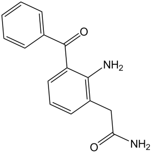
奈帕芬是一种单羧酸酰胺,是安芬酸的羧酸基团转化为相应的羧酰胺基团后的化合物。它是安芬酸的前体药物,用于滴眼液中治疗白内障手术后的疼痛和炎症。它具有多种药理作用,包括作为前体药物、环氧合酶-2抑制剂、环氧合酶-1抑制剂、非甾体抗炎药和非麻醉性镇痛药。
奈帕芬是一种非甾体抗炎药前体药物(NSAID),通常作为处方滴眼液出售。它用于治疗白内障手术相关的疼痛和炎症。 奈帕芬是一种非甾体抗炎药。奈帕芬的作用机制是作为环氧合酶抑制剂。 奈帕芬是一种局部用非甾体类抗炎药,用于滴眼液中治疗眼痛和肿胀。 药物适应症 用于治疗白内障手术相关的疼痛和炎症。 FDA标签 奈帕芬适用于:,,,预防和治疗白内障手术相关的术后疼痛和炎症;,降低糖尿病患者白内障手术后黄斑水肿的风险。,, 预防白内障手术相关的术后疼痛和炎症 作用机制 奈帕芬是一种前体药物。奈帕芬酸穿透角膜后,会迅速生物活化为安芬酸,后者是一种强效的非甾体抗炎药,可均匀抑制COX-1和COX-2的活性。 奈帕芬酸是一种前药,由于其亲脂性结构,其眼部渗透性优于其活性代谢物安芬酸,因此适用于眼部炎症的局部眼用给药[3]。 - 奈帕芬酸(0.1%眼用混悬液)的主要临床适应症是治疗白内障手术相关的疼痛和炎症,这已通过其在动物模型中抑制眼部PGE₂生成和前房炎症的能力得到证实[3]。 - 与选择性COX-2抑制剂不同,奈帕芬酸(通过安芬酸)可同时抑制COX-1和COX-2,这有助于其在全身(例如,爪水肿)和局部(例如眼部)炎症模型[1, 2] - 在小鼠棉球肉芽肿模型中,奈帕芬不仅减轻了肉芽肿重量,而且抑制了胶原沉积(通过羟脯氨酸测定法测量),表明其在抑制慢性炎症组织重塑中发挥作用[2] |
| 分子式 |
C15H14N2O2
|
|
|---|---|---|
| 分子量 |
254.28
|
|
| 精确质量 |
254.105
|
|
| 元素分析 |
C, 70.85; H, 5.55; N, 11.02; O, 12.58
|
|
| CAS号 |
78281-72-8
|
|
| 相关CAS号 |
Nepafenac-d5;1246814-53-8
|
|
| PubChem CID |
151075
|
|
| 外观&性状 |
Light yellow to yellow solid powder
|
|
| 密度 |
1.3±0.1 g/cm3
|
|
| 沸点 |
562.5±50.0 °C at 760 mmHg
|
|
| 熔点 |
177-181ºC
|
|
| 闪点 |
294.0±30.1 °C
|
|
| 蒸汽压 |
0.0±1.5 mmHg at 25°C
|
|
| 折射率 |
1.641
|
|
| LogP |
0.73
|
|
| tPSA |
86.18
|
|
| 氢键供体(HBD)数目 |
2
|
|
| 氢键受体(HBA)数目 |
3
|
|
| 可旋转键数目(RBC) |
4
|
|
| 重原子数目 |
19
|
|
| 分子复杂度/Complexity |
337
|
|
| 定义原子立体中心数目 |
0
|
|
| InChi Key |
QEFAQIPZVLVERP-UHFFFAOYSA-N
|
|
| InChi Code |
InChI=1S/C15H14N2O2/c16-13(18)9-11-7-4-8-12(14(11)17)15(19)10-5-2-1-3-6-10/h1-8H,9,17H2,(H2,16,18)
|
|
| 化学名 |
2-(2-amino-3-benzoylphenyl)acetamide
|
|
| 别名 |
|
|
| HS Tariff Code |
2934.99.9001
|
|
| 存储方式 |
Powder -20°C 3 years 4°C 2 years In solvent -80°C 6 months -20°C 1 month |
|
| 运输条件 |
Room temperature (This product is stable at ambient temperature for a few days during ordinary shipping and time spent in Customs)
|
| 溶解度 (体外实验) |
|
|||
|---|---|---|---|---|
| 溶解度 (体内实验) |
注意: 如下所列的是一些常用的体内动物实验溶解配方,主要用于溶解难溶或不溶于水的产品(水溶度<1 mg/mL)。 建议您先取少量样品进行尝试,如该配方可行,再根据实验需求增加样品量。
注射用配方
注射用配方1: DMSO : Tween 80: Saline = 10 : 5 : 85 (如: 100 μL DMSO → 50 μL Tween 80 → 850 μL Saline)(IP/IV/IM/SC等) *生理盐水/Saline的制备:将0.9g氯化钠/NaCl溶解在100 mL ddH ₂ O中,得到澄清溶液。 注射用配方 2: DMSO : PEG300 :Tween 80 : Saline = 10 : 40 : 5 : 45 (如: 100 μL DMSO → 400 μL PEG300 → 50 μL Tween 80 → 450 μL Saline) 注射用配方 3: DMSO : Corn oil = 10 : 90 (如: 100 μL DMSO → 900 μL Corn oil) 示例: 以注射用配方 3 (DMSO : Corn oil = 10 : 90) 为例说明, 如果要配制 1 mL 2.5 mg/mL的工作液, 您可以取 100 μL 25 mg/mL 澄清的 DMSO 储备液,加到 900 μL Corn oil/玉米油中, 混合均匀。 View More
注射用配方 4: DMSO : 20% SBE-β-CD in Saline = 10 : 90 [如:100 μL DMSO → 900 μL (20% SBE-β-CD in Saline)] 口服配方
口服配方 1: 悬浮于0.5% CMC Na (羧甲基纤维素钠) 口服配方 2: 悬浮于0.5% Carboxymethyl cellulose (羧甲基纤维素) 示例: 以口服配方 1 (悬浮于 0.5% CMC Na)为例说明, 如果要配制 100 mL 2.5 mg/mL 的工作液, 您可以先取0.5g CMC Na并将其溶解于100mL ddH2O中,得到0.5%CMC-Na澄清溶液;然后将250 mg待测化合物加到100 mL前述 0.5%CMC Na溶液中,得到悬浮液。 View More
口服配方 3: 溶解于 PEG400 (聚乙二醇400) 请根据您的实验动物和给药方式选择适当的溶解配方/方案: 1、请先配制澄清的储备液(如:用DMSO配置50 或 100 mg/mL母液(储备液)); 2、取适量母液,按从左到右的顺序依次添加助溶剂,澄清后再加入下一助溶剂。以 下列配方为例说明 (注意此配方只用于说明,并不一定代表此产品 的实际溶解配方): 10% DMSO → 40% PEG300 → 5% Tween-80 → 45% ddH2O (或 saline); 假设最终工作液的体积为 1 mL, 浓度为5 mg/mL: 取 100 μL 50 mg/mL 的澄清 DMSO 储备液加到 400 μL PEG300 中,混合均匀/澄清;向上述体系中加入50 μL Tween-80,混合均匀/澄清;然后继续加入450 μL ddH2O (或 saline)定容至 1 mL; 3、溶剂前显示的百分比是指该溶剂在最终溶液/工作液中的体积所占比例; 4、 如产品在配制过程中出现沉淀/析出,可通过加热(≤50℃)或超声的方式助溶; 5、为保证最佳实验结果,工作液请现配现用! 6、如不确定怎么将母液配置成体内动物实验的工作液,请查看说明书或联系我们; 7、 以上所有助溶剂都可在 Invivochem.cn网站购买。 |
| 制备储备液 | 1 mg | 5 mg | 10 mg | |
| 1 mM | 3.9327 mL | 19.6634 mL | 39.3267 mL | |
| 5 mM | 0.7865 mL | 3.9327 mL | 7.8653 mL | |
| 10 mM | 0.3933 mL | 1.9663 mL | 3.9327 mL |
1、根据实验需要选择合适的溶剂配制储备液 (母液):对于大多数产品,InvivoChem推荐用DMSO配置母液 (比如:5、10、20mM或者10、20、50 mg/mL浓度),个别水溶性高的产品可直接溶于水。产品在DMSO 、水或其他溶剂中的具体溶解度详见上”溶解度 (体外)”部分;
2、如果您找不到您想要的溶解度信息,或者很难将产品溶解在溶液中,请联系我们;
3、建议使用下列计算器进行相关计算(摩尔浓度计算器、稀释计算器、分子量计算器、重组计算器等);
4、母液配好之后,将其分装到常规用量,并储存在-20°C或-80°C,尽量减少反复冻融循环。
计算结果:
工作液浓度: mg/mL;
DMSO母液配制方法: mg 药物溶于 μL DMSO溶液(母液浓度 mg/mL)。如该浓度超过该批次药物DMSO溶解度,请首先与我们联系。
体内配方配制方法:取 μL DMSO母液,加入 μL PEG300,混匀澄清后加入μL Tween 80,混匀澄清后加入 μL ddH2O,混匀澄清。
(1) 请确保溶液澄清之后,再加入下一种溶剂 (助溶剂) 。可利用涡旋、超声或水浴加热等方法助溶;
(2) 一定要按顺序加入溶剂 (助溶剂) 。
| NCT Number | Recruitment | interventions | Conditions | Sponsor/Collaborators | Start Date | Phases |
| NCT03406689 | Completed | Drug: Nepafenac 0.1% Oph Susp Drug: Nepafenac 0.3% Oph Susp |
Pain | University Hospital of Patras | September 1, 2017 | Not Applicable |
| NCT02752646 | Completed | Drug: nepafenac 0.3% Drug: ketorolac |
Cataract | MDbackline, LLC | April 2016 | Not Applicable |
| NCT02821390 | Completed | Drug: Nepafenac Eye Drops Drug: Placebo (Artificial Tears) |
Pain | University Hospital of Patras | June 2016 | Not Applicable |
| NCT01995890 | Completed | Drug: nepafenac | Intraocular Pressure | Dr T V Patel Eye Institute | December 2012 | Phase 4 |
|
|---|
|
InvivoChem的所有产品仅用于作科学研究,不面向患者销售
Copyright 2020 InvivoChem LLC | All Rights Reserved 粤ICP备20063088号-1


 COA
COA


 463611831
