| 规格 | 价格 | |
|---|---|---|
| 500mg | ||
| 1g | ||
| Other Sizes |

| 靶点 |
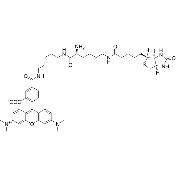
Fluorescent Dye
|
|---|---|
| 体内研究 (In Vivo) |
可以使用 TMR Biocytin [2] 研究血脑屏障 (BBB) 通透性变化。指南(本协议仅作为指南;应对其进行调整以满足您的独特要求。以下是我们建议的协议)[2]。 1. 对于每只小鼠,将 1 毫克 TMR Biocytin 稀释在 100 μL PBS 中,然后将混合物注射到尾静脉中。 2.注射后半小时麻醉并灌注动物。 3. 取下脊髓,然后制作深度冷冻的连续 10 μm 纵向切片。 4. 使用 DAPI 进行核复染。 5. 对每个队列使用相同的激光强度、曝光时间和放大倍数,使用 ×10 物镜和 ×10 目镜拍摄整个切片的照片。 6. 使用来自注射示踪剂和未注射示踪剂的动物的肝脏样本来设定上述值。
TMR生物细胞素的初始纤维转运速度高达5.4mm/h。TMR生物胞素可以与AM钙染料结合使用,从几毫米的距离标记神经元胞体,并在几个小时内记录钙瞬变。TMR生物细胞蛋白的细胞外应用导致快速顺行运输,并在10分钟内标记局部突触。TMR生物细胞素是可固定的,在水杨酸甲酯清除过程中是稳定的,可以在神经组织深处观察到。 与现有方法相比:TMR生物细胞蛋白的逆行标记仅需几个小时即可实现远程神经元可视化和并发钙成像,这比其他基于荧光的示踪剂快得多。发绿光的Atto 488生物素也被吸收和逆向运输,但它与标准发绿光的钙染料不兼容。 结论:TMR生物细胞素是一种有吸引力的神经元示踪剂。它可以长距离快速标记神经元,并且可以与钙染料结合使用,以报告逆行标记的活神经元中的神经元活动。[1] 电休克疗法的脑刺激对神经精神疾病的有效机制尚不清楚。小胶质细胞毒性在神经精神、神经炎症和退行性疾病中起着关键作用。我们研究了电惊厥发作(ECS)调节小胶质细胞表型和对刺激反应的机制。通过形态学分析、Iba1和细胞因子表达检查小胶质细胞反应。ECS不影响静息小胶质细胞的表型或形态,但通过脂多糖刺激调节其活化。在幼年小鼠的ECS或假疗程后分离小胶质细胞进行转录组分析。RNA测序鉴定出141个差异表达基因。ECS调节多个免疫相关基因家族,并减弱神经毒性相关基因的表达。通过注射Biocytin TMR示踪剂检查血脑屏障。血脑屏障没有破裂,外周单核细胞的基因特征也没有增加,这表明ECS的作用主要是对驻留的小胶质细胞。对调控序列的无偏见分析确定了小胶质细胞视黄酸受体α(RARα)基因表达的诱导,以及多个ECS上调基因中可能存在的共同RARα结合基序。在体外研究了选择性RARα激动剂AM580对小胶质细胞对LPS反应的影响。AM580可抑制LPS诱导的细胞因子表达和活性氧的产生。利用慢性小鼠实验性自身免疫性脑脊髓炎(EAE)来证实RARα信号传导作为ECS诱导的转录途径的介质在调节小胶质细胞毒性中的作用。连续侧脑室内注射AM580可有效减轻EAE的严重程度。总之,ECS通过激活小胶质细胞视黄酸受体α途径调节中枢神经系统先天免疫系统反应,这标志着慢性神经炎症、神经精神和神经退行性疾病的一种新的治疗方法[2]。 |
| 动物实验 |
将标本放入安装在奥林巴斯 BX51 显微镜(立式,改装为固定载物台,安装在 XY 平台上)上的记录室后 15-150 分钟开始进行光学记录。记录室容积为 2 ml,温度为 29.5°C,并以 2 ml/min 的流速持续灌注预热的充氧(95% O2,5% CO2)人工脑脊液 (ACSF),其成分(单位:mM)为:129 NaCl、3 KCl、5 KH2PO4、25 NaHCO3、30 d-(+)-葡萄糖、0.4 MgSO4 和 0.7 CaCl2。为了检测荧光信号,将金属卤化物光源 PhotoFluor II 或 LED 光源 M470L2(Thorlabs,新泽西州,美国)通过液体导光管和相应的光学滤光片(单位:nm)耦合到 Olympus BX51 立体显微镜(正置式,改装为固定载物台,安装在 XY 平台上):Biocytin-TMR、Atto 565 Biotin、Atto 565、Atto 550 Biotin、TMR;Olympus U-MWIG2,激发波长:BP520-550、DM565,发射波长:BA580IF 和 Atto 488 Biotin、Fluo-8 AM;改装的 Olympus U-MWIB,激发波长:BP457-487、DM505,发射波长:515-550。图像由 SOLIS 软件控制的 sCMOS 相机采集。成像采用×10、×20、×40和×63水浸物镜,帧速率为1–10帧/秒,成像时间为1–20秒。部分注射了Biocytin-TMR的样本在0.1 M Sorensen磷酸盐缓冲液中的4%多聚甲醛中固定过夜,然后用浓度递增的乙醇溶液(20%、30%、50%、70%、95%、100%,每次10分钟)脱水,用100%水杨酸甲酯透明化,并使用蔡司LSM 710共聚焦显微镜成像(激发波长:561 nm,发射波长:623 nm)。[1]
脑血屏障通透性[2] 评估了四组Biozzi ABH小鼠(每组3只小鼠);本研究纳入未免疫小鼠(阴性对照)、处于首次复发高峰期的EAE小鼠(免疫后第17天,阳性对照)、单次ECS治疗后1小时以及连续3天每日ECS治疗后24小时的小鼠。每只小鼠尾静脉注射1 mg溶于100 μL PBS的5-(和6)-四甲基罗丹明生物素(Biocytin-TMR)。注射后数秒内,小鼠耳部出现粉红色,表明药物已进入血液循环。注射30分钟后,对小鼠进行麻醉和灌注,取出脊髓,进行深度冷冻,并按上述方法制备10 μm厚的连续纵切片。使用DAPI(Vector Laboratories)进行核复染。所有组均采用相同的激光强度、曝光时间和放大倍数,在物镜10倍、目镜10倍的条件下获取全切片图像。为了设定这些参数,我们使用了注射示踪剂的小鼠和未注射示踪剂的小鼠的肝脏。 |
| 参考文献 | |
| 其他信息 |
生物素的长距离运输已被用于在体内或体外条件下追踪神经束(Kobbert et al., 2000; Thomson and Armstrong, 2011)。共注射NMDA或KCl可以促进生物素的摄取(Chang et al., 2000; Jiang et al., 1993; Tarras-Wahlberg and Rekling, 2009; Zheng et al., 1998),但胞外沉积的生物素是如何被摄取的尚不完全清楚,负责神经束内主动运输的机制也尚未明确。然而,该分子似乎对转运机制中的某些组分具有亲和力,我们利用这一原理证明,通过长间隔臂与生物素偶联的荧光团TMR可作为一种快速逆行示踪剂,在数小时内标记长距离的活神经元。[1]
在我们测试的六种化合物中,TMR生物素在脊髓束中的转运速度最高(5.4 mm/h)。未偶联的TMR无法被纤维吸收,表明生物素部分对于吸收和纤维转运至关重要。其他几种生物素偶联的荧光化合物也被吸收和转运,特别是Atto 488生物素,其转运速度接近TMR生物素。生物素-TMR和Atto 488生物素分别具有红色和绿色发射光谱,因此可用于双重标记实验。然而,Atto 550 生物素的吸收率极低,转运速度也很慢。我们推测,至少有两种化学性质可能影响所测试的共轭化合物的吸收和转运特性。首先,最新数据显示,受试化合物的荧光团与脂质双层膜的相互作用差异显著。例如,运输速度较慢的Atto 550生物素的荧光团与脂质双层膜的相互作用较强(MIF:33),而生物胞素-TMR的荧光团与脂质双层膜的相互作用较弱(MIF:0.35,Hughes等人,2014)。这种关系表明,与脂质双层膜相互作用较强的化合物可能被脂质捕获,从而无法被轴突运输机制募集。其次,受试化合物中荧光团与生物素/生物胞素部分之间的间隔基长度也不同。例如,TMR生物胞素和Atto 488生物素分别具有20个和18个碳氮原子的脂肪族间隔基(图4),而运输速度较慢的Atto 565生物素则具有13个碳氮原子的脂肪族间隔基。脂肪族间隔基。因此,间隔基的长度也可能是影响化合物转运特性的一个因素,因为它决定了转运所需的分子相互作用的空间大小。我们选择在幼年出生后的动物身上进行实验,以便在体外条件下保持大块神经组织的活性。在最年长的动物(P15.5)中,髓鞘形成已经开始,因此我们预期TMR生物胞素在老年动物中也有效,但这仍需在单独的实验中验证。[1] TMR生物胞素的几个特性使其成为一种极具吸引力的神经元示踪剂。该荧光团发射可见光谱红色端的光,这是一个优势,因为较长的发射波长意味着在组织中的散射更少,从而提高了深层结构的显像效果。因此,我们使用共聚焦显微镜对经水杨酸甲酯透明化的组织进行观察,观察到了深度超过0.5毫米的生物胞素-TMR标记的纤维束(图7)。一些基于合成的、性能最佳的钙传感器小分子有机染料,例如 Fluo-4、Fluo-8、俄勒冈绿和钙绿,其发射光谱(Em:506–531 nm)可以与 TMR 生物胞素的发射光谱(Em:581 nm)有效分离。最近改进的基因编码钙传感器,例如 GCaMP 家族,也具有更短波长的发射峰(Em:∼512 nm (Badura 等,2014))。因此,可以使用钙传感器和 TMR 生物胞素对神经元进行双重标记,从而同时进行钙成像和胞体树突可视化。在 Fluo-8 AM/TRM 生物胞素标记的 IO 神经元中,自发钙瞬变的持续时间不受 TMR 生物胞素的影响,并且在 Fluo-8 AM/TRM 生物胞素标记的器官型切片培养物中,双重标记的神经元显示出自发振荡。我们认为这表明在本文使用的照明参数下,神经元具有良好的存活率,即使用 TMR 时预计光毒性较低。生物素。TMR生物素的高转运速度使得这类实验可以在数小时内完成,如本文所示,通过对含有呼吸神经元的器官型切片培养物中振荡的连合神经元进行成像(图5)。类似的实验,例如在含有振荡连合神经元的急性制备的脑干切片中线注射钙绿-1 AM,则需要过夜孵育(Koshiya和Smith,1999)。逆行运输的TMR生物素也能进入树突(图1,图7),这为涉及靶向树突记录和使用AM染料进行双重标记的实验提供了可能,在某些情况下,这些染料还能标记近端树突(Del Negro等人,2011)。胞旁电穿孔导入生物素-TMR可导致强烈的胞体树突标记,并顺行运输至局部轴突。 10分钟内即可观察到具有清晰突触轮廓的树突状结构(图7)。这种顺行运输可用于揭示局部回路基序、靶向突触前实验以及形态学和电生理学联合表征。TMR生物胞素易于被颅神经根吸收(图7),因此有望成为发育神经生物学中常用的碳菁染料(如DiO和DiI)的有效替代品,用于标记特定的颅神经和脊髓区域。此外,TMR生物胞素中赖氨酸残基的存在使其能够被多聚甲醛和戊二醛固定,这有助于在固定和透明化的组织中进行形态学可视化。[1] 基于类似的原理(Mishra等人,2011),他们开发了一种生物胞素衍生的MRI造影剂,并证明了这种功能化示踪剂的长程逆行运输。不难想象,进一步发展这一原理将带来怎样的前景。生物素/生物素部分与功能性荧光团偶联,可形成可在神经束中高速运输的报告分子,并报告突触、树突、胞体或轴突中钙、钠或氯离子的浓度。然而,功能性荧光团的化学性质可能对成功运输至关重要。[1] 先前的研究表明,ECS可能导致血脑屏障(BBB)的破坏(Ito等人,2017),这提示CD11b+细胞表达特征的变化可能代表血液单核细胞的流入。因此,我们使用Biocytin-TMR示踪剂研究了ECS对BBB的影响。首先,我们证实全身给药的Biocytin-TMR能够渗透到急性EAE(一种神经炎症模型)的脑实质中(图4A-D)。注射在未经处理的小鼠中,电休克治疗(ECS)后1小时和连续3天ECS治疗后24小时的生物胞素-TMR染色显示血脑屏障(BBB)完整(图4E-H)。我们还检测了差异表达基因是否与已报道的代表浸润性单核细胞/巨噬细胞的基因相对应。在98个上调基因中,仅有2个与急性实验性自身免疫性脑脊髓炎(EAE)中定义浸润性单核细胞的基因相匹配(Yamasaki等人,2014)(图4I),而仅有3个与病毒诱导的神经炎症中定义浸润性单核细胞/巨噬细胞的基因相匹配(DePaula-Silva等人,2019)。尽管浸润性单核细胞的标志物是在神经炎症状态下而非健康中枢神经系统(CNS)中描述的,但代表浸润性单核细胞的基因数量如此之少表明,ECS诱导了驻留中枢神经系统来源细胞的表型转变。 CD11b+细胞,而不是诱导大量外周单核细胞的浸润。单细胞分析可能进一步揭示中枢神经系统单核细胞小亚群的变化。[2] |
| 分子式 |
C46H60N8O7S
|
|---|---|
| 分子量 |
869.1
|
| 精确质量 |
868.43056
|
| CAS号 |
749247-49-2
|
| PubChem CID |
165412506
|
| 外观&性状 |
Typically exists as solid at room temperature
|
| LogP |
1.1
|
| tPSA |
235 Ų
|
| 氢键供体(HBD)数目 |
6
|
| 氢键受体(HBA)数目 |
10
|
| 可旋转键数目(RBC) |
20
|
| 重原子数目 |
12
|
| 分子复杂度/Complexity |
1710
|
| 定义原子立体中心数目 |
4
|
| SMILES |
CN(C)C1=CC2=C(C=C1)C(=C3C=CC(=[N+](C)C)C=C3O2)C4=C(C=C(C=C4)C(=O)NCCCCCNC(=O)[C@H](CCCCNC(=O)CCCC[C@H]5[C@@H]6[C@H](CS5)NC(=O)N6)N)C(=O)[O-]
|
| InChi Key |
RRJNRPHRYXQIAB-VSZNSILHSA-N
|
| InChi Code |
InChI=1S/C46H60N8O7S/c1-53(2)29-16-19-32-37(25-29)61-38-26-30(54(3)4)17-20-33(38)41(32)31-18-15-28(24-34(31)45(58)59)43(56)49-22-9-5-10-23-50-44(57)35(47)12-8-11-21-48-40(55)14-7-6-13-39-42-36(27-62-39)51-46(60)52-42/h15-20,24-26,35-36,39,42H,5-14,21-23,27,47H2,1-4H3,(H5-,48,49,50,51,52,55,56,57,58,59,60)/t35-,36-,39-,42-/m0/s1
|
| 化学名 |
5-[5-[[(2S)-6-[5-[(3aS,4S,6aR)-2-oxo-1,3,3a,4,6,6a-hexahydrothieno[3,4-d]imidazol-4-yl]pentanoylamino]-2-aminohexanoyl]amino]pentylcarbamoyl]-2-[3-(dimethylamino)-6-dimethylazaniumylidenexanthen-9-yl]benzoate
|
| 别名 |
TMR Biocytin; 749247-49-2;
|
| HS Tariff Code |
2934.99.9001
|
| 存储方式 |
Powder -20°C 3 years 4°C 2 years In solvent -80°C 6 months -20°C 1 month |
| 运输条件 |
Room temperature (This product is stable at ambient temperature for a few days during ordinary shipping and time spent in Customs)
|
| 溶解度 (体外实验) |
May dissolve in DMSO (in most cases), if not, try other solvents such as H2O, Ethanol, or DMF with a minute amount of products to avoid loss of samples
|
|---|---|
| 溶解度 (体内实验) |
注意: 如下所列的是一些常用的体内动物实验溶解配方,主要用于溶解难溶或不溶于水的产品(水溶度<1 mg/mL)。 建议您先取少量样品进行尝试,如该配方可行,再根据实验需求增加样品量。
注射用配方
注射用配方1: DMSO : Tween 80: Saline = 10 : 5 : 85 (如: 100 μL DMSO → 50 μL Tween 80 → 850 μL Saline)(IP/IV/IM/SC等) *生理盐水/Saline的制备:将0.9g氯化钠/NaCl溶解在100 mL ddH ₂ O中,得到澄清溶液。 注射用配方 2: DMSO : PEG300 :Tween 80 : Saline = 10 : 40 : 5 : 45 (如: 100 μL DMSO → 400 μL PEG300 → 50 μL Tween 80 → 450 μL Saline) 注射用配方 3: DMSO : Corn oil = 10 : 90 (如: 100 μL DMSO → 900 μL Corn oil) 示例: 以注射用配方 3 (DMSO : Corn oil = 10 : 90) 为例说明, 如果要配制 1 mL 2.5 mg/mL的工作液, 您可以取 100 μL 25 mg/mL 澄清的 DMSO 储备液,加到 900 μL Corn oil/玉米油中, 混合均匀。 View More
注射用配方 4: DMSO : 20% SBE-β-CD in Saline = 10 : 90 [如:100 μL DMSO → 900 μL (20% SBE-β-CD in Saline)] 口服配方
口服配方 1: 悬浮于0.5% CMC Na (羧甲基纤维素钠) 口服配方 2: 悬浮于0.5% Carboxymethyl cellulose (羧甲基纤维素) 示例: 以口服配方 1 (悬浮于 0.5% CMC Na)为例说明, 如果要配制 100 mL 2.5 mg/mL 的工作液, 您可以先取0.5g CMC Na并将其溶解于100mL ddH2O中,得到0.5%CMC-Na澄清溶液;然后将250 mg待测化合物加到100 mL前述 0.5%CMC Na溶液中,得到悬浮液。 View More
口服配方 3: 溶解于 PEG400 (聚乙二醇400) 请根据您的实验动物和给药方式选择适当的溶解配方/方案: 1、请先配制澄清的储备液(如:用DMSO配置50 或 100 mg/mL母液(储备液)); 2、取适量母液,按从左到右的顺序依次添加助溶剂,澄清后再加入下一助溶剂。以 下列配方为例说明 (注意此配方只用于说明,并不一定代表此产品 的实际溶解配方): 10% DMSO → 40% PEG300 → 5% Tween-80 → 45% ddH2O (或 saline); 假设最终工作液的体积为 1 mL, 浓度为5 mg/mL: 取 100 μL 50 mg/mL 的澄清 DMSO 储备液加到 400 μL PEG300 中,混合均匀/澄清;向上述体系中加入50 μL Tween-80,混合均匀/澄清;然后继续加入450 μL ddH2O (或 saline)定容至 1 mL; 3、溶剂前显示的百分比是指该溶剂在最终溶液/工作液中的体积所占比例; 4、 如产品在配制过程中出现沉淀/析出,可通过加热(≤50℃)或超声的方式助溶; 5、为保证最佳实验结果,工作液请现配现用! 6、如不确定怎么将母液配置成体内动物实验的工作液,请查看说明书或联系我们; 7、 以上所有助溶剂都可在 Invivochem.cn网站购买。 |
| 制备储备液 | 1 mg | 5 mg | 10 mg | |
| 1 mM | 1.1506 mL | 5.7531 mL | 11.5062 mL | |
| 5 mM | 0.2301 mL | 1.1506 mL | 2.3012 mL | |
| 10 mM | 0.1151 mL | 0.5753 mL | 1.1506 mL |
1、根据实验需要选择合适的溶剂配制储备液 (母液):对于大多数产品,InvivoChem推荐用DMSO配置母液 (比如:5、10、20mM或者10、20、50 mg/mL浓度),个别水溶性高的产品可直接溶于水。产品在DMSO 、水或其他溶剂中的具体溶解度详见上”溶解度 (体外)”部分;
2、如果您找不到您想要的溶解度信息,或者很难将产品溶解在溶液中,请联系我们;
3、建议使用下列计算器进行相关计算(摩尔浓度计算器、稀释计算器、分子量计算器、重组计算器等);
4、母液配好之后,将其分装到常规用量,并储存在-20°C或-80°C,尽量减少反复冻融循环。
计算结果:
工作液浓度: mg/mL;
DMSO母液配制方法: mg 药物溶于 μL DMSO溶液(母液浓度 mg/mL)。如该浓度超过该批次药物DMSO溶解度,请首先与我们联系。
体内配方配制方法:取 μL DMSO母液,加入 μL PEG300,混匀澄清后加入μL Tween 80,混匀澄清后加入 μL ddH2O,混匀澄清。
(1) 请确保溶液澄清之后,再加入下一种溶剂 (助溶剂) 。可利用涡旋、超声或水浴加热等方法助溶;
(2) 一定要按顺序加入溶剂 (助溶剂) 。
 Apotracker Red
 18:1-18:1-C11 BODIPY 505/515 TG
 18:1-6:0 DNP-C11 BODIPY 505/515 TG
 Anisoin
InvivoChem的所有产品仅用于作科学研究,不面向患者销售
Copyright 2020 InvivoChem LLC | All Rights Reserved 粤ICP备20063088号-1


 463611831
