| 规格 | 价格 | 库存 | 数量 |
|---|---|---|---|
| 1mg |
|
||
| Other Sizes |
|
| 靶点 |
iron chelator; α-synuclein aggregation
Iron and alpha-synuclein. PBT434 (ATH434) is a novel, brain-penetrant small molecule that acts as a modulator of iron metabolism. It reduces iron accumulation and iron-mediated redox activity, which in turn inhibits the aggregation of alpha-synuclein protein, lowers its tissue levels, and prevents its associated neurotoxicity in preclinical models. This dual mechanism is designed to halt the progression of diseases like Multiple System Atrophy (MSA). |
|---|---|
| 体外研究 (In Vitro) |
PBT434甲磺酸盐(0–20 µM;3 小时)可显著抑制铁生成的H2O2,并显著降低铁介导的α-突触核蛋白聚集速率[1]。PBT434甲磺酸盐(0–100 µM;24 小时)对脑微血管内皮细胞无细胞毒性作用[2]。PBT434甲磺酸盐(20 µM;24 小时)可增加人脑微血管内皮细胞(hBMVEC)中总转铁蛋白受体(TfR)和Cp蛋白的表达水平[2]。本研究发现,目前正在开发用于治疗帕金森病和多系统萎缩的铁螯合剂PBT434,可通过螯合细胞外Fe2+来调节人脑微血管内皮细胞(hBMVEC)对铁的摄取。用 PBT434 处理 hBMVEC 细胞后,转铁蛋白受体 (TfR) 和铜蓝蛋白 (Cp) 的转录本丰度增加。Western blot 和 ELISA 分析也显示这些蛋白的表达量相应增加。在细胞内,PBT434 可提高可螯合的不稳定 Fe2+ 的检测水平;数据表明,这些 Fe2+ 是从铁蛋白中释放出来的。此外,PBT434 可能由于胞质中亚铁离子(铁输出蛋白——铁转运蛋白的底物)的增加而增强铁的外排。PBT434 能快速且双向地穿过 hBMVEC 细胞与血脑屏障。这些结果表明,PBT434-铁复合物并非hBMVEC摄取的底物,因此支持这样一种模型:PBT434螯合间质铁,抑制血脑屏障内皮细胞对铁的再摄取,并抑制神经血管单元其他细胞对铁的摄取。总而言之,这为治疗性铁螯合提供了一种新颖且有前景的机制。[2]
体外实验表明,PBT434(ATH434)能够预防细胞模型中的α-突触核蛋白毒性。它通过螯合和重新分配铁,降低氧化应激和毒性α-突触核蛋白聚集体的形成。这种神经保护作用是其在突触核蛋白病治疗中应用的关键所在。PBT434是一种能够穿透血脑屏障的小分子α-突触核蛋白聚集抑制剂。 |
| 体内研究 (In Vivo) |
PBT434甲磺酸盐(30 mg/kg;口服;每日一次,持续21天)在左旋多巴模型中显著减少了旋转行为,在MPTP模型中显著减少了黑质致密部(SNpc)神经元的丢失,并在6-羟基多巴胺(6-OHDA)毒性模型中显著维持了神经元数量[1]。
在体内,PBT434不会耗竭正常啮齿动物的组织铁储备,但却能阻止黑质致密部(SNpc)神经元的丢失,降低黑质α-突触核蛋白的积累,并改善暴露于帕金森毒素6-OHDA和MPTP的小鼠以及帕金森病转基因动物模型(hA53T α-突触核蛋白)的运动功能。这些改善与氧化损伤标志物的减少以及铁转运蛋白(一种铁输出蛋白)和DJ-1水平的升高相关。研究人员得出结论,针对组织中未以高亲和力复合物形式存在的病理性铁池而设计的化合物可以维持黑质致密部神经元的存活,并可能对帕金森病起到疾病修饰作用。[1] 在针对多系统萎缩症 (MSA) 的 II 期临床试验中,ATH434 被证实安全且耐受性良好。该试验显示出显著的临床疗效,接受治疗的受试者疾病进展显著减缓。具体而言,在一项为期 12 个月的安慰剂对照研究中,ATH434 治疗使中度 MSA 患者的疾病进展减缓了近 50%,其中 30% 的患者症状稳定。这些积极结果表明 ATH434 具有强大的疾病改善潜力。 |
| 酶活实验 |
α-突触核蛋白聚集测定[1]
每批合成的重组α-突触核蛋白均经过蛋白质测序和质谱分析以确保纯度。将冻干纯化的野生型重组α-突触核蛋白用pH 7.4的Tris缓冲盐溶液(TBS)复溶。合并等分试样,在4℃下以100,000 g离心30分钟,以去除预先形成的聚集体/种子。收集含有单体形式的上清液用于测定。采用BCA法测定蛋白质浓度。称取硝酸铁并溶解于TBS溶液中。将PBT434溶解于100% DMSO中,然后用超纯水稀释至储备液。依次向每个试管中加入等浓度的TBS、Fe、化合物/载体和α-突触核蛋白。 α-突触核蛋白、Fe和化合物的最终浓度为186.6 μM。 所有溶液加入试管后,涡旋振荡2秒,然后进行加样。在ThT(20 μM)存在下进行样品测定。使用Perkin-Elmer Enspire多功能酶标仪在37°C下进行测定,每30分钟(1800秒)读取一次,每次读取之间以800 rpm的转速振荡(1800秒),持续42小时。在450 nm发射波长和485 nm激发波长下测量ThT荧光强度随时间的变化。将RFU值标准化至TBS ThT空白孔,并绘制成随时间变化的曲线。报告延迟时间和最大相对荧光单位(RFU)作为化合物动力学特征的指标。 电位滴定法[1] 肽的电位滴定在MettlerTitrando 907/Dosino 800滴定系统上进行,使用InLab 422复合玻璃-Ag/AgCl电极,该电极每日用硝酸滴定进行校准。滴定剂为0.1 M NaOH(无二氧化碳)。样品体积为1.2–1.5 ml。样品通常含有0.8 mM的PBT434,溶于4 mM HNO3/96 mM KNO3溶液中。Fe(II)和Fe(III)络合物的形成通过加入过量2.5–4倍的硝酸盐形式的化合物来研究。所有实验均在氩气保护下于25 °C、pH范围为2.3至12.2的条件下进行。收集的数据使用HYPERQUAD程序[1]进行分析。计算中同时纳入了三到五次滴定,分别针对质子化、Fe(II)和Fe(III)络合。 紫外-可见光谱在25 °C下使用Cary 50或Perkin Elmer分光光度计记录,光谱范围为230–800 nm。所有实验的光程均为1 cm。将含有PBT434(单独或与Fe(II)、Fe(III)、Cu(II)或Zn(II)离子混合)的样品用NaOH溶液在pH 2.0–12.0范围内进行滴定,通过小心地手动添加极少量浓缩碱溶液来实现。对于Fe(III)和Fe(II),使用的PBT434浓度为0.1 mM,配体与金属的比例为4:1,以保持与获得良好电位滴定结果的条件一致。对于 Cu(II),所用 PBT434 浓度为 0.1 mM,配体与金属的比例在 1:1 至 4:1 之间变化。对于 Zn(II),为了避免沉淀,光谱滴定在较低的浓度下进行,PBT434 浓度为 0.04 mM,Zn(II) 浓度为 0.02 mM。Fe(II) 样品在 Coy 手套箱中,于氮气保护下制备,并转移至分光光度计。 用于检测 PBT434 (ATH434) 的无细胞分析主要关注其金属结合特性。为了评估其结合铁的能力,可以使用比色法或荧光法。将 10 uM 三价铁 (Fe3⁺) 溶液与不同浓度的 ATH434 (1-100 uM) 在 50 mM HEPES 缓冲液 (pH 7.0) 中孵育。待混合物达到平衡后,可以使用分光光度法或金属敏感荧光探针(例如钙黄绿素)来测量游离或铁结合的 ATH434 的量。钙黄绿素在与铁结合后荧光会发生猝灭。由此可以确定铁-ATH434 复合物的结合亲和力 (Kd) 和化学计量比。此外,还可以进行 α-突触核蛋白聚集实验,以测量其在无细胞体系中的抗聚集活性。 |
| 细胞实验 |
细胞毒性试验[2]
细胞类型: hBMVEC 测试浓度: 1、10、20、50、100 µM 孵育时间: 24 小时 实验结果: 表明对脑微血管内皮细胞无细胞毒性作用。 蛋白质印迹分析[2] 细胞类型: hBMVEC 测试浓度: 20 μM 孵育时间: 24 h 实验结果: 总转铁蛋白受体 (TfR) 和 Cp 蛋白水平升高。 MTT 法[2] 将 hBMVEC 培养至 24 孔板中汇合,然后在细胞培养基中加入指定浓度的 PBT434,于 37°C 下处理 24 小时。第二天,移除培养基,将细胞置于含0.5 mg/ml MTT的RPMI+血清培养基中,于37℃孵育2小时。随后,加入10% SDS/0.01 N HCl溶液,于37℃继续孵育16小时,以溶解MTT甲臜晶体。溶解后,将溶液转移至96孔板中,每个样品设三个复孔,并在酶标仪上于570 nm处读取吸光度值。所有数值均已进行空白校正,并以未处理的对照组进行标准化。用 0.1% Triton X-100 处理的细胞用作细胞死亡的阳性对照。 14C-PBT434 积累和外排测定[2] 对于 14C-PBT434 的摄取,将 hBMVEC 单层细胞在含血清的 RPMI1640 培养基中用 20 μM 14C-PBT434 孵育,于 37°C 下孵育至多 3 小时。反应用冰冷的淬灭缓冲液终止,如前所述,并在裂解缓冲液中裂解细胞。使用 Beckman LS6500 闪烁计数器测定细胞裂解液中的 14C 计数,并以 BCA 法测定的蛋白质含量进行标准化。 对于 14C-PBT434 外排实验,将 hBMVEC 单层细胞在含 20 μM 14C-PBT434 的 RPMI 培养基(含血清)中于 37°C 孵育 30 分钟,然后用预热的含柠檬酸盐的 RPMI 培养基洗涤两次,并在含血清的 RPMI 外排培养基中继续孵育 2.5 小时。每隔 30 分钟,用冰冷的淬灭缓冲液淬灭细胞,裂解细胞,并按上述步骤进行处理。细胞相关的14C计数已根据蛋白质含量进行标准化。 对于14C-PBT434轨迹分析,将hBMVEC细胞培养于Transwell小室的上室,并在上室(RPMI+血清培养基)或下室(RPMI-血清培养基)中加入20 μM14C-PBT434。在指定时间点分别从上室和下室收集培养基样品,3小时后,用冰冷的淬灭缓冲液淬灭细胞,裂解细胞,并按上述相同步骤进行处理。为了粗略估算摄取和外排测定终点时细胞内 PBT434 的浓度,根据初始接种密度估计汇合时细胞数量为 20 万至 25 万,以及近似内皮细胞体积为 10,000 μm3,利用细胞中剩余的 14C-PBT434 的 pmol 计算出浓度。 在细胞实验中,用柠檬酸铁铵处理人神经母细胞瘤细胞(例如SH-SY5Y),以诱导铁过载和氧化应激,模拟多系统萎缩症(MSA)的病理条件。细胞预先用不同浓度的PBT434(0.1-10 uM)处理24小时,然后进行铁过载处理。采用Western blot法检测α-突触核蛋白的表达水平,并使用荧光染料(例如DCFH-DA)评估氧化应激标志物(例如活性氧(ROS)水平)。α-突触核蛋白的聚集可通过免疫细胞化学法进行可视化。该化合物预防铁诱导神经毒性的能力通过MTT细胞活力测定法进行评估。 |
| 动物实验 |
动物/疾病模型: 12 周龄,25 克,雄性 C57BL/6 J 小鼠(6-OHDA 中毒模型)[1]
剂量: 30 mg/kg 给药途径: 口服;每日一次,持续 21 天(损伤诱导后 3 天开始) 实验结果: 可预防 6-OHDA 引起的神经元丢失,在细胞死亡的初始阶段后,可保留高达 75% 的 SNpc 神经元(尼氏染色阳性和酪氨酸羟化酶 (TH) 阳性神经元)。 动物/疾病模型: 12 周,25 克,雄性 C57BL/6J 小鼠(MPTP 模型)[1] 剂量: 1、3、10、30、80 mg/kg 给药途径: 口服;每日一次,持续21天(损伤诱导后24小时开始) 实验结果: SNpc细胞存活率增加,旋转行为有改善的趋势,轴突膨体数量显著增加,突触前标记物突触素(SYNP)水平呈剂量依赖性下降,从而阻止了其下降。 6-OHDA中毒模型[1] 将小鼠用2.5-3%异氟烷麻醉后,固定于立体定位仪上,并按先前所述,向右侧SNpc注射3.0 μg 6-OHDA。在6-OHDA损伤三天后,使用自动旋转计数器系统测量苯丙胺(5 mg/kg)诱导的旋转行为。损伤后一天即可观察到明显的旋转行为。仅纳入第3天每小时旋转次数在200至450次之间的小鼠进行试验。随后,将小鼠随机分配至PBT434治疗组或假手术-载体(VEH)治疗组。PBT434治疗组在损伤诱导后3天开始灌胃,剂量为30 mg/kg/天。实验人员对各组的治疗分配情况不知情。小鼠在6-OHDA损伤后21天进行复测,然后处死。MPTP模型[1] 小鼠接受急性给药方案,即每隔两小时注射四次MPTP。每个实验组均包含经MPTP损伤的动物,这些动物被随机分为假手术组(仅注射赋形剂)和药物治疗组(30 mg/kg/天的PBT434,从MPTP损伤后24小时开始给药,直至第21天处死)。实验人员对各组的治疗分配情况不知情。其中一组动物的小鼠接受了PBT434类似物(PBT434-met,30 mg/kg/天)的治疗,该类似物不具备金属结合能力,作为对照。 犬脑脊液采集[1] 在对10月龄比格犬进行为期28天的毒理学研究结束后,采集了脑脊液(CSF)。 PBT434每日一次经口灌胃给药,持续28天,剂量如下:载体对照组(0 mg/kg/天)、10 mg/kg/天、30 mg/kg/天和50 mg/kg/天。每个治疗组包括3只雄性犬和3只雌性犬。在尸检时,将脑脊液提取到含有10 μL丁基羟基甲苯的收集管中,用干冰冷冻,并在−80 °C下保存直至分析。任何出现溶血迹象的样本均被排除,因为血液中可能存在α-突触核蛋白污染[52]。 大鼠脑脊液采集[1] 通过立体定位手术将套管插入野生型大鼠的侧脑室。使用啮齿动物微透析杯(BASi,n = 8)进行脑脊液采样。采集基线脑脊液样本后,以30 mg/kg的剂量灌胃给予动物PBT434。分别于灌胃后1小时和4小时采集脑脊液样本。采用Western blot法分析样本中α-突触核蛋白的存在情况,具体方法如前所述。 本研究采用过表达α-突触核蛋白的多系统萎缩(MSA)转基因小鼠模型,研究了PBT434(ATH434)的体内疗效。PBT434配制成0.5%甲基纤维素悬浮液,每日一次灌胃给药,剂量为10-30 mg/kg,持续3个月。对照组动物给予赋形剂。采用旋转杆试验(测量跌倒潜伏期)和爬杆试验(评估运动迟缓)评估运动功能。研究结束时,对动物进行灌注,并收集其脑组织。采用免疫组织化学和蛋白质印迹法定量脑干和纹状体中聚集的α-突触核蛋白水平。采用电感耦合等离子体质谱法(ICP-MS)测定特定脑区铁含量。 |
| 药代性质 (ADME/PK) |
PBT434甲磺酸盐(ATH434甲磺酸盐)是一种口服生物利用度高的小分子药物。其设计旨在使其能够穿透血脑屏障,从而到达中枢神经系统的靶点。在临床前模型中,口服给药后吸收良好,可达到足够的脑浓度以调节铁水平。其I期研究的详细人体药代动力学数据已完成,支持其进入II期临床试验。
|
| 毒性/毒理 (Toxicokinetics/TK) |
临床前毒理学研究表明,PBT434(ATH434)安全且耐受性良好。在为期12个月的多系统萎缩症(MSA)II期临床试验中,该药物被报道具有良好的安全性,并取得了积极的临床疗效。已发表的试验结果中未报告严重不良事件。该化合物展现出良好的安全性,在动物模型中未观察到明显的体内毒性。
|
| 参考文献 |
|
| 其他信息 |
遗传学和实验证据强烈表明,α-突触核蛋白与帕金森病(PD)的病因密切相关,因此该蛋白被认为是疾病修饰疗法的潜在靶点。随着我们对铁在PD发病机制中作用的理解不断加深,越来越多的证据表明,选择性靶向这种普遍存在的生物金属可以调节α-突触核蛋白的水平。PBT434的研发正是为了利用这一治疗契机。除了其潜在的临床应用价值外,它还将成为研究金属在调节α-突触核蛋白水平中的作用、氧化应激作为黑质病变起始和持续因素的作用以及神经元铁转运机制其他成分参与情况的重要工具。目前针对PD和非典型帕金森综合征的疗法充其量只能提供有限的症状缓解,而无法改变疾病的进展。在三种不同的帕金森病动物模型中,PBT434 对运动功能、神经病理学和疾病状态生化标志物的有益作用表明其具有疾病改善的潜力。[1] 总之,我们提供的体外证据表明,PBT434 可以穿过血脑屏障并进入细胞间隙,这与早期临床试验的结果一致。此外,我们发现,虽然 PBT434 对 LIP 及其下游铁依赖性蛋白的表达调控具有中等程度的影响,但与高亲和力铁螯合剂不同,它不会显著干扰正常的细胞生理功能。此外,PBT434 能够结合并重新分布细胞外离子 Fe2+,从而限制与这种促氧化剂及其在细胞毒性蛋白聚集中的作用相关的下游氧化应激。这种新的作用机制为继续开发 PBT434 作为治疗与金属积累相关的神经退行性疾病的药物提供了强有力的理由。[2]
PBT434甲磺酸盐(ATH434甲磺酸盐)是一种口服、可穿透血脑屏障的金属分子伴侣,目前正处于多系统萎缩症(MSA)的临床开发阶段。它已获得美国FDA的快速通道资格认定。已公布的II期临床试验结果显示,该药物能显著延缓MSA患者的疾病进展。其作用机制是通过调节铁稳态、减少α-突触核蛋白聚集以及提供神经保护。该药物尚未获得FDA批准,目前仅供研究使用。CAS号:2387898-69-1。 |
| 分子式 |
C13H17CL2N3O5S
|
|---|---|
| 分子量 |
398.262180089951
|
| 精确质量 |
397.026
|
| 元素分析 |
C, 39.21; H, 4.30; Cl, 17.80; N, 10.55; O, 20.09; S, 8.05
|
| CAS号 |
2387898-69-1
|
| 相关CAS号 |
1232840-87-7; 2387898-69-1 (mesylate); 1232841-78-9 (HBr)
|
| PubChem CID |
139593496
|
| 外观&性状 |
White to off-white solid powder
|
| tPSA |
128Ų
|
| 氢键供体(HBD)数目 |
3
|
| 氢键受体(HBA)数目 |
7
|
| 可旋转键数目(RBC) |
3
|
| 重原子数目 |
24
|
| 分子复杂度/Complexity |
484
|
| 定义原子立体中心数目 |
0
|
| SMILES |
S(O)(=O)(=O)C.O=C1N(C(CNCC)=NC2=C(C(Cl)=CC(Cl)=C12)O)C
|
| InChi Key |
UBTJWJNTOFSHON-UHFFFAOYSA-N
|
| InChi Code |
InChI=1S/C12H13Cl2N3O2.CH4O3S/c1-3-15-5-8-16-10-9(12(19)17(8)2)6(13)4-7(14)11(10)18;1-5(2,3)4/h4,15,18H,3,5H2,1-2H3;1H3,(H,2,3,4)
|
| 化学名 |
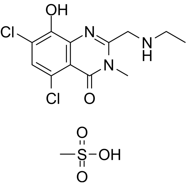
5,7-dichloro-2-(ethylaminomethyl)-8-hydroxy-3-methylquinazolin-4-one;methanesulfonic acid
|
| 别名 |
PBT434 MESYLATE; PBT434 (methanesulfonate); 2387898-69-1; ATH434 MESYLATE; ATH-434 MESYLATE; ATH434; ATH-434; ATH 434; PBT-434 MESYLATE; 826P1VAG3U; EX-A8324;
|
| HS Tariff Code |
2934.99.9001
|
| 存储方式 |
Powder -20°C 3 years 4°C 2 years In solvent -80°C 6 months -20°C 1 month |
| 运输条件 |
Room temperature (This product is stable at ambient temperature for a few days during ordinary shipping and time spent in Customs)
|
| 溶解度 (体外实验) |
May dissolve in DMSO (in most cases), if not, try other solvents such as H2O, Ethanol, or DMF with a minute amount of products to avoid loss of samples
|
|---|---|
| 溶解度 (体内实验) |
注意: 如下所列的是一些常用的体内动物实验溶解配方,主要用于溶解难溶或不溶于水的产品(水溶度<1 mg/mL)。 建议您先取少量样品进行尝试,如该配方可行,再根据实验需求增加样品量。
注射用配方
注射用配方1: DMSO : Tween 80: Saline = 10 : 5 : 85 (如: 100 μL DMSO → 50 μL Tween 80 → 850 μL Saline)(IP/IV/IM/SC等) *生理盐水/Saline的制备:将0.9g氯化钠/NaCl溶解在100 mL ddH ₂ O中,得到澄清溶液。 注射用配方 2: DMSO : PEG300 :Tween 80 : Saline = 10 : 40 : 5 : 45 (如: 100 μL DMSO → 400 μL PEG300 → 50 μL Tween 80 → 450 μL Saline) 注射用配方 3: DMSO : Corn oil = 10 : 90 (如: 100 μL DMSO → 900 μL Corn oil) 示例: 以注射用配方 3 (DMSO : Corn oil = 10 : 90) 为例说明, 如果要配制 1 mL 2.5 mg/mL的工作液, 您可以取 100 μL 25 mg/mL 澄清的 DMSO 储备液,加到 900 μL Corn oil/玉米油中, 混合均匀。 View More
注射用配方 4: DMSO : 20% SBE-β-CD in Saline = 10 : 90 [如:100 μL DMSO → 900 μL (20% SBE-β-CD in Saline)] 口服配方
口服配方 1: 悬浮于0.5% CMC Na (羧甲基纤维素钠) 口服配方 2: 悬浮于0.5% Carboxymethyl cellulose (羧甲基纤维素) 示例: 以口服配方 1 (悬浮于 0.5% CMC Na)为例说明, 如果要配制 100 mL 2.5 mg/mL 的工作液, 您可以先取0.5g CMC Na并将其溶解于100mL ddH2O中,得到0.5%CMC-Na澄清溶液;然后将250 mg待测化合物加到100 mL前述 0.5%CMC Na溶液中,得到悬浮液。 View More
口服配方 3: 溶解于 PEG400 (聚乙二醇400) 请根据您的实验动物和给药方式选择适当的溶解配方/方案: 1、请先配制澄清的储备液(如:用DMSO配置50 或 100 mg/mL母液(储备液)); 2、取适量母液,按从左到右的顺序依次添加助溶剂,澄清后再加入下一助溶剂。以 下列配方为例说明 (注意此配方只用于说明,并不一定代表此产品 的实际溶解配方): 10% DMSO → 40% PEG300 → 5% Tween-80 → 45% ddH2O (或 saline); 假设最终工作液的体积为 1 mL, 浓度为5 mg/mL: 取 100 μL 50 mg/mL 的澄清 DMSO 储备液加到 400 μL PEG300 中,混合均匀/澄清;向上述体系中加入50 μL Tween-80,混合均匀/澄清;然后继续加入450 μL ddH2O (或 saline)定容至 1 mL; 3、溶剂前显示的百分比是指该溶剂在最终溶液/工作液中的体积所占比例; 4、 如产品在配制过程中出现沉淀/析出,可通过加热(≤50℃)或超声的方式助溶; 5、为保证最佳实验结果,工作液请现配现用! 6、如不确定怎么将母液配置成体内动物实验的工作液,请查看说明书或联系我们; 7、 以上所有助溶剂都可在 Invivochem.cn网站购买。 |
| 制备储备液 | 1 mg | 5 mg | 10 mg | |
| 1 mM | 2.5109 mL | 12.5546 mL | 25.1092 mL | |
| 5 mM | 0.5022 mL | 2.5109 mL | 5.0218 mL | |
| 10 mM | 0.2511 mL | 1.2555 mL | 2.5109 mL |
1、根据实验需要选择合适的溶剂配制储备液 (母液):对于大多数产品,InvivoChem推荐用DMSO配置母液 (比如:5、10、20mM或者10、20、50 mg/mL浓度),个别水溶性高的产品可直接溶于水。产品在DMSO 、水或其他溶剂中的具体溶解度详见上”溶解度 (体外)”部分;
2、如果您找不到您想要的溶解度信息,或者很难将产品溶解在溶液中,请联系我们;
3、建议使用下列计算器进行相关计算(摩尔浓度计算器、稀释计算器、分子量计算器、重组计算器等);
4、母液配好之后,将其分装到常规用量,并储存在-20°C或-80°C,尽量减少反复冻融循环。
计算结果:
工作液浓度: mg/mL;
DMSO母液配制方法: mg 药物溶于 μL DMSO溶液(母液浓度 mg/mL)。如该浓度超过该批次药物DMSO溶解度,请首先与我们联系。
体内配方配制方法:取 μL DMSO母液,加入 μL PEG300,混匀澄清后加入μL Tween 80,混匀澄清后加入 μL ddH2O,混匀澄清。
(1) 请确保溶液澄清之后,再加入下一种溶剂 (助溶剂) 。可利用涡旋、超声或水浴加热等方法助溶;
(2) 一定要按顺序加入溶剂 (助溶剂) 。
 MK-7337
 Synucleozid-2.0
 α-synuclein-IN-16
 Squalamine phosphate
InvivoChem的所有产品仅用于作科学研究,不面向患者销售
Copyright 2020 InvivoChem LLC | All Rights Reserved 粤ICP备20063088号-1


 463611831
