| 规格 | 价格 | 库存 | 数量 |
|---|---|---|---|
| 5mg |
|
||
| 10mg |
|
||
| 50mg |
|
||
| Other Sizes |
|

| 靶点 |
Doxorubicin prodrug; Topoisomerase
|
|---|---|
| 体外研究 (In Vitro) |
在这项研究中,研究人员合成了异基因DOX作为生物正交前药(补充图23-26)。阿霉素(DOX)在临床上用于癌症治疗;它通过结合DNA并引发酶介导的链断裂来起作用。通过将伯胺与烯丙基氨基甲酸酯基团封合,如荧光滴定法所示(图4a、b和补充图27,Kalloc-DOX和KDOX分别表示异基因DOX和DOX与ctDNA相互作用的结合常数),异基因DOX(Kalloc-DOX=7.96×10⁴ M-1)对小牛胸腺DNA的结合亲和力远低于DOX(KDOX=9.36×10⁵ M-1)。异基因DOX的这种较弱的DNA结合能力是因为柔红霉素部分正电荷的减少使DNA-药物嵌入复合物的稳定性降低。同时,高效液相色谱(HPLC)分析证明了溶液中Pd-TNS催化的合金DOX的衰变(补充图28)。这种掩蔽策略可以提供活性较低的前药,同时允许通过生物正交催化进行恢复。[1]
|
| 体内研究 (In Vivo) |
同时,通过测量小鼠的体重变化并与DOX治疗的小鼠进行比较,确定了异基因DOX的体内毒性(补充图36)。每三天腹腔注射高达150mg kg-1的异基因DOX,导致体重减轻约7.7%;同时,当剂量超过10mg/kg-1时,DOX会导致严重的体重减轻和死亡,这表明前药在体内毒性较低。根据毒性耐受数据,为了强调前药毒性较小的特点,同时避免过高剂量的意外副作用,在以下抗肿瘤研究中应用了等毒性剂量的异基因DOX(100 mg kg–1)和DOX(5 mg kg–l),差异为20倍。[2]
接下来,将携带B16-F10肿瘤的小鼠随机分为五组,每三天用同种异体DOX/PT-MN组合、DOX、同种异体DOX、PT-MNs或PBS治疗一次。DOX、异基因DOX和PBS通过腹腔注射给药。在注射前药之前,将PT-MNs从表面皮肤插入肿瘤部位并固定一小时,以使针头彻底膨胀。记录生物发光成像和肿瘤体积,以观察肿瘤生长并评估治疗效率(图5a)。如图5a-c所示,与用PBS治疗的对照组一样,单独用同种异体DOX或PT-MNs治疗的组没有观察到延迟的肿瘤生长,表明前药和单独的催化装置都没有抗肿瘤作用。在DOX治疗组中观察到中度抑制结果,但在停止DOX给药后肿瘤生长迅速。相比之下,同种异体DOX/PT-MN组合更好地抑制了肿瘤生长,导致小鼠在四次治疗后肿瘤最小。此外,各组小鼠的体重均未出现明显波动(图5d),表明前药和PT-MN装置的副作用较低。重要的是,TUNEL检测仅在用同种异体DOX/PT-MNs和DOX治疗的组中验证了凋亡信号,这暗示了类似的肿瘤细胞杀伤机制(图5e)。[2] |
| 酶活实验 |
DOX或alloc-DOX与ctDNA的相互作用[2]
进行荧光滴定以研究ctDNA与DOX或alloc-DOX之间的相互作用。溶液在波长λEx=490 nm下激发,在25°C下收集520至720 nm的光谱。滴定是通过将越来越多的ctDNA直接加入含有2µM DOX或alloc-DOX溶液的试管中进行的。在平衡过程中,避免了光照。所有样品均在pH 7.5、含有1mM乙二胺四乙酸二钠的10mM Tris缓冲液中制备。[2] Pd-TNS激活前药[2] 与同种异体RH 110激活过程类似,首先将Pd TNS(1 mg)分散在0.9 ml PBS缓冲液中,然后与100µl在DMSO(1 mM)中制备的alloc-DOX储备溶液混合。在37°C下在黑暗中搅拌混合物。在指定的时间点,取出5µl反应介质并与50µl甲醇混合。在21100g下离心5分钟后,通过HPLC分析上层溶液。HPLC分析在Waters Alliance e2695系统上进行,使用Kromasil C18 HPLC柱(粒径,5μm;孔径300Å;长度与内径之比为250 mm×4.6 mm),流速为1.0 ml min-1,柱温为40°C。流动相由水/乙腈(68:32)和0.1%三氟乙酸组成。通过测量波长λ=254 nm的紫外吸收记录所有色谱图。 |
| 细胞实验 |
细胞毒性试验[2]
将三种细胞系,即B16-F10、4T1和Hep G2细胞,以每孔200µl培养基中10000个细胞的密度接种在96孔板中。孵育24小时后,用不同浓度的DOX和同种异体DOX处理细胞,一式三份。对于alloc-DOX/PT-MN组合组,将带有阵列的PT-MN切成足够大的小块,覆盖一个孔,每个小块约58针浸入培养基中。培养基的体积约为每孔350µl。孵育48小时后,向每个孔中加入20µl 3-(4,5-二甲基噻唑-2-基)-2,5-二苯基溴化四唑(MTT)溶液,并将平板再孵育4小时。之后,通过加入100µl DMSO裂解细胞。在微孔板读数器中,以660nm为参考波长,在570nm处测量吸光度。对于流式细胞术的凋亡研究,根据制造商的说明,使用凋亡检测试剂盒(Invitrogen,Thermal Fisher Scientific)进行Annexin V-APC(别藻蓝蛋白)和SYTOX Green双重染色。对于不同处理后DNA断裂的激光扫描共聚焦显微镜分析,根据制造商的说明进行TUNEL分析;同时,用Hoechst 33342对细胞核进行共定位染色。为了证明DOX在细胞外空间的产生,取出培养基并与甲醇/氯仿(3:2)充分混合。在21100g下离心30分钟后,分离有机相并在氮气下干燥。将残余物重新溶解在乙腈中,并通过HPLC进行分析。为了分析潜在的催化剂泄漏,将上层培养基移至试管中,同时用150µl浓硝酸消化底部的细胞。收集每个孔的所有溶液,与1ml王水混合并进一步消化。在21100g下离心30分钟后,稀释上清液并通过电感耦合等离子体质谱法进行分析。 |
| 动物实验 |
DOX 和 alloc-DOX 的体内毒性[2]
alloc-DOX 和 DOX 溶液的制备方法是将 alloc-DOX 溶解于含 2% Tween 80 的 PBS 缓冲液中。为了确定可用于抗肿瘤研究的剂量,通过每三天腹腔注射一次 alloc-DOX 后测量小鼠的体重变化来确定其毒性。同时测量 DOX 治疗小鼠的体重变化以作比较。体重变化在 10% 以内的可逆性变化被认为是安全的。[2] 生物正交催化介导的体内抗肿瘤活性[2] 将 1 × 10⁶ 个荧光素酶标记的 B16-F10 黑色素瘤细胞皮下注射到 C57BL/6J 小鼠的右侧腹部。当肿瘤体积达到约 50–100 mm³ 时,将小鼠随机分为不同组,分别接受 alloc-DOX (100 mg kg⁻¹)/PT-MN 组合、DOX (5 mg kg⁻¹)、alloc-DOX (100 mg kg⁻¹)、PT-MN 或 PBS 治疗,每三天一次,共四次。药物、前药和 PBS 均采用腹腔注射给药。对于 PT-MN 治疗组,调整 PT-MN 的大小以覆盖肿瘤区域,并在前药给药前一小时将 PT-MN 插入肿瘤部位,并用 Liquivet Rapid 组织粘合剂固定,以使针头充分膨胀。通过腹腔注射荧光素(150 mg kg⁻¹,货号 LUCK-100,Gold Biotechnology)监测肿瘤生长,并使用 IVIS Lumina 成像系统获取图像。使用数字游标卡尺测量肿瘤大小的变化,并根据以下公式计算肿瘤体积(mm³):长径 × 短径² × 0.5。如果小鼠出现健康受损的迹象,或肿瘤体积超过1500 mm³,则使用二氧化碳对小鼠实施安乐死。为了评估潜在的毒性,测量小鼠的体重变化,并在治疗结束时对肿瘤和主要器官(即心脏、肝脏、脾脏、肺和肾脏)进行苏木精-伊红染色组织学分析。对于细胞凋亡分析,根据制造商的方案,使用TUNEL BrdU-Red试剂盒对固定的肿瘤切片进行染色,并用Hoechst 33342对细胞核进行染色。然后使用蔡司LSM 880共聚焦显微镜对染色后的肿瘤切片进行成像。对于接受 DOX-MNs 治疗或瘤内注射 DOX 的组,所给予的 DOX 剂量与接受 alloc-DOX/PT-MNs 治疗的组在每个相应治疗时间点产生的平均 DOX 量相似,该平均 DOX 量是通过收集肿瘤、血液和主要器官中测得的所有 DOX 来确定的。[2] 血浆和组织中 DOX、alloc-DOX 和 Pd 分布的分析[2] 将 DOX 或 alloc-DOX 腹腔注射到荷瘤体积约为 200 mm3 的小鼠体内后,使用 BD 微量毛细管采血器和 BD 微盖封口器通过眼眶后穿刺采集血液。将血液以 1000g 离心 10 分钟分离血浆。取出 50 µl 血浆,并与 150 µl 乙腈混合以提取 DOX 和 alloc-DOX。所有组织(心脏、肝脏、脾脏、肺脏、肾脏和肿瘤)表面用抹布擦干后称重,并在200 µl乙腈中用Cole-Parmer Tissue Tearor 985370-07组织匀浆器充分匀浆。所有样品在室温下避光振荡24小时后,将含有血浆或匀浆组织的试管以21,100 g离心30分钟。然后取出30 µl上清液,与已知量的DOX和内标物alloc-DOX混合,并按照上述相同的HPLC方法进行分析。DOX-MNs治疗组或瘤内注射DOX组也采用类似方法进行分析。为了分析血浆和组织中的钯含量,首先将血浆(50 µl)和匀浆组织溶解于500 µl浓硝酸中1天。然后,向每个样品中加入500 µl王水,并将混合物继续孵育一天。离心(21,100g,30分钟)去除组织碎片后,取出上清液,并用电感耦合等离子体质谱法测定钯含量。 |
| 参考文献 | |
| 其他信息 |
钯催化剂因其高效性和安全性而被广泛应用于有机合成和各种工业领域,但其在生物体内的应用至今仍十分有限。本文表明,纳米包封钯是一种通过体内催化靶向治疗疾病的有效手段。我们筛选了不同的钯化合物,并将双[三(2-呋喃基)膦]二氯化钯(II)包封在生物相容性聚乳酸-羟基乙酸共聚物-聚乙二醇(PLGA-b-PEG)载体中,制备了钯纳米颗粒(Pd-NPs)。利用小鼠癌症模型,我们发现Pd-NPs能够高效地在肿瘤中积累,并激活不同的模型前药。纵向研究证实,与标准阿霉素制剂相比,Pd-NP激活前药能够抑制肿瘤生长,延长荷瘤小鼠的生存期,并降低其毒性。因此,我们在此证明了钯化合物在哺乳动物体内安全有效的催化活性。[1]
过渡金属介导的生物正交催化催生了人工化学的一个新子领域,它与酶促反应互为补充,能够通过非天然过程选择性地标记生物分子或原位合成生物活性物质。然而,生物正交催化在体内的有效应用仍然面临挑战,金属毒性的安全隐患以及催化剂给药程序的复杂性都阻碍了其应用。本文介绍了一种生物正交催化装置,该装置由集成了沉积在二氧化钛纳米片上的钯纳米颗粒的微针阵列贴片组成。该装置坚固耐用且可移除,能够在高水平的生物系统中介导笼状底物局部转化为其活性状态。我们特别指出,这种贴片可以促进皮下肿瘤部位前药的活化,从而恢复其母体药物的抗癌治疗特性。这种原位应用装置可增强局部治疗效果,并消除健康器官或远处组织中的脱靶前药激活和剂量依赖性副作用。[2] |
| 分子式 |
C31H33NO13
|
|---|---|
| 分子量 |
627.59
|
| 精确质量 |
627.195
|
| CAS号 |
561022-65-9
|
| PubChem CID |
171714282
|
| 外观&性状 |
Typically exists as solid at room temperature
|
| LogP |
2.6
|
| tPSA |
218Ų
|
| 氢键供体(HBD)数目 |
6
|
| 氢键受体(HBA)数目 |
13
|
| 可旋转键数目(RBC) |
9
|
| 重原子数目 |
45
|
| 分子复杂度/Complexity |
1160
|
| 定义原子立体中心数目 |
6
|
| SMILES |
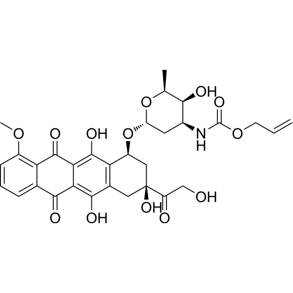
OC1=C2C(C3=CC=CC(OC)=C3C(=O)C2=C(O)C2[C@H](C[C@](O)(C(=O)CO)CC1=2)O[C@]1([H])O[C@@H](C)[C@@H](O)[C@@H](NC(=O)OCC=C)C1)=O
|
| InChi Key |
MYTMRYOJAKUVBH-BPZXBQJLSA-N
|
| InChi Code |
InChI=1S/C31H33NO13/c1-4-8-43-30(40)32-16-9-20(44-13(2)25(16)35)45-18-11-31(41,19(34)12-33)10-15-22(18)29(39)24-23(27(15)37)26(36)14-6-5-7-17(42-3)21(14)28(24)38/h4-7,13,16,18,20,25,33,35,37,39,41H,1,8-12H2,2-3H3,(H,32,40)/t13-,16-,18-,20-,25+,31-/m0/s1
|
| 化学名 |
prop-2-enyl N-[(2S,3S,4S,6R)-3-hydroxy-2-methyl-6-[[(1S,3S)-3,5,12-trihydroxy-3-(2-hydroxyacetyl)-10-methoxy-6,11-dioxo-2,4-dihydro-1H-tetracen-1-yl]oxy]oxan-4-yl]carbamate
|
| 别名 |
N-Alloc doxorubicin
|
| HS Tariff Code |
2934.99.9001
|
| 存储方式 |
Powder -20°C 3 years 4°C 2 years In solvent -80°C 6 months -20°C 1 month |
| 运输条件 |
Room temperature (This product is stable at ambient temperature for a few days during ordinary shipping and time spent in Customs)
|
| 溶解度 (体外实验) |
May dissolve in DMSO (in most cases), if not, try other solvents such as H2O, Ethanol, or DMF with a minute amount of products to avoid loss of samples
|
|---|---|
| 溶解度 (体内实验) |
注意: 如下所列的是一些常用的体内动物实验溶解配方,主要用于溶解难溶或不溶于水的产品(水溶度<1 mg/mL)。 建议您先取少量样品进行尝试,如该配方可行,再根据实验需求增加样品量。
注射用配方
注射用配方1: DMSO : Tween 80: Saline = 10 : 5 : 85 (如: 100 μL DMSO → 50 μL Tween 80 → 850 μL Saline)(IP/IV/IM/SC等) *生理盐水/Saline的制备:将0.9g氯化钠/NaCl溶解在100 mL ddH ₂ O中,得到澄清溶液。 注射用配方 2: DMSO : PEG300 :Tween 80 : Saline = 10 : 40 : 5 : 45 (如: 100 μL DMSO → 400 μL PEG300 → 50 μL Tween 80 → 450 μL Saline) 注射用配方 3: DMSO : Corn oil = 10 : 90 (如: 100 μL DMSO → 900 μL Corn oil) 示例: 以注射用配方 3 (DMSO : Corn oil = 10 : 90) 为例说明, 如果要配制 1 mL 2.5 mg/mL的工作液, 您可以取 100 μL 25 mg/mL 澄清的 DMSO 储备液,加到 900 μL Corn oil/玉米油中, 混合均匀。 View More
注射用配方 4: DMSO : 20% SBE-β-CD in Saline = 10 : 90 [如:100 μL DMSO → 900 μL (20% SBE-β-CD in Saline)] 口服配方
口服配方 1: 悬浮于0.5% CMC Na (羧甲基纤维素钠) 口服配方 2: 悬浮于0.5% Carboxymethyl cellulose (羧甲基纤维素) 示例: 以口服配方 1 (悬浮于 0.5% CMC Na)为例说明, 如果要配制 100 mL 2.5 mg/mL 的工作液, 您可以先取0.5g CMC Na并将其溶解于100mL ddH2O中,得到0.5%CMC-Na澄清溶液;然后将250 mg待测化合物加到100 mL前述 0.5%CMC Na溶液中,得到悬浮液。 View More
口服配方 3: 溶解于 PEG400 (聚乙二醇400) 请根据您的实验动物和给药方式选择适当的溶解配方/方案: 1、请先配制澄清的储备液(如:用DMSO配置50 或 100 mg/mL母液(储备液)); 2、取适量母液,按从左到右的顺序依次添加助溶剂,澄清后再加入下一助溶剂。以 下列配方为例说明 (注意此配方只用于说明,并不一定代表此产品 的实际溶解配方): 10% DMSO → 40% PEG300 → 5% Tween-80 → 45% ddH2O (或 saline); 假设最终工作液的体积为 1 mL, 浓度为5 mg/mL: 取 100 μL 50 mg/mL 的澄清 DMSO 储备液加到 400 μL PEG300 中,混合均匀/澄清;向上述体系中加入50 μL Tween-80,混合均匀/澄清;然后继续加入450 μL ddH2O (或 saline)定容至 1 mL; 3、溶剂前显示的百分比是指该溶剂在最终溶液/工作液中的体积所占比例; 4、 如产品在配制过程中出现沉淀/析出,可通过加热(≤50℃)或超声的方式助溶; 5、为保证最佳实验结果,工作液请现配现用! 6、如不确定怎么将母液配置成体内动物实验的工作液,请查看说明书或联系我们; 7、 以上所有助溶剂都可在 Invivochem.cn网站购买。 |
| 制备储备液 | 1 mg | 5 mg | 10 mg | |
| 1 mM | 1.5934 mL | 7.9670 mL | 15.9340 mL | |
| 5 mM | 0.3187 mL | 1.5934 mL | 3.1868 mL | |
| 10 mM | 0.1593 mL | 0.7967 mL | 1.5934 mL |
1、根据实验需要选择合适的溶剂配制储备液 (母液):对于大多数产品,InvivoChem推荐用DMSO配置母液 (比如:5、10、20mM或者10、20、50 mg/mL浓度),个别水溶性高的产品可直接溶于水。产品在DMSO 、水或其他溶剂中的具体溶解度详见上”溶解度 (体外)”部分;
2、如果您找不到您想要的溶解度信息,或者很难将产品溶解在溶液中,请联系我们;
3、建议使用下列计算器进行相关计算(摩尔浓度计算器、稀释计算器、分子量计算器、重组计算器等);
4、母液配好之后,将其分装到常规用量,并储存在-20°C或-80°C,尽量减少反复冻融循环。
计算结果:
工作液浓度: mg/mL;
DMSO母液配制方法: mg 药物溶于 μL DMSO溶液(母液浓度 mg/mL)。如该浓度超过该批次药物DMSO溶解度,请首先与我们联系。
体内配方配制方法:取 μL DMSO母液,加入 μL PEG300,混匀澄清后加入μL Tween 80,混匀澄清后加入 μL ddH2O,混匀澄清。
(1) 请确保溶液澄清之后,再加入下一种溶剂 (助溶剂) 。可利用涡旋、超声或水浴加热等方法助溶;
(2) 一定要按顺序加入溶剂 (助溶剂) 。
InvivoChem的所有产品仅用于作科学研究,不面向患者销售
Copyright 2020 InvivoChem LLC | All Rights Reserved 粤ICP备20063088号-1


 463611831
