| 规格 | 价格 | |
|---|---|---|
| 500mg | ||
| 1g | ||
| Other Sizes |
| 靶点 |
Endogenous Metabolite
|
|---|---|
| 体外研究 (In Vitro) |
原卟啉 IX 二钠 (3.6 nmol/g 组织) 在光照下可诱导正常大鼠结肠粘膜坏死[4]。原卟啉 IX 二钠 (6.6 nmol/g 肿瘤) 可延缓大鼠肿瘤照射后的肿瘤生长[4]。原卟啉 IX 二钠 (0-25 μM, 2 小时) 可迅速导致 Hela 细胞核 G4 水平升高[5]。
Olivo等人报告称,经尿道切除更高级别和更高阶段的膀胱肿瘤后,ALA滴注后可产生和积累更高水平的原卟啉IX。他们的发现支持我们的结果。Miyake及其同事表明,癌症尿路上皮中铁螯合酶的低表达或分子缺陷与细胞内原卟啉IX的积累有关。Hagiya及其同事报告称,肽转运蛋白1和人ATP结合盒转运蛋白2的表达水平在ALA诱导的原卟啉IX积累中起着关键作用。Ogino等人报道了铁螯合酶抑制剂和人ATP结合盒转运蛋白2抑制剂刺激ALA诱导的T24细胞中原卟啉IX的积累。多个因素似乎与高级别肿瘤中原卟啉IX积累增加的机制有关。哪个因素起主导作用是未来研究的课题。[1] 哺乳动物卟啉是由δ-氨基酮戊酸(ALA)通过几个连续的酶促反应生成原卟啉IX(PpIX),后者进一步与铁阳离子络合,产生血红素或其衍生物氯化血红素[5]。 |
| 体内研究 (In Vivo) |
给予氨基乙酰丙酸(300 mg/kg,单次静脉注射)三小时后,在肝脏和肠道中发现原卟啉 IX 二钠的浓度最高(约 6.3 nmol/g 组织),其次是主动脉(4.3 nmol/g 组织)和食道(2.1 nmol/g 组织)[4]。
有限的穿透深度显著限制了局部使用δ(5)-氨基酮戊酸(ALA)对结节性基底细胞癌(BCC)的光动力治疗。为了证明口服ALA诱导BCC内源性原卟啉IX(PpIX)产生的安全性和有效性,13名BCC患者按照剂量递增方案摄入了ALA。所有剂量范围(10、20或40mg/kg单次剂量)均导致人体皮肤和基底细胞癌中形成Protoporphyrin IX/PpIX,可通过体内荧光分光光度法测量。摄入ALA后1至3小时,肿瘤中的PpIX荧光在正常邻近皮肤之前达到峰值。离体标本的总荧光成像显示,仅在40mg/kg剂量下,肿瘤中的PpIX荧光比正常皮肤更大。荧光显微镜证实了这一发现,仅在给予40mg/kg的ALA后,所有BCC亚型都显示出明显的全层PpIX荧光。副作用是剂量依赖性和自限性的。在20和40mg/kg剂量下,光敏性持续时间小于24小时,恶心与皮肤PpIX荧光峰值一致。40mg/kg ALA后,血清肝酶水平在24小时内升至最高值,然后在1-3周内消失。两名患者出现短暂性胆红素尿。[3] 在这项研究中,研究了5-氨基酮戊酸(ALA)在大鼠体内的生物分布和原卟啉IX(PpIX)的积累。两组21只WAG/Rij大鼠口服或静脉注射200mg/kg ALA。六只老鼠作为对照。在ALA给药后1、2、3、4、6、12和24小时,测量18种组织和液体中的ALA和卟啉浓度。测量肝酶和肾功能测试以确定ALA毒性。在两组中,肾脏、膀胱和尿液中的ALA浓度最高。口服给药后,十二指肠吸出物和空肠中也发现了高浓度。两个治疗组均出现轻度、短暂的肌酐升高。卟啉,尤其是PpIX,主要在十二指肠吸出物、空肠、肝脏和肾脏中积累(>10 nmol/g组织),在食管、胃、结肠、脾脏、膀胱、心脏、肺和神经中积累较少(2-10 nmol/g组),在血浆、肌肉、脂肪、皮肤和脑中仅少量积累(<2 nmol/g)。卟啉的原位合成而不是肠肝循环有助于Protoporphyrin IX/PpIX的积累。共聚焦激光扫描显微镜显示上皮层中的选择性卟啉荧光。口服和静脉注射ALA后,卟啉的峰值水平和总产量相等。 总之:200 mg/kg ALA给药后1至6小时,除肌肉、脂肪、皮肤和脑外,所有组织中都会积累光敏浓度的Protoporphyrin IX/PpIX。了解时间-浓度关系应有助于选择ALA光动力疗法的剂量、给药途径和时间[4]。 |
| 细胞实验 |
原卟啉IX在膀胱癌症脱落细胞中的光动力学检测[1]
在适当的设置下(激发波长405 nm,发射波长550-700 nm,增益160),使用分光光度计测试每个腔室中ALA处理和ALA未处理的尿液沉积物的原卟啉IX荧光。当样品强度超出范围时,调整分光光度计的增益。用来自膀胱癌症患者的ALA处理的样品的光谱在635nm处显示出峰值,而ALA对照样品的光谱没有显示出这样的峰值(图2a)。对照组患者的样本中未检测到峰值(图2b)。 原卟啉IX在脱落的膀胱癌症细胞中的评价[1] 为了评估635nm处的峰值,计算了ALA处理和ALA未处理样品的强度差异。该分析的曲线下面积、敏感性和特异性分别为0.68、60%和65%。如上所述,当样品的强度超出测量范围时,调整增益。因此,将计算出的差异除以635nm处未经ALA处理的样品的强度,以调整不同增益设置的数据。调整后的曲线下面积、敏感性和特异性分别为0.74、73%和63%。由于ALA处理和未处理的样品之间存在差异,一些在635nm处没有峰值的对照病例被诊断为阳性;在这些样品中,在550nm至700nm的范围内检测到635nm处的差异(图3b)。为了解决这个问题,从ALA处理和ALA未处理样品在635 nm处的强度差中减去ALA处理与ALA未治疗样品在600 nm下的强度差(图3a和b)。之后,将调整后的差值除以ALA未处理样品在635nm处的强度。这种调整后的变化率用于诊断膀胱癌症(ALA诱导荧光细胞学方法)。 |
| 动物实验 |
卟啉分析[4]
该分析按照Chisolm和Brown的方法进行,并作如下修改:将组织悬浮于无菌水中(1:10 wt./vol.),并在组织研磨器中匀浆[2 11]。取100 μL匀浆液,加入700 μL 2 mmol/L HCl和800 μL乙酸乙酯/冰醋酸(3:1)。在1800 g下离心10分钟后,去除含有部分蛋白质的乙酸乙酯相,再次在1800 g下离心5分钟后,使用LS 5B荧光分光光度计,在激发波长410 nm和发射波长650 nm下,用红色敏感光电倍增管测量HCl相。尽管卟啉通常通过 601 nm 处的发射光谱进行测定,但在所述条件下,原卟啉 IX (PpIX) 在 650 nm 处也具有特异性发射,其强度约为 601 nm 处发射强度的 80%,并且可以使用红敏光电倍增管轻松检测。根据我们的经验,十二指肠抽吸物中的胆汁酸(以及其他组织中的胆汁酸含量较少)含有在较低波长处具有高发射强度的荧光团,这会干扰 601 nm 处卟啉的定量分析。如上所述的直接提取法中,650 nm 处的发射强度与通过有限次数的 HPLC 卟啉分离测定的原卟啉 IX (PpIX) 的定量结果成正比。使用紫外光谱法和摩尔消光系数 (δd07 = 0.275 L mol⁻¹ cm⁻¹) 对卟啉标准品的实际浓度进行了单独分析。通过向样品中添加标准品原卟啉IX/PpIX来检测卟啉的回收率。回收率达到90%至100%。 |
| 药代性质 (ADME/PK) |
本研究聚焦于三种临床应用的光敏剂(PpIX 和 PF)以及用于体内实验的光敏剂(PPa)。我们的团队提出将 PPa 与叶酸偶联,用于光动力疗法(PDT)治疗卵巢转移瘤(专利号 WO/2019/016397)。
通过分析这三种光敏剂在不同条件下的光物理性质,我们发现每种光敏剂都具有独特性,其反应会根据化学结构和浓度而发生显著变化。 介质极性的变化对 PF 的紫外-可见吸收光谱影响不大,但对 PpIX 和 PPa 的影响则非常显著。文献中通常认为 PpIX 在体外或体内应在 630 nm 处激发。该激发波长是基于其在乙醇中的吸收光谱确定的。在更接近生理介质的水性介质 FBS 和 PBS 中,其 QI 带位于 641 nm 处。 根据光敏剂在细胞中的定位,局部黏度可能存在显著差异。我们还观察到,改变溶剂粘度对PpIX和PF中QI的最大吸收波长影响不大,但对PPa而言,其吸收波长发生了10 nm的蓝移(从678 nm到668 nm)。 温度变化对PpIX和PF的紫外-可见吸收光谱影响甚微,但在10至40 °C范围内,对PPa的紫外-可见吸收光谱影响显著。 最后,改变pH值也使PPa的QI吸收带发生了25 nm的蓝移(从704 nm到679 nm)。 或许最有趣的结果是不同溶剂中ΦΔ值的测定。根据溶剂的不同,ΦΔ值也截然不同。在甲苯中,我们未检测到任何1O2,而PpIX和PPa的ΦΔ值分别为0.68和0.49,表现相当不错。在乙醇中,PpIX、PPa 和 PF 的 ΦΔ 值分别为 0.92、0.53 和 0.80。如果改用 D2O,则无法检测到 PpIX 或 PPa 的 1O2,而 PF 的 ΦΔ 值则降至 0.15。此外,在实际应用中,光敏剂理想情况下处于细胞环境中。蛋白质、脂质和其他生物分子的存在也会影响光敏剂的光物理性质。这就引出了一个问题:在进行体外研究时,应该采用何种类型的实验以及在溶液中使用何种溶剂。[2] |
| 毒性/毒理 (Toxicokinetics/TK) |
71484 大鼠静脉注射 LD50 240 mg/kg,《日本药物》,6(729),1982
71484 小鼠口服 LD50 >5 gm/kg,《日本药物》,-(1192),1995 71484 小鼠腹腔注射 LD50 1029 mg/kg,《日本药物》,6(729),1982 71484 小鼠皮下注射 LD50 1147 mg/kg,《日本药物》,6(729),1982 71484 小鼠静脉注射 LD50 484 mg/kg,《日本药物》,6(729),1982 |
| 参考文献 |
|
| 其他信息 |
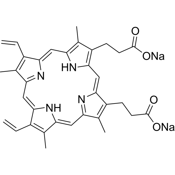
原卟啉是一种环状四吡咯,由卟啉环组成,其3、8、13和17位分别连接四个甲基,7和12位分别连接两个乙烯基,2和18位分别连接两个2-羧乙基。它是原卟啉类化合物的母体。它可用作光敏剂、代谢物、大肠杆菌代谢物和鼠代谢物。它是原卟啉酸和原卟啉(2-)的共轭酸。
原卟啉IX是存在于大肠杆菌(K12菌株、MG1655菌株)中或由其产生的代谢产物。 已有报道称,在智人和鸫中也发现了原卟啉IX,并有相关数据。 原卟啉IX是一种四吡咯,含有4个甲基、2个丙酸和2个乙烯基侧链,是血红素、细胞色素c和叶绿素的代谢前体。原卟啉IX是由原卟啉原氧化酶催化原卟啉原亚甲基桥氧化生成的。 原卟啉IX是酿酒酵母(Saccharomyces cerevisiae)中发现或产生的代谢产物。 背景:我们评估了通过对体外用5-氨基乙酰丙酸(ALA)处理后排出的尿液样本进行分光光度分析,从而进行膀胱癌光动力诊断的可行性。 方法:我们招募了61例经经尿道膀胱肿瘤切除术后组织学确诊的膀胱癌患者作为膀胱癌组,并招募了50例无尿路上皮癌病史或癌症相关表现的门诊患者作为对照组。一半的排出尿液样本与ALA孵育(ALA处理组),另一半不进行处理(ALA未处理组)。为了检测细胞内原卟啉IX水平,我们使用分光光度计测量了样品在405 nm激发波长下的吸光度。计算了ALA处理组和未处理组样品在635 nm处的吸光度差值。 结果:膀胱癌组的吸光度差值显著高于对照组(p < 0.001)。高级别肿瘤患者的吸光度差值也显著高于低级别肿瘤患者(p = 0.004),浸润性膀胱癌患者的吸光度差值也显著高于非浸润性膀胱癌患者(p = 0.007)。曲线下面积为0.84。该方法的灵敏度和特异性分别为82%和80%。 结论:我们证明,在膀胱癌患者中,使用分光光度计可以定量检测ALA处理后尿液细胞内的原卟啉IX水平。因此,这种癌症检测系统具有临床应用潜力。[1] 光动力疗法(PDT)是一种治疗恶性或病变组织的创新方法。PDT 的疗效取决于光剂量、氧气供应和光敏剂(PS)的性质。根据介质的不同,PS 的光物理性质会发生变化,导致荧光发射增加或减少,并产生活性氧(ROS),尤其是单线态氧(1O2)。本研究采用紫外-可见吸收光谱、荧光发射光谱、单线态氧发射光谱和时间分辨荧光光谱,研究了溶剂极性、粘度、浓度、温度和pH值对原卟啉IX、焦脱镁叶绿酸a和Photofrin®光物理性质的影响。[2] 背景:G-四链体(G4)是一种独特的非经典核酸二级结构,被认为能够与转录因子和染色质重塑因子发生物理相互作用,从而调控细胞类型特异性转录组并塑造染色质结构。 结果:基于G4与天然卟啉的直接相互作用,我们建立了全基因组方法来分析铁配位卟啉血红素在染色质中的结合位点。血红素促进全基因组范围内的G4形成,抑制转录起始,并改变染色质结构,包括降低启动子区域的H3K27ac和H3K4me3修饰水平。有趣的是,G4状态并不参与经典的血红素-BACH1-NRF2介导的增强子激活过程,这揭示了一种前所未有的G4依赖性转录代谢调控机制。此外,血红素处理可诱导肝细胞中特定的基因表达谱,强调卟啉在体内代谢调控基因转录的潜力。 结论:这些研究表明,G4在细胞内作为天然卟啉代谢物的传感器发挥作用,揭示了一种G4依赖性基因转录和染色质结构代谢调控机制,这将加深我们对G4生物学以及细胞代谢物对基因调控贡献的理解。[5] |
| 分子式 |
C34H32N4NA2O4
|
|---|---|
| 分子量 |
606.62
|
| 精确质量 |
606.22
|
| 元素分析 |
C, 67.32; H, 5.32; N, 9.24; Na, 7.58; O, 10.55
|
| CAS号 |
50865-01-5
|
| 相关CAS号 |
553-12-8
|
| PubChem CID |
71484
|
| 外观&性状 |
Typically exists as solids at room temperature
|
| 沸点 |
1128.9ºC at 760 mmHg
|
| 闪点 |
636.6ºC
|
| LogP |
1.371
|
| tPSA |
136.56
|
| 氢键供体(HBD)数目 |
2
|
| 氢键受体(HBA)数目 |
6
|
| 可旋转键数目(RBC) |
6
|
| 重原子数目 |
44
|
| 分子复杂度/Complexity |
995
|
| 定义原子立体中心数目 |
0
|
| SMILES |
C=CC1=C(C)C2=NC1=CC3=C(C)C(=C(C=C4C(=C(CCC(=O)[O-])C(=N4)C=C5C(=C(C)C(=C2)N5)CCC(=O)[O-])C)N3)C=C.[Na+].[Na+]
|
| InChi Key |
GPRXGEKBQVXWAQ-UHFFFAOYSA-L
|
| InChi Code |
InChI=1S/C34H34N4O4.2Na/c1-7-21-17(3)25-13-26-19(5)23(9-11-33(39)40)31(37-26)16-32-24(10-12-34(41)42)20(6)28(38-32)15-30-22(8-2)18(4)27(36-30)14-29(21)35-25;;/h7-8,13-16,35-36H,1-2,9-12H2,3-6H3,(H,39,40)(H,41,42);;/q;2*+1/p-2
|
| 化学名 |
disodium;3-[18-(2-carboxylatoethyl)-8,13-bis(ethenyl)-3,7,12,17-tetramethyl-22,23-dihydroporphyrin-2-yl]propanoate
|
| 别名 |
Protoporphyrin IX (disodium); 50865-01-5; protoporphyrin disodium; Protoporphyrin IX disodium salt; Disodium protoporphyrin IX; Palepron; Protoporphyrin, disodium salt; Protoporphyrin disodium [JAN]; Dojin PM;
|
| HS Tariff Code |
2934.99.9001
|
| 存储方式 |
Powder -20°C 3 years 4°C 2 years In solvent -80°C 6 months -20°C 1 month |
| 运输条件 |
Room temperature (This product is stable at ambient temperature for a few days during ordinary shipping and time spent in Customs)
|
| 溶解度 (体外实验) |
May dissolve in DMSO (in most cases), if not, try other solvents such as H2O, Ethanol, or DMF with a minute amount of products to avoid loss of samples
|
|---|---|
| 溶解度 (体内实验) |
注意: 如下所列的是一些常用的体内动物实验溶解配方,主要用于溶解难溶或不溶于水的产品(水溶度<1 mg/mL)。 建议您先取少量样品进行尝试,如该配方可行,再根据实验需求增加样品量。
注射用配方
注射用配方1: DMSO : Tween 80: Saline = 10 : 5 : 85 (如: 100 μL DMSO → 50 μL Tween 80 → 850 μL Saline)(IP/IV/IM/SC等) *生理盐水/Saline的制备:将0.9g氯化钠/NaCl溶解在100 mL ddH ₂ O中,得到澄清溶液。 注射用配方 2: DMSO : PEG300 :Tween 80 : Saline = 10 : 40 : 5 : 45 (如: 100 μL DMSO → 400 μL PEG300 → 50 μL Tween 80 → 450 μL Saline) 注射用配方 3: DMSO : Corn oil = 10 : 90 (如: 100 μL DMSO → 900 μL Corn oil) 示例: 以注射用配方 3 (DMSO : Corn oil = 10 : 90) 为例说明, 如果要配制 1 mL 2.5 mg/mL的工作液, 您可以取 100 μL 25 mg/mL 澄清的 DMSO 储备液,加到 900 μL Corn oil/玉米油中, 混合均匀。 View More
注射用配方 4: DMSO : 20% SBE-β-CD in Saline = 10 : 90 [如:100 μL DMSO → 900 μL (20% SBE-β-CD in Saline)] 口服配方
口服配方 1: 悬浮于0.5% CMC Na (羧甲基纤维素钠) 口服配方 2: 悬浮于0.5% Carboxymethyl cellulose (羧甲基纤维素) 示例: 以口服配方 1 (悬浮于 0.5% CMC Na)为例说明, 如果要配制 100 mL 2.5 mg/mL 的工作液, 您可以先取0.5g CMC Na并将其溶解于100mL ddH2O中,得到0.5%CMC-Na澄清溶液;然后将250 mg待测化合物加到100 mL前述 0.5%CMC Na溶液中,得到悬浮液。 View More
口服配方 3: 溶解于 PEG400 (聚乙二醇400) 请根据您的实验动物和给药方式选择适当的溶解配方/方案: 1、请先配制澄清的储备液(如:用DMSO配置50 或 100 mg/mL母液(储备液)); 2、取适量母液,按从左到右的顺序依次添加助溶剂,澄清后再加入下一助溶剂。以 下列配方为例说明 (注意此配方只用于说明,并不一定代表此产品 的实际溶解配方): 10% DMSO → 40% PEG300 → 5% Tween-80 → 45% ddH2O (或 saline); 假设最终工作液的体积为 1 mL, 浓度为5 mg/mL: 取 100 μL 50 mg/mL 的澄清 DMSO 储备液加到 400 μL PEG300 中,混合均匀/澄清;向上述体系中加入50 μL Tween-80,混合均匀/澄清;然后继续加入450 μL ddH2O (或 saline)定容至 1 mL; 3、溶剂前显示的百分比是指该溶剂在最终溶液/工作液中的体积所占比例; 4、 如产品在配制过程中出现沉淀/析出,可通过加热(≤50℃)或超声的方式助溶; 5、为保证最佳实验结果,工作液请现配现用! 6、如不确定怎么将母液配置成体内动物实验的工作液,请查看说明书或联系我们; 7、 以上所有助溶剂都可在 Invivochem.cn网站购买。 |
| 制备储备液 | 1 mg | 5 mg | 10 mg | |
| 1 mM | 1.6485 mL | 8.2424 mL | 16.4848 mL | |
| 5 mM | 0.3297 mL | 1.6485 mL | 3.2970 mL | |
| 10 mM | 0.1648 mL | 0.8242 mL | 1.6485 mL |
1、根据实验需要选择合适的溶剂配制储备液 (母液):对于大多数产品,InvivoChem推荐用DMSO配置母液 (比如:5、10、20mM或者10、20、50 mg/mL浓度),个别水溶性高的产品可直接溶于水。产品在DMSO 、水或其他溶剂中的具体溶解度详见上”溶解度 (体外)”部分;
2、如果您找不到您想要的溶解度信息,或者很难将产品溶解在溶液中,请联系我们;
3、建议使用下列计算器进行相关计算(摩尔浓度计算器、稀释计算器、分子量计算器、重组计算器等);
4、母液配好之后,将其分装到常规用量,并储存在-20°C或-80°C,尽量减少反复冻融循环。
计算结果:
工作液浓度: mg/mL;
DMSO母液配制方法: mg 药物溶于 μL DMSO溶液(母液浓度 mg/mL)。如该浓度超过该批次药物DMSO溶解度,请首先与我们联系。
体内配方配制方法:取 μL DMSO母液,加入 μL PEG300,混匀澄清后加入μL Tween 80,混匀澄清后加入 μL ddH2O,混匀澄清。
(1) 请确保溶液澄清之后,再加入下一种溶剂 (助溶剂) 。可利用涡旋、超声或水浴加热等方法助溶;
(2) 一定要按顺序加入溶剂 (助溶剂) 。
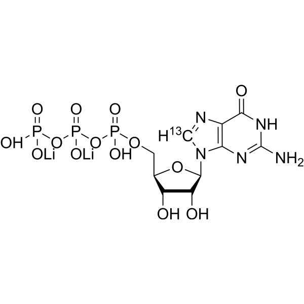 Guanosine triphosphate-13C (dilithium)
 γ-Glutamyltranspeptidase, Equine
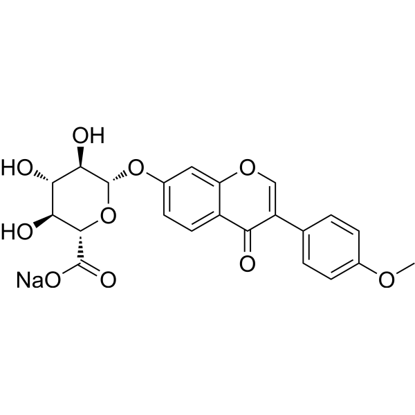 Formononetin 7-O-β-D-glucuronide sodium
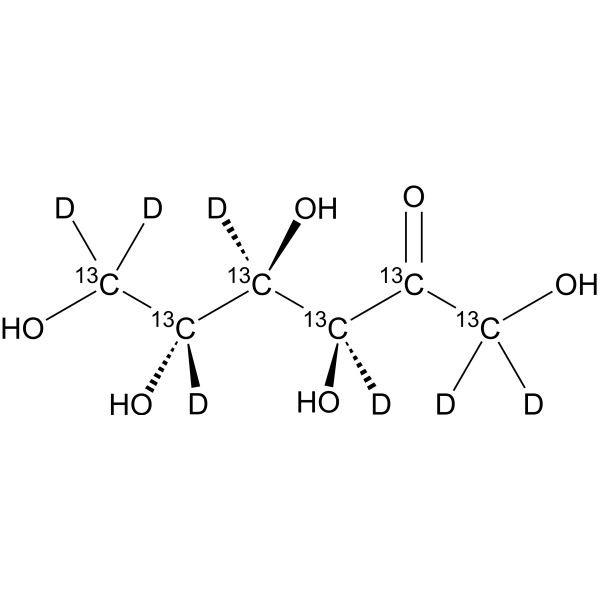 D-Fructose-13C6,d7
InvivoChem的所有产品仅用于作科学研究,不面向患者销售
Copyright 2020 InvivoChem LLC | All Rights Reserved 粤ICP备20063088号-1


 463611831
