| 规格 | 价格 | |
|---|---|---|
| 500mg | ||
| 1g | ||
| Other Sizes |

| 靶点 |
AMPK; Autophagy; Mitophagy
|
|---|---|
| 体外研究 (In Vitro) |
二甲双胍抑制 Bcl-2 和 Bcl-xl,上调 BAX 激活,促进 BIM、BAD 和 PUMA,并诱导细胞色素 c 从线粒体释放到细胞质中,直接诱导 caspase-9 介导的线粒体凋亡。 [4]
二甲双胍激活肝细胞中的AMPK;结果,乙酰辅酶a羧化酶(ACC)活性降低,脂肪酸氧化被诱导,脂肪生成酶的表达被抑制。二甲双胍或腺苷类似物激活AMPK会抑制SREBP-1的表达,SREBP-1是一种关键的脂肪生成转录因子。在二甲双胍治疗的大鼠中,SREBP-1(和其他脂肪生成)mRNA和蛋白质的肝脏表达降低;AMPK靶标ACC的活性也降低了。使用一种新型AMPK抑制剂,我们发现二甲双胍对肝细胞葡萄糖产生的抑制作用需要AMPK激活。在离体大鼠骨骼肌中,二甲双胍刺激葡萄糖摄取,同时AMPK激活。AMPK的激活为该药物的多效有益作用提供了统一的解释;这些结果还表明,调节AMPK的替代方法可用于治疗代谢性疾病。[1] 二甲双胍被广泛推荐用于治疗2型糖尿病,通过5'-AMP活化蛋白激酶(AMPK)发挥其多效性作用;然而,它对线粒体吞噬的影响仍然难以捉摸。最近的证据表明,外周血单核细胞(PBMCs)表达胰岛素受体和人类有机阳离子转运蛋白,它们被广泛用作检测2型糖尿病线粒体功能的替代品。二甲双胍治疗增加了酸性囊泡和丝裂吞噬体的形成,上调了丝裂吞噬标记物,并增强了丝裂噬通量,如LC3-II表达增加和p62蛋白水平降低所示。此外,用化合物C(一种AMPK抑制剂)预处理显著降低了二甲双胍处理细胞中自噬标志物的表达,表明二甲双胍通过AMPK途径诱导自噬。总之,二甲双胍诱导的线粒体吞噬可能通过恢复正常的线粒体表型来改善细胞功能,包括β细胞,这可能对2型糖尿病和其他线粒体相关疾病患者有益。此外,PBMCs可以用作鉴定线粒体疾病的新型诊断生物标志物。[2] 在这项研究中,我们首次揭示了二甲双胍治疗导致人肺癌细胞系A549和NCI-H1299细胞凋亡增加,并以剂量和时间依赖的方式显著抑制细胞增殖,从裸鼠A549肿瘤异种移植物获得的数据进一步证明了这一点。我们还发现,二甲双胍治疗可以激活AMP活化蛋白激酶、JNK/p38 MAPK信号通路和半胱氨酸天冬氨酸蛋白酶,并上调生长停滞和DNA损伤诱导基因153(GADD153)的表达。阻断JNK/p38 MAPK通路或敲除GADD153基因都会消除二甲双胍的凋亡诱导作用。总之,我们的数据表明,二甲双胍通过激活JNK/p38 MAPK途径和GADD153抑制肺癌细胞的生长并诱导细胞凋亡。[3] 这项研究表明,二甲双胍通过诱导凋亡减少了活化HSC的数量,但不影响肝细胞的数量。二甲双胍上调BAX激活,促进BIM、BAD和PUMA;下调Bcl-2和Bcl-xl,但不影响Mcl-1。此外,二甲双胍诱导细胞色素c从线粒体释放到细胞质中,直接触发胱天蛋白酶-9介导的线粒体凋亡。线粒体膜电位(ΔΨm)的下降和线粒体中超氧化物的沉积加速了线粒体膜完整性的破坏。此外,我们验证了二甲双胍在非酒精性脂肪性肝炎(NASH)相关肝纤维化小鼠模型中的治疗效果,其中二甲双胍改善了肝功能、NASH病变和纤维化。总之,本研究表明,二甲双胍通过诱导HSC凋亡对NASH衍生的肝纤维化具有显著的治疗价值,但不影响肝细胞的增殖[4]。 |
| 体内研究 (In Vivo) |
二甲双胍在非酒精性脂肪性肝炎 (NASH) 相关肝纤维化小鼠模型中表现出治疗作用,从而改善肝功能、NASH 病变和纤维化。 [4]
评估二甲双胍的体内效应。[1] 为了评估上述二甲双胍的选定效应是否也在体内发生,对SD大鼠进行了研究(表1)。大鼠口服二甲双胍或赋形剂(H2O)5天。大鼠饥饿20小时,然后在最后一次给药前重新喂食2小时。最后一次给药后4小时,采集组织和血液样本进行分析(见方法)。在饥饿期间,应该很少有脂质合成。重新进食后,应显著诱导肝脏脂质合成。在再喂养条件下检查了二甲双胍的作用。除了血浆胰岛素和甘油三酯的适度降低外,β-羟基丁酸酯也有小幅但显著的增加,这表明二甲双胍治疗的大鼠肝脏脂肪酸氧化是诱导的。此外,二甲双胍治疗显著降低了SREBP-1、FAS和S14的mRNA在肝脏的表达,这与细胞中记录的效果一致(表1)。 使用抗SREBP-1抗体检测大鼠肝细胞核提取物中的成熟SREBP-1蛋白(图5b)。正如预期的那样,在饥饿动物的肝核提取物中没有检测到SREBP-1成熟形式的蛋白质。在再喂养的动物中,成熟的SREBP-1蛋白的积累与这种情况下脂质合成的增加相一致。用二甲双胍治疗可以防止这种积聚。使用AICAR(500mg/kg/天)治疗后再喂养大鼠的肝核提取物获得的其他结果也表明,SREBP-1成熟形式蛋白的存在被消除。 在离体肝脏中测量AMPK活化是困难的,因为已知短暂的缺氧会导致酶的显著活化。因此,我们使用二甲双胍治疗大鼠的肝组织来确定在几种测试的柠檬酸盐浓度下ACC活性显著降低(图6)。柠檬酸盐浓度为1 mM时ACC活性降低最大(从54.6±11.8降至35.6±7.7 nmol/mg/min;P<0.01)。这些结果与二甲双胍在体内产生AMPK激活和ACC失活的结果一致。 二甲双胍缓解了非酒精性脂肪性肝炎(NASH)相关纤维化小鼠模型的肝脏病变[4] 建立了NASH相关纤维化小鼠模型(图7a),以探索二甲双胍是否能在体内疾病状态下保护肝脏。每隔一周和每三周分别测量每只小鼠的体重和生化指标(ALT和AST)。结果显示,与MCS饮食组或AIN93饮食组相比,HFMCD饮食组的体重没有显著增加(图S2a)。此外,与MCS饮食组或AIN93组相比,在HFMCD饮食治疗组中,二甲双胍降低了AST和ALT水平以及肝脏指数(图7b、图S2b)。总的来说,二甲双胍改善了NASH相关纤维化小鼠模型的肝功能。 从NASH纤维化模型中获得的石蜡包埋的肝组织样本用苏木精和伊红染色,以观察二甲双胍对肝损伤的影响。HFMCD组新鲜肝组织颜色偏深(图S2c)。结果显示,AIN93或MCS组的肝脏没有明显病变,也没有受到二甲双胍的影响(图S2d)。然而,在HFMCD预防和治疗组中,使用二甲双胍后,脂肪变性面积和炎性细胞数量均减少(图7c),表明NASH病变的严重程度减轻。此外,通过V-G染色观察纤维化程度。结果显示,HFMCD治疗组的胶原蛋白积累远少于HFMCD饮食盐水组(图7d)。Masson染色也观察到了相同的结果(图S2e)。此外,免疫组织化学染色显示,二甲双胍治疗组HSC切割半胱氨酸天冬氨酸蛋白酶-3的阳性率为38.7±5.2%,高于生理盐水组(7.69±0.61%)、空白对照组(0.58±0.03%)和二甲双胍治疗组(1.82±0.05%),表明二甲双胍诱导HSC凋亡(图7e,表3)。因此,二甲双胍延缓了小鼠模型的纤维化。 |
| 酶活实验 |
这项研究表明,二甲双胍通过诱导细胞凋亡减少了活化的HSC的数量,但不影响肝细胞的数量。二甲双胍通过促进BIM、BAD和PUMA上调BAX活化;下调Bcl-2和Bcl-xl,但不影响Mcl-1。此外,二甲双胍诱导细胞色素c从线粒体释放到细胞质中,直接触发胱天蛋白酶-9介导的线粒体凋亡。线粒体膜电位(ΔΨm)的下降和超氧化物在线粒体中的沉积加速了线粒体膜完整性的破坏。此外,我们验证了二甲双胍在与非酒精性脂肪性肝炎(NASH)相关的肝纤维化小鼠模型中的治疗效果,在该模型中,二甲双胍改善了肝功能、NASH病变和纤维化。总之,本研究表明,二甲双胍通过诱导HSC凋亡,对NASH衍生的肝纤维化具有显著的治疗价值,但不影响肝细胞的增殖[4]。
免疫沉淀AMPK测定。[1] 使用针对AMPKα1(NH2-DFYLATSPPDSFLDDHHLTR-OH)或AMPKα2(NH2-MDSAMHIPPGLKPH-OH)的多克隆抗体对来自AICAR或二甲双胍处理的大鼠肝细胞的10微克35%硫酸铵沉淀物(含AMPK)进行免疫沉淀,然后进行AMPK测定。 肌肉AMPK活性和葡萄糖摄取的测量。[1] 将分离的大鼠耳蜗上肌与二甲双胍(2 mM)或对照培养基一起孵育3小时,然后如上所述测量AMPKα1或AMPKα2活性。对于葡萄糖摄取,在3小时温育的最后30分钟,胰岛素(300 nM)存在。然后,如前所述,在二甲双胍和/或胰岛素存在或不存在的情况下,使用10分钟的温育来测量3-0-甲基葡萄糖摄取。 半胱天冬酶酶活性测定[4] 按照生产说明,用胱天蛋白酶活性测定试剂盒测量胱天蛋白酶1、3、8和9的酶活性。将2×10⁵个细胞在冰上裂解15分钟,然后依次加入底物。酶活性用酶标仪(λ=405nm)测量。 |
| 细胞实验 |
将大鼠 HSC T6、人肝细胞 L02 和大鼠肝细胞 BRL-3A 维持在 DMEM 中,在 37°C 的湿润环境中补充有 10% (v/v) FBS 和 1% (v/v) 青霉素/链霉素硫酸盐。 5% CO2 培养箱。人肝星状细胞 (HSC) LX-2 维持在 RPMI 1640 培养基中。通过孔径为 0.22 m 的微孔膜过滤器过滤二甲双胍和 AICAR 的溶液。 CCK8 测定用于测量不同浓度的二甲双胍和 AICAR 对肝星状细胞生长的影响。用不同剂量的二甲双胍和 AICAR 治疗 24 小时后,使用蛋白质印迹发现胶原蛋白和 -SMA 蛋白的表达。给予细胞10 mM二甲双胍和0.5 mM AICAR 24小时后,使用Western blot鉴定胶原蛋白和-SMA蛋白的表达。 RT-qPCR 用于检测 I 型胶原蛋白和 -SMA 的 mRNA 水平。
原代肝细胞中AMPK、ACC和脂肪酸氧化的测量。[1] 通过胶原酶消化从雄性Sprague-Dawley(SD)大鼠中分离肝细胞。对于AMPK测定,将细胞以1.5×10⁶个细胞/孔的速度接种在含有100 U/ml青霉素、100μg/ml链霉素、10%FBS、100 nM胰岛素、100 nM-地塞米松和5μg/ml转铁蛋白的DMEM中的六孔板上,持续4小时。然后将细胞在无血清DMEM中培养16小时,然后用对照培养基、5-氨基咪唑甲酰胺核苷(AICAR)或指定浓度的二甲双胍处理1小时或7小时。在39小时的治疗中,对照组和二甲双胍(10或20μM)组的细胞在DMEM加5%FBS和100 nM胰岛素中培养,每12小时更换一次新鲜对照组和含二甲双胍的培养基(最后一次更换培养基是在收获前3小时)。处理后,细胞直接在含有洋地黄皂苷和磷酸酶抑制剂的缓冲液A中裂解,然后用35%饱和度的硫酸铵沉淀。通过测量合成肽底物SAMS(HMRSAMSGLHLVKRR)的磷酸化来测定AMPK活性。对于ACC测定,使用来自洋地黄皂苷裂解肝细胞的35%硫酸铵沉淀(每个4μg)在20mM柠檬酸盐存在下通过14CO2固定测定ACC活性,如前所述。对于脂肪酸氧化,14C-油酸盐氧化为酸溶性产物的过程与之前一样进行,但在没有白蛋白的培养基M199中进行。 |
| 动物实验 |
雄性C57BL/6N小鼠
10-200 mg/kg;65 mg/kg 腹腔注射 采用灌胃法,向雄性SD大鼠(300-350 g,n = 7-8)灌胃1 ml二甲双胍(100 mg/ml)或仅灌胃水。大鼠每天给药一次(见表1和图5b)或两次(见图6),连续5天。在最后一次给药前,大鼠禁食20小时,然后恢复喂食2小时;最后一次给药4小时后,对动物进行麻醉,并通过冷冻钳夹迅速取出肝脏,随后采集血液。使用Ultraspec RNA分离试剂从冷冻钳夹的肝脏中提取RNA。从7只大鼠的肝脏中制备核提取物。使用标准葡萄糖氧化酶测定试剂盒测定葡萄糖水平;采用标准试剂盒测定NAD还原为NADH的量来测定β-羟基丁酸浓度。采用试剂盒测定游离脂肪酸(FFA)水平。采用试剂盒测定甘油三酯水平。采用酶联免疫吸附测定法测定胰岛素浓度。[1] 雄性C57BL/6N小鼠(6-8周龄,体重16-20 g)随机分为三组(每组n=10):(a)高脂蛋氨酸/胆碱缺乏(HFMCD)饮食组(NASH-纤维化模型组);(b)蛋氨酸/胆碱充足(MCS)饮食组(模型对照组);(c)AIN93饮食组(空白对照组)。每组再分为两个亚组(每组n=5):一组腹腔注射二甲双胍,另一组腹腔注射生理盐水。小鼠饲养于温度、湿度和光照可控的环境中(温度 25 ± 2 °C,12/12 小时光暗循环,湿度 55 ∼ 60%)。所有小鼠均可自由摄取食物和水,食物和水每天下午固定时间更换。正常对照组和模型对照组小鼠与 HFMCD 组小鼠的饲喂时间相同。 根据二甲双胍和生理盐水的给药时间,所有小鼠分为预防组(从第一周开始腹腔注射)、治疗组(从第八周开始腹腔注射)和生理盐水组。小鼠接受的二甲双胍浓度梯度为 10 mg kg⁻¹ 至 200 mg kg⁻¹,以获得合适的浓度。二甲双胍溶于生理盐水中。小鼠分别在适应后 8 天(预防组)或 8 周 HFMCD 饮食诱导的肝损伤(治疗组)后,每隔一天接受二甲双胍或生理盐水(65 mg kg−1 ip)给药,持续 11 周或 4 周。 |
| 毒性/毒理 (Toxicokinetics/TK) |
二甲双胍是治疗2型糖尿病(T2D)的一线药物,通常与其他药物联合使用,以控制患者的血糖水平并达到糖化血红蛋白(HbA1c)目标值。T2D的新治疗方案很可能包括二甲双胍的固定剂量复方制剂,这可能需要进行临床前联合用药毒理学研究。迄今为止,已发表的评估二甲双胍单药毒性的报告很少,这不利于此类研究的设计。因此,为了解二甲双胍单药的毒性,本研究采用灌胃法,连续13周,每天分别给予Crl:CD(SD)大鼠0、200、600、900或1200 mg/kg的二甲双胍。结果表明,每天给予≥900 mg/kg的剂量会导致大鼠濒死/死亡,并出现毒性临床症状。其他不良反应包括:雄性大鼠每日接受 1200 mg/kg 剂量治疗后,腮腺出现轻微坏死和轻度炎症;大鼠每日接受 ≥ 600 mg/kg 剂量治疗后,出现体重下降和临床症状。二甲双胍还与轻度代谢性酸中毒(血清乳酸和β-羟基丁酸升高,血清碳酸氢盐和尿液 pH 值降低)相关,该现象在 ≥ 600 mg/kg/天剂量下出现。平均 AUC(0-24) 和 C(max) 值在性别间无显著差异,重复给药与单次给药相比,平均 AUC(0-24) 和 C(max) 值也无显著差异。未观察到不良反应剂量 (NOAEL) 为 200 mg/kg/天(基于第 13 周性别平均值,平均 AUC(0-24) = 41.1 μg·h/mL;平均 C(max) = 10.3 μg/mL)。在评估二甲双胍固定剂量组合的潜在毒性时,应考虑这些影响。[9]
|
| 参考文献 |
|
| 其他信息 |
背景:有研究提出二甲双胍对缺血性心脏具有保护作用。本研究旨在评估二甲双胍对异丙肾上腺素诱导的心肌梗死(MI)模型中心脏功能、血流动力学参数及组织病理学改变的影响。方法:将雄性Wistar大鼠随机分为六组(n=6):对照组、异丙肾上腺素组(100 mg/kg;MI模型)、二甲双胍单药组(100 mg/kg;假手术组)以及二甲双胍(25、50、100 mg/kg)联合异丙肾上腺素组。随后,连续两天皮下注射异丙肾上腺素,并同时每日两次口服二甲双胍。结果:异丙肾上腺素可使心电图ST段抬高,R波振幅降低。所有剂量的二甲双胍均能显著改善心电图表现。异丙肾上腺素还导致严重的心肌坏死,同时动脉血压指标、左心室收缩力(LVdP/dt(max))和舒张力(LVdP/dt(min))显著下降,左心室舒张末期压力(LVEDP)升高。组织病理学分析显示,所有二甲双胍治疗组的心肌细胞坏死均显著减轻(p < 0.001)。二甲双胍50 mg/kg可显著(p < 0.01)提高心肌梗死组的LVdP/dt(max),从2988 ± 439 mmHg/s升高至4699 ± 332 mmHg/s。同样,在心肌梗死大鼠中,使用 50 mg/kg 二甲双胍治疗可将升高的左心室舒张末期压力 (LVEDP) 从 27 ± 8 mmHg 降至正常值 5 ± 1.4 mmHg (p < 0.01),并将作为心肌水肿指标的心体重比从 4.14 ± 0.13 降至 3.75 ± 0.08 (p < 0.05)。结论:本研究结果表明,短期服用二甲双胍可显著保护心肌免受异丙肾上腺素诱导的梗死,提示心肌缺血患者可能受益于二甲双胍治疗。[8]
子宫腺肌症是一种与痛经和月经量过多相关的疾病,与 PI3K/AKT 信号通路过度激活有关。本研究旨在探讨二甲双胍对子宫腺肌症患者正常子宫内膜间质细胞(ESCs)生长的影响,并探究AMP活化蛋白激酶(AMPK)和PI3K/AKT信号通路的参与情况。我们分别从正常子宫内膜(正常子宫内膜间质细胞,N-ESCs)和子宫腺肌症正常子宫内膜(子宫腺肌症子宫内膜间质细胞,A-ESCs)中分离培养人ESCs。采用免疫细胞化学和Western blot分析检测AMPK的表达。使用MTT法检测二甲双胍和化合物C对ESCs的影响,并检测ESCs的生长和增殖。最后,采用Western blot检测AMPK和PI3K/AKT信号通路。与N-ESCs相比,A-ESCs表现出更高的AMPK表达水平。二甲双胍以浓度依赖的方式抑制ESCs的增殖。A-ESCs的IC50为2.45 mmol/L,N-ESCs的IC50为7.87 mmol/L。二甲双胍使A-ESCs中AMPK的激活水平(p-AMPK/AMPK)增加2.0±0.3倍,在分泌期A-ESCs中增加2.3倍,在增殖期A-ESCs中增加1.6倍。与未用17β-雌二醇处理的对照组相比,17β-雌二醇对增殖期A-ESCs的平均抑制率为2.1±0.8倍,对分泌期A-ESCs的平均抑制率为2.5±0.5倍。二甲双胍对AKT活化(p-AKT/AKT)的抑制作用在分泌期A-ESCs中比在增殖期A-ESCs中更为显著(分泌期抑制倍数为3.2倍,而增殖期A-ESCs的抑制倍数为2.3倍)。选择性AMPK抑制剂Compound C可消除二甲双胍对细胞生长和PI3K/AKT信号通路的影响。二甲双胍通过激活 AMPK 并随后抑制 PI3K/AKT 信号通路来抑制 A-ESC 的细胞生长,尤其是在分泌期,这表明二甲双胍对分泌期 A-ESC 的影响更大。[10] 目的:检测预先用二甲双胍(0-500 μM)孵育 24 小时的培养大鼠肝细胞中糖原合成以及葡萄糖和乳酸的生成。方法:将细胞与 [1-13C]-葡萄糖和 [1-13C]-乳酸孵育,以便我们利用核磁共振波谱法详细研究二甲双胍对糖原分解和糖异生产生葡萄糖的影响。采用流动注射技术记录了经过滤的1H和13C1H-NMR谱图。结果:二甲双胍以剂量依赖的方式降低糖原合成,IC50值为196.5 μM。糖原磷酸化酶抑制剂DAB的存在不能逆转这种作用,表明糖原分解未受影响。观察到葡萄糖生成量与糖原含量(0.97 < R < 0.99;p < 0.001)以及乳酸生成量与糖原含量(0.97 < R < 0.99;p < 0.001)之间存在显著相关性。此外,在用 350 μM 二甲双胍处理的细胞中,观察到乳酸/丙酮酸(3 mm/0.3 mm)生成葡萄糖的显著抑制(62%,p < 0.001)。结论:在临床相关浓度的二甲双胍存在下预孵育 24 小时的肝细胞,其糖原合成和糖异生均受损。[11] |
| 分子式 |
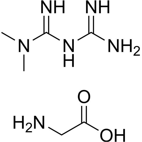
C6H16N6O2
|
|---|---|
| 分子量 |
204.23
|
| 精确质量 |
204.133
|
| CAS号 |
121369-64-0
|
| PubChem CID |
44520175
|
| 外观&性状 |
Typically exists as solids at room temperature
|
| LogP |
0
|
| tPSA |
152.31
|
| 氢键供体(HBD)数目 |
5
|
| 氢键受体(HBA)数目 |
4
|
| 可旋转键数目(RBC) |
3
|
| 重原子数目 |
14
|
| 分子复杂度/Complexity |
175
|
| 定义原子立体中心数目 |
0
|
| SMILES |
OC(CN)=O.N(C(=N)/N=C(\N)/N)(C)C
|
| InChi Key |
KOFRCHIVYXPUNL-UHFFFAOYSA-N
|
| InChi Code |
InChI=1S/C4H11N5.C2H5NO2/c1-9(2)4(7)8-3(5)6;3-1-2(4)5/h1-2H3,(H5,5,6,7,8);1,3H2,(H,4,5)
|
| 化学名 |
2-aminoacetic acid;3-(diaminomethylidene)-1,1-dimethylguanidine
|
| 别名 |
1,1-Dimethylbiguanide (glycinate)
|
| HS Tariff Code |
2934.99.9001
|
| 存储方式 |
Powder -20°C 3 years 4°C 2 years In solvent -80°C 6 months -20°C 1 month |
| 运输条件 |
Room temperature (This product is stable at ambient temperature for a few days during ordinary shipping and time spent in Customs)
|
| 溶解度 (体外实验) |
May dissolve in DMSO (in most cases), if not, try other solvents such as H2O, Ethanol, or DMF with a minute amount of products to avoid loss of samples
|
|---|---|
| 溶解度 (体内实验) |
注意: 如下所列的是一些常用的体内动物实验溶解配方,主要用于溶解难溶或不溶于水的产品(水溶度<1 mg/mL)。 建议您先取少量样品进行尝试,如该配方可行,再根据实验需求增加样品量。
注射用配方
注射用配方1: DMSO : Tween 80: Saline = 10 : 5 : 85 (如: 100 μL DMSO → 50 μL Tween 80 → 850 μL Saline)(IP/IV/IM/SC等) *生理盐水/Saline的制备:将0.9g氯化钠/NaCl溶解在100 mL ddH ₂ O中,得到澄清溶液。 注射用配方 2: DMSO : PEG300 :Tween 80 : Saline = 10 : 40 : 5 : 45 (如: 100 μL DMSO → 400 μL PEG300 → 50 μL Tween 80 → 450 μL Saline) 注射用配方 3: DMSO : Corn oil = 10 : 90 (如: 100 μL DMSO → 900 μL Corn oil) 示例: 以注射用配方 3 (DMSO : Corn oil = 10 : 90) 为例说明, 如果要配制 1 mL 2.5 mg/mL的工作液, 您可以取 100 μL 25 mg/mL 澄清的 DMSO 储备液,加到 900 μL Corn oil/玉米油中, 混合均匀。 View More
注射用配方 4: DMSO : 20% SBE-β-CD in Saline = 10 : 90 [如:100 μL DMSO → 900 μL (20% SBE-β-CD in Saline)] 口服配方
口服配方 1: 悬浮于0.5% CMC Na (羧甲基纤维素钠) 口服配方 2: 悬浮于0.5% Carboxymethyl cellulose (羧甲基纤维素) 示例: 以口服配方 1 (悬浮于 0.5% CMC Na)为例说明, 如果要配制 100 mL 2.5 mg/mL 的工作液, 您可以先取0.5g CMC Na并将其溶解于100mL ddH2O中,得到0.5%CMC-Na澄清溶液;然后将250 mg待测化合物加到100 mL前述 0.5%CMC Na溶液中,得到悬浮液。 View More
口服配方 3: 溶解于 PEG400 (聚乙二醇400) 请根据您的实验动物和给药方式选择适当的溶解配方/方案: 1、请先配制澄清的储备液(如:用DMSO配置50 或 100 mg/mL母液(储备液)); 2、取适量母液,按从左到右的顺序依次添加助溶剂,澄清后再加入下一助溶剂。以 下列配方为例说明 (注意此配方只用于说明,并不一定代表此产品 的实际溶解配方): 10% DMSO → 40% PEG300 → 5% Tween-80 → 45% ddH2O (或 saline); 假设最终工作液的体积为 1 mL, 浓度为5 mg/mL: 取 100 μL 50 mg/mL 的澄清 DMSO 储备液加到 400 μL PEG300 中,混合均匀/澄清;向上述体系中加入50 μL Tween-80,混合均匀/澄清;然后继续加入450 μL ddH2O (或 saline)定容至 1 mL; 3、溶剂前显示的百分比是指该溶剂在最终溶液/工作液中的体积所占比例; 4、 如产品在配制过程中出现沉淀/析出,可通过加热(≤50℃)或超声的方式助溶; 5、为保证最佳实验结果,工作液请现配现用! 6、如不确定怎么将母液配置成体内动物实验的工作液,请查看说明书或联系我们; 7、 以上所有助溶剂都可在 Invivochem.cn网站购买。 |
| 制备储备液 | 1 mg | 5 mg | 10 mg | |
| 1 mM | 4.8964 mL | 24.4822 mL | 48.9644 mL | |
| 5 mM | 0.9793 mL | 4.8964 mL | 9.7929 mL | |
| 10 mM | 0.4896 mL | 2.4482 mL | 4.8964 mL |
1、根据实验需要选择合适的溶剂配制储备液 (母液):对于大多数产品,InvivoChem推荐用DMSO配置母液 (比如:5、10、20mM或者10、20、50 mg/mL浓度),个别水溶性高的产品可直接溶于水。产品在DMSO 、水或其他溶剂中的具体溶解度详见上”溶解度 (体外)”部分;
2、如果您找不到您想要的溶解度信息,或者很难将产品溶解在溶液中,请联系我们;
3、建议使用下列计算器进行相关计算(摩尔浓度计算器、稀释计算器、分子量计算器、重组计算器等);
4、母液配好之后,将其分装到常规用量,并储存在-20°C或-80°C,尽量减少反复冻融循环。
计算结果:
工作液浓度: mg/mL;
DMSO母液配制方法: mg 药物溶于 μL DMSO溶液(母液浓度 mg/mL)。如该浓度超过该批次药物DMSO溶解度,请首先与我们联系。
体内配方配制方法:取 μL DMSO母液,加入 μL PEG300,混匀澄清后加入μL Tween 80,混匀澄清后加入 μL ddH2O,混匀澄清。
(1) 请确保溶液澄清之后,再加入下一种溶剂 (助溶剂) 。可利用涡旋、超声或水浴加热等方法助溶;
(2) 一定要按顺序加入溶剂 (助溶剂) 。
Link: https://clinicaltrials.gov/ct2/show/NCT04943692
Conditions:Type 2 DiabetesLink: https://clinicaltrials.gov/ct2/show/NCT04626089
Conditions:Severe Acute Respiratory Syndrome Coronavirus 2|Metabolic Syndrome|Type 2 DiabetesLink: https://clinicaltrials.gov/ct2/show/NCT04625985
Conditions:Severe Acute Respiratory Syndrome Coronavirus 2
Title:Efficacy Study of Metformin Glycinate on Postprandial Lipemia
Status:Unknown status
updateDate:2018-01-30
Ctid:NCT02064881
Link: https://clinicaltrials.gov/ct2/show/NCT02064881
Conditions:Type 2 DiabetesLink: https://clinicaltrials.gov/ct2/show/NCT01386671
Conditions:Type 2 DiabetesLink: https://clinicaltrials.gov/ct2/show/NCT00960882
Conditions:HealthyLink: https://clinicaltrials.gov/ct2/show/NCT00940472
Conditions:Diabetes Type 2Link: https://clinicaltrials.gov/ct2/show/NCT00940797
Conditions:Diabetes Type 2 PARL-IN-2
 SHS206
 Rosolutamide
 T-271
T-271
InvivoChem的所有产品仅用于作科学研究,不面向患者销售
Copyright 2020 InvivoChem LLC | All Rights Reserved 粤ICP备20063088号-1


 463611831
