| 规格 | 价格 | |
|---|---|---|
| 500mg | ||
| 1g | ||
| Other Sizes |
| 靶点 |
Tyk2 JH2 (IC50 = 0.2 nM); JAK1 JH2 (IC50 = 1 nM)
|
|---|---|
| 体外研究 (In Vitro) |
小分子JAK抑制剂已成为治疗自身免疫性疾病的主要治疗进展。发现传统上靶向该激酶家族催化活性位点的异构体选择性JAK抑制剂是一个巨大的挑战。我们实现TYK2高选择性的策略依赖于靶向TYK2假激酶(JH2)结构域。在此,我们报道了后期的优化工作,包括结构导向设计和水置换策略,从而发现了BMS-986165(11)作为高亲和力JH2配体和TYK2的强效变构抑制剂。除了前所未有的JAK同种型和kinome选择性外,11还显示出优异的药代动力学特性,具有最小的分析责任,并在几种自身免疫性疾病的小鼠模型中有效。基于这些发现,11似乎与所有其他报道的JAK抑制剂不同,并已成为临床开发中第一种以假激酶为导向的治疗方法,作为自身免疫性疾病的口服治疗[1]。
药物化合物包括碳、氢和其他元素的稳定重同位素,在药物开发过程中主要作为定量示踪剂。由于氘化可能会影响药物的药代动力学和代谢特性,因此值得关注[1]。氘代化合物的潜在好处:生物体内的半衰期更长。氘代化合物可能能够增加化合物的药代动力学特性或体内半衰期。这可以促进给药并增强化合物的安全性、有效性和耐受性。第二,提高口服生物利用度。大量未代谢的药物能够达到其作用目标,因为氘化物质减少了肝脏和肠壁中不需要的代谢(首过代谢)量。低剂量下更好的耐受性和活性取决于高生物利用度。 (3)增强新陈代谢功能。氘代物质可以增强药物代谢并减少有害或反应性代谢物的产生。 (四)增强药品安全性。氘代化学物质是无害的,可以减轻或消除药物的不良副作用。 (5) 保持治疗效果。根据早期研究,氘化分子应保持与氢类似物相当的生物效力和选择性。 |
| 体内研究 (In Vivo) |
Deucravacitinib (BMS-986165)治疗的 NZB/W 小鼠中狼疮样疾病得到强烈抑制。Deucravacitinib (BMS-986165) 是安全的且总体耐受性良好。活性药物组(75%)和安慰剂组(76%)没有出现严重不良事件,且非严重不良事件发生的频率相似。口服后,Deucravacitinib (BMS-986165)被迅速吸收,表观消除半衰期为 8-15 小时[1]。
在小鼠中,在IL-23驱动的棘皮病皮肤炎症(银屑病样)模型中评估了11/Deucravatinib(BMS-986165),其中向小鼠耳朵重复皮内注射IL-23诱导了由Th17细胞和IL-22介导的深度表皮增生(棘皮病)和炎性细胞浸润,类似于银屑病的潜在机制(图6)。在该模型中,11/Deucravatinib(BMS-986165)剂量依赖性地保护小鼠免受IL-23诱导的棘皮病,每天两次口服15mg/kg剂量的11,持续9天,证明与阳性对照的抗IL-23附件素一样有效(图6a)。30mg/kg每日两次口服剂量在提供保护方面比抗IL-23附件更有效。组织学评估表明,表皮增生和炎性细胞浸润也以剂量依赖的方式受到抑制,每天两次30mg/kg的高剂量比抗IL-23附件素阳性对照更有效地提供保护(图6b)。皮肤活检的定量聚合酶链式反应(PCR)分析显示,11种方法在阻断炎症细胞因子表达方面非常有效,包括IL-17A、IL-21以及IL-12和IL-23的亚基(图6c)。对研究动物的PK测量表明,7.5、15和30 mg/kg每日两次的剂量分别在19、21和24小时内提供了等于或高于100 nM(IFNα诱导的pSTAT1)的体外小鼠全血IC50值的药物水平。除了银屑病的临床前模型外,11也被证明在结肠炎和狼疮的小鼠模型中非常有效。这些结果表明,在严重联合免疫缺陷病(SCID)小鼠的抗CD40诱导的结肠炎模型中,与抗p40单克隆抗体对照组相比,11只每天两次服用50mg/kg,可抑制99%的峰值体重减轻(IL-12驱动)和70%的组织学评分。此外,在使用NZB/W狼疮易感小鼠的三个月狼疮疾病模型中,口服最高30mg/kg每日一次的剂量时,11具有良好的耐受性,在预防肾炎方面非常有效。在后一项研究中,疗效与抑制研究小鼠全血和肾脏中I型IFN依赖性基因表达密切相关,至少与阻断抗IFNαR抗体一样有效[1]。 干扰素α-2a攻击后干扰素反应基因的表达[2] 在早晨服用Deucravatinib(BMS-986165)或安慰剂2小时后,用临床剂量的IFNα-2a进行体内攻击约3小时后,开始诱导IRG,这可以通过诱导寡腺苷酸合成酶样(OASL)基因来证明,这是一种典型的IRG(数据未显示)。在服用安慰剂的志愿者中,IRG诱导很强,大多数基因的诱导倍数超过10倍。在评估的53个靶基因中,有52个被纳入分析,因为发现补体成分1q(C1Q)不会立即被干扰素诱导,而是在26小时的时间点被诱导,因此被认为不太可能是干扰素诱导的基因表达的直接靶点。与安慰剂组相比,预先服用去克拉伐替尼以剂量依赖的方式强烈抑制了聚集中包含的所有52个基因的表达(图3);攻击后26小时,C1Q表达受到抑制。IFNα-2a的暴露量以剂量依赖的方式增加,因此,阻断信号转导可以抑制IFNα-1a的消耗。IFNα-2a的这种反常增加似乎是deucravacitinib的另一种PD测量方法。 |
| 酶活实验 |
使用均相时间分辨荧光(HTRF)测定法测定所有生物化学效价和选择性,其中化合物显示与荧光探针竞争结合人重组JAK1、JAK2、JAK3和TYK2 JH1结构域蛋白以及TYK2和JAK1 JH2蛋白结构域。生成剂量-反应曲线,以确定通过非线性回归分析得出的抑制50%HTRF信号(IC50)所需的浓度。使用稳定整合的STAT依赖性萤光素酶报告基因测定法测定T细胞中的细胞潜能和选择性,使用IFNα-刺激测定TYK2/JAK1依赖性信号传导,使用IL-23刺激测定TYK2/JAK1依存性信号传导。使用GM-CSF刺激在TF-1细胞中测量JAK2依赖性信号传导。生成剂量-反应曲线,以确定通过非线性回归分析得出的抑制50%细胞反应(IC50)所需的浓度。还使用特异性细胞因子刺激在人和小鼠全血中测量了JAK依赖性信号传导的效力和选择性,并通过细胞染色和流式细胞术测量了特异性STAT蛋白的磷酸化。先前已经报道了所有测定的实验细节。对所有在生物测定中具有活性的化合物进行电子过滤,以获得泛测定干扰化合物(PAINS)常见的结构属性,并发现其为阴性[1]。
|
| 细胞实验 |
使用T细胞中稳定整合的STAT依赖性萤光素酶报告子测定法测定细胞效力和选择性,使用IFNα刺激测量TYK2/JAK1依赖性信号传导,使用IL-23刺激测量TYK2/JAK1依存性信号传导。使用GM-CSF刺激在TF-1细胞中测量JAK2依赖性信号传导。生成剂量-反应曲线,以确定通过非线性回归分析得出的抑制50%细胞反应所需的浓度(IC50)。还使用特定的细胞因子刺激在人和小鼠全血中测量了JAK依赖性信号传导的效力和选择性,并通过细胞染色和流式细胞术测量了特定STAT蛋白的磷酸化。所有测定的实验细节之前都有报道。所有在生物测定中具有活性的化合物都经过了电子过滤,以获得泛测定干扰化合物(PAINS)共有的结构属性,结果为阴性[1]。
|
| 动物实验 |
IL-23诱导小鼠棘层增厚[1]
在6-8周龄的C57BL/6雌性小鼠(平均体重19-20 g,杰克逊实验室)右耳皮内注射双链重组人IL-23,诱导棘层增厚。从研究的第0天到第9天,每隔一天注射一次IL-23。每个治疗组包含8只小鼠。化合物11/Deucravacitinib(BMS-986165)以7.5、15和30 mg/kg的剂量,每日两次(BID)溶于溶剂(EtOH:TPGS:PEG300,5:5:90)中,以及单独使用溶剂,均通过灌胃法给药,首次给药在首次IL-23注射前一晚进行。在首次注射IL-23前约1小时,皮下注射抗IL-23抗体adnectin(3 mg/kg)和PBS对照,之后每周两次。使用Mitutoyo游标卡尺测量耳厚度,并计算每只动物在首次注射IL-23前(第0天)的耳厚度变化百分比。研究结束时,从每组4只动物中采集注射IL-23的耳和未注射IL-23的对照耳,用于组织学检查和基因表达分析。通过眶后静脉丛采集终末血样,用于药代动力学(PK)测定。统计分析采用Student's t检验或ANOVA方差分析,并进行Dunnett事后检验。研究结束时,取出耳,固定于10%中性缓冲福尔马林溶液中24-48小时。然后将固定后的耳纵向切开,并将两块平行包埋制成石蜡块。将石蜡包埋的组织块切片后置于载玻片上,进行苏木精-伊红(H&E)染色,用于组织学评估。耳部炎症的严重程度采用基于以下参数的客观评分系统进行评分:病变范围、角化过度程度、脓疱的数量和大小、表皮增生高度(棘层增厚,测量于毛囊间表皮)以及真皮和软组织中的炎症浸润量。后两项参数,即棘层增厚和炎症浸润,分别采用0至4分的评分标准进行独立评分:0分,无;1分,极轻微;2分,轻度;3分,中度;4分,重度。组织学变化由病理学家进行盲法评估。统计分析采用单因素方差分析(ANOVA),并使用Dunnett检验比较各治疗组与载体对照组的差异。 |
| 药代性质 (ADME/PK) |
吸收、分布和排泄
在健康受试者中,口服给药后,deucravacitinib 的血浆 Cmax 和 AUC 在 3 mg 至 36 mg 的剂量范围内(相当于批准推荐剂量的 0.5 至 6 倍)呈比例增加。每日一次服用 6 mg deucravacitinib 后,其稳态 Cmax 和 AUC24 分别为 45 ng/mL 和 473 ng·hr/mL。每日一次服用 6 mg deucravacitinib 的活性代谢物 BMT-153261 后,其稳态 Cmax 和 AUC24 分别为 5 ng/mL 和 95 ng·hr/mL。deucravacitinib 的绝对口服生物利用度为 99%,中位 Tmax 为 2 至 3 小时。高脂肪、高热量膳食使地克拉伐替尼的Cmax和AUC分别降低了24%和11%,并使Tmax延长了1小时;然而,这对药物的吸收和暴露量有显著的临床影响。 单次服用放射性标记的地克拉伐替尼后,分别约有13%和26%的剂量以原形从尿液和粪便中排出。尿液和粪便中分别检测到约 6% 和 12% 的剂量为 BMT-153261。 地克拉伐替尼的稳态分布容积为 140 L。 地克拉伐替尼的肾清除率范围为 27 至 54 mL/分钟。 代谢/代谢物 地克拉伐替尼经细胞色素 P-450 (CYP) 1A2 介导的 N-去甲基化作用生成主要代谢物 BMT-153261,其药理活性与母体药物相当。然而,BMT-153261 的循环暴露量约占药物相关成分总系统暴露量的 20%。 Deucravacitinib 也通过 CYP2B6、CYP2D6、羧酸酯酶 (CES) 2 和尿苷葡萄糖醛酸转移酶 (UGT) 1A9 代谢。 生物半衰期 Deucravacitinib 的末端半衰期为 10 小时。 三唑 11/Deucravacitinib (BMS-986165) 显示出极低的代谢缺陷、优异的药代动力学特性,并且在炎症和自身免疫性疾病模型中具有很高的疗效[1] 11/Deucravacitinib (BMS-986165) 已显示出对 TYK2 依赖性反应的优异效力和功能选择性,并进一步进行了体外分析,结果显示其代谢缺陷极低,且具有可接受的药代动力学特性,适合进一步开发(表 7)。在肝微粒体中孵育后,该药物在包括人、小鼠、大鼠、猴、犬和兔在内的多种物种中均表现出优异的稳定性(T1/2 > 120 分钟)。在 Caco-2 细胞实验中也观察到良好的渗透性,并伴有中等程度的外排(顶端至基底侧的 Pc = 73 nm/s;基底侧至顶端的 Pc = 740 nm/s;外排比约为 10)。药物相互作用 (DDI) 的评估表明总体风险较低,因为在最高测试浓度 (40 μM) 下,未观察到对多种细胞色素 P450 (CYP) 同工酶(1A2、2C9、2C19、2D6 和 3A4)的显著抑制或对 CYP3A4 的诱导。在hERG钾通道膜片钳实验中,化合物11的抑制率较低(10 μM时为26 ± 11%),提示其QTc间期延长相关的心血管风险较低。在包括人、猴和小鼠在内的多种物种中,其蛋白结合率处于中等水平(游离态为12%–15%)。化合物11结晶游离碱形式的水溶性较低(5.2 μg/mL),但足以用于临床前研究。在多种物种的临床前研究中,化合物11的总体药代动力学参数良好(表8)。与微粒体代谢实验中观察到的低代谢率(T1/2 > 120 min)一致,化合物11在小鼠、犬和猴体内的清除率分别为13.2、6.8和4.8 mL min–1 kg–1,处于较低至中等水平。该化合物的分布容积(Vss)较低,在 2–3 L/kg 范围内,半衰期适中,约为 4–5 小时(跨物种)。口服 10 mg/kg 时,化合物 11 在小鼠、犬和猴体内吸收良好,暴露量高,生物利用度高(%F > 85)。在这些研究中,由化合物 11 的氘甲基酰胺的 N-去烷基化生成的循环伯酰胺代谢物的浓度接近检测下限 (<2 nM),这与先前在该系列研究中报道的氘代作用有效阻断该代谢途径的结果一致。 健康志愿者药代动力学[2] 给药后,Deucravacitinib (BMS-986165) 被迅速吸收,单次给药后表观消除半衰期 (t1/2) 为 7.9–15.0 小时,多次给药后平均有效半衰期 (t1/2) 为 7.5–13.1 小时(图 1,表 1)。单次给药后(SAD 组最高剂量为 10 mg),Cmax 和 AUCINF 随剂量增加呈非比例性增加,但在剂量 ≥10 mg 时,则呈表观剂量比例性增加。在MAD组中,多次给药后观察到类似的药代动力学特征,剂量≥4 mg bid时呈剂量比例关系。多次给药后观察到适度的药物蓄积(1.4-1.9倍),并在第5天达到稳态,即首次采集Cmin样本的当天。仅在SAD组中评估了未代谢的deucravacitinib的尿液回收率,所有剂量组的回收率范围为10%至15%;deucravacitinib的肾清除率在整个剂量范围内为27.5至54.2 ml/min。 |
| 毒性/毒理 (Toxicokinetics/TK) |
肝毒性
在纳入1519例受试者数据的deucravacitinib预注册临床试验中,仅有1.8%的患者出现血清ALT或AST升高超过正常值上限5倍的情况,且这些升高均被认为不太可能是药物引起的肝损伤,其中大部分升高是由肌炎所致,少数升高是由潜在的酒精性或非酒精性脂肪肝所致。在一项为期24周的试验中,deucravacitinib组有1.1%至1.3%的患者出现ALT水平升高超过正常值上限3倍的情况,而安慰剂组的这一比例为1.2%。虽然接受deucravacitinib治疗的患者中未出现乙型肝炎病毒再激活的病例,但既往血清中存在HBsAg的患者被排除在入组之外,且大多数疗程的持续时间有限。自获批并广泛应用于临床以来,尚未有因服用地克拉伐替尼而导致临床上明显的肝损伤的报告,但该药上市时间有限。 可能性评分:E(疑似但未经证实的临床上明显的肝损伤原因,包括乙型肝炎病毒再激活)。 妊娠和哺乳期用药 ◉ 哺乳期用药概述 目前尚无关于哺乳期服用地克拉伐替尼的信息。由于该药与血浆蛋白的结合率超过80%,因此其在乳汁中的含量可能较低。然而,该药口服吸收良好。如果较大婴儿的母亲需要服用地克拉伐替尼,这并非停止母乳喂养的理由,但在获得更多数据之前,可能更倾向于选择其他药物,尤其是在哺乳新生儿或早产儿期间。 ◉ 对母乳喂养婴儿的影响 截至修订日期,未找到相关的已发表信息。 ◉ 对泌乳和母乳的影响 截至修订日期,未找到相关的已发表信息。 ◈ 什么是地克拉伐替尼? 地克拉伐替尼是一种已获批准用于治疗中重度斑块状银屑病的药物。MotherToBaby 网站提供关于银屑病和银屑病关节炎的资料单,网址为:https://mothertobaby.org/fact-sheets/psoriasis-and-pregnancy/。地克拉伐替尼的商品名为 SOTYKTU™。有时,当人们发现自己怀孕后,会考虑改变服药方式,甚至完全停药。然而,在改变服药方式之前,务必咨询您的医疗保健提供者。您的医疗保健提供者可以与您讨论治疗您病情的益处以及怀孕期间未治疗疾病的风险。 ◈ 我正在服用地克拉伐替尼。它会影响我怀孕吗? 目前尚无研究表明地克拉伐替尼会影响怀孕。 ◈ 服用地克拉伐替尼会增加流产的风险吗? 流产很常见,任何妊娠都可能发生,原因多种多样。目前尚无研究确定地克拉伐替尼是否会增加流产风险。 ◈服用地克拉伐替尼会增加胎儿出生缺陷的风险吗? 每次怀孕都有3-5%的胎儿出生缺陷风险,这被称为背景风险。目前尚未开展地克拉伐替尼与人类妊娠相关的研究。制造商报告的动物实验研究并未发现出生缺陷风险增加。 ◈ 孕期服用 deucravacitinib 是否会增加其他妊娠相关问题的风险? 目前尚无研究确定 deucravacitinib 是否会增加早产(妊娠 37 周前分娩)或低出生体重(出生体重低于 2500 克)等妊娠相关问题的风险。 ◈ 孕期服用 deucravacitinib 是否会影响孩子未来的行为或学习? 目前尚无研究确定 deucravacitinib 是否会导致孩子出现行为或学习问题。 ◈ 服用 deucravacitinib 期间哺乳: 尚未研究 deucravacitinib 在哺乳期间的使用情况。请务必就所有关于母乳喂养的问题咨询您的医疗保健提供者。 ◈ 如果男性服用地克拉伐替尼,是否会影响生育能力(使伴侣怀孕的能力)或增加伴侣妊娠中胎儿畸形的风险? 目前尚无研究探讨地克拉伐替尼是否会影响男性生育能力或增加伴侣妊娠中胎儿畸形的风险。一般来说,父亲或精子捐赠者接触到的药物不太可能增加妊娠风险。更多信息,请参阅 MotherToBaby 网站上的“父亲暴露”情况说明书,网址为 https://mothertobaby.org/fact-sheets/paternal-exposures-pregnancy/。 蛋白质结合 地克拉伐替尼的蛋白质结合率为 82% 至 90%,血药浓度与血浆浓度之比为 1.26。 毒性概述目前尚无地克拉伐替尼过量用药的临床数据。如果任何患者出现过量用药,建议联系中毒救助热线,以便根据药物过量处理方案进行进一步处理。通过血液透析治疗清除地克拉伐替尼的程度很小,每次透析治疗最多只能清除剂量的5.4%,因此限制了血液透析在治疗地克拉伐替尼过量或中毒中的应用。 多项采用速率模型的研究表明,在雄性或雌性大鼠中,口服剂量高达15 mg/kg/天(根据AUC比较,该剂量是最大推荐人用剂量的51倍)的地克拉伐替尼均未观察到致癌性。在雌性大鼠中,口服剂量高达50 mg/kg/天(根据AUC比较,该剂量是最大推荐人用剂量的171倍)的地克拉伐替尼对交配、生育力或胚胎早期发育等生殖参数没有影响。在雄性大鼠中,口服剂量高达 50 mg/kg/天的 deucravacitinib 对交配、精子形态、生育能力或其后代的早期胚胎参数均无影响,该剂量是根据 AUC 比较得出的 MRHD 的 224 倍。在 SAD 队列中,11 名接受 deucravacitinib 治疗的志愿者(36.7%)和 4 名接受安慰剂治疗的志愿者(40%)报告了不良事件;按首选术语报告的最常见不良事件是头痛(deucravacitinib 组:5 名志愿者,16.7%;安慰剂组:2 名志愿者,20.0%)。接受 deucravacitinib 治疗的志愿者共报告了 7 例(23.3%)胃肠道疾病不良事件(按系统顺序分类),而安慰剂组未报告此类不良事件,其中最常见的是消化不良,3 名志愿者(10%)报告了此类不良事件。除安慰剂组出现1例中度晕厥前兆事件外,所有不良事件的严重程度均为轻度。SAD队列中最常见的全因不良事件总结于表2。[2] 在MAD队列中,所有不良事件的严重程度均为轻度至中度,且地克拉伐替尼组(37名志愿者,82%)和安慰剂组(13名志愿者,87%)的不良事件总体发生率相似。在地克拉伐替尼组和安慰剂组中,按首选术语报告的最常见不良事件分别为:头痛(11名志愿者,24.4%;5名志愿者,33.3%)、皮疹(9名志愿者,20%;2名志愿者,13.3%)、上呼吸道感染(8名志愿者,17.8%;3名志愿者,20.0%)、痤疮(6名志愿者,13.3%;0名志愿者)和恶心(6名志愿者,13.3%;2名志愿者,13.3%)。MAD队列中最常见的所有原因不良事件汇总于表2。除1例中度荨麻疹外,所有不良事件均已消退。该荨麻疹发生于地克拉伐替尼6 mg bid组,被认为与研究治疗无关。共有 7 名志愿者因不良事件而中止试验:6 名志愿者在服用 deucravacitinib 后中止试验(2 mg bid:1 名志愿者,胸痛;4 mg bid:1 名志愿者,皮疹;6 mg bid:2 名志愿者,荨麻疹;12 mg bid:1 名志愿者,扁桃体炎;12 mg qd:1 名志愿者,疖),1 名志愿者在安慰剂组中中止试验(意识下降)。[2] 与安慰剂相比,Deucravacitinib 会增加皮疹以及痤疮和荨麻疹样皮肤反应的发生率,尤其是在最高剂量 24 mg/天(12 mg bid 剂量组)时。皮肤皮疹/痤疮不良事件的严重程度为轻度或中度,必要时可采用局部治疗(荨麻疹样皮疹使用皮质类固醇乳膏,痤疮使用过氧化苯甲酰乳膏、克林霉素溶液或氯己定软膏)治疗,且很少导致停药。[2] 采用标准自动细胞计数法监测白细胞计数,并使用TBNK流式细胞术进一步计数主要白细胞群,结果显示,在接受deucravacitinib治疗后,T细胞、B细胞或自然杀伤(NK)细胞亚群均未出现异常(数据未显示)。[2] 在研究的任何阶段,均未发现任何临床实验室显著异常或心电图异常(包括超出正常范围的心电图间期)的剂量相关性趋势,也未观察到deucravacitinib对心率或体温的影响。[2] |
| 参考文献 |
|
| 其他信息 |
药效学
Deucravacitinib 是一种酪氨酸激酶 2 (TYK2) 抑制剂,可抑制炎症性疾病(例如斑块状银屑病)中的免疫信号通路。在包含银屑病患者的临床研究中,Deucravacitinib 以剂量依赖的方式降低了银屑病皮肤中银屑病相关基因的表达,包括 IL-23 通路和 I 型干扰素通路调控基因的减少。每日一次治疗 16 周后,Deucravacitinib 使 IL-17A、IL-19 和 β-防御素等炎症标志物分别降低了 47% 至 50%、72% 和 81% 至 84%。Deucravacitinib 不影响 JAK2 依赖的造血功能。 小分子 JAK 抑制剂已成为治疗自身免疫性疾病的一项重大突破。传统上,JAK激酶家族的同工酶选择性抑制剂主要靶向其催化活性位点,而发现此类抑制剂一直是巨大的挑战。我们实现对TYK2高选择性的策略是靶向TYK2假激酶(JH2)结构域。本文报道了后期优化工作,包括结构导向设计和水置换策略,最终发现了BMS-986165 (11),它是一种高亲和力的JH2配体和有效的TYK2变构抑制剂。除了前所未有的JAK同工酶和激酶组选择性外,化合物11还表现出优异的药代动力学性质,具有极低的药代动力学缺陷,并且在多种自身免疫性疾病小鼠模型中有效。基于这些发现,化合物11与其他已报道的JAK抑制剂截然不同,并作为首个靶向假激酶的口服自身免疫性疾病治疗药物进入临床开发阶段。 [1] 这项随机、双盲、单次和多次递增剂量研究评估了选择性强效酪氨酸激酶2小分子抑制剂deucravacitinib(Sotyktu™)在100名健康志愿者(75名活性药物组,25名安慰剂组)中的药代动力学(PK)、药效学和安全性(NCT02534636)。Deucravacitinib吸收迅速,半衰期为8-15小时,多次给药后药物蓄积量为1.4-1.9倍。体外实验表明,Deucravacitinib以剂量和浓度依赖的方式抑制白细胞介素(IL)-12/IL-18诱导的干扰素(IFN)γ产生。体内实验表明,在IFNα-2a刺激后,Deucravacitinib以剂量依赖的方式抑制淋巴细胞计数下降和53个IFN调节基因的表达。未发生严重不良事件(AE);deucravacitinib组(64%)和安慰剂组(68%)的AE总体发生率相似。在这项首次人体试验中,deucravacitinib在健康志愿者中抑制了IL-12/IL-23和I型干扰素通路,且具有良好的药代动力学和安全性。Deucravacitinib有望成为治疗免疫介导疾病(包括克罗恩病、银屑病、银屑病关节炎和系统性红斑狼疮)的一种有效疗法。[2] |
| 分子式 |
C20H23CLN8O3
|
|---|---|
| 分子量 |
461.91990685463
|
| 精确质量 |
461.177
|
| CAS号 |
1609392-28-0
|
| 相关CAS号 |
1609392-27-9
|
| PubChem CID |
146048026
|
| 外观&性状 |
Typically exists as solids at room temperature
|
| tPSA |
136
|
| 氢键供体(HBD)数目 |
4
|
| 氢键受体(HBA)数目 |
8
|
| 可旋转键数目(RBC) |
7
|
| 重原子数目 |
32
|
| 分子复杂度/Complexity |
648
|
| 定义原子立体中心数目 |
0
|
| SMILES |
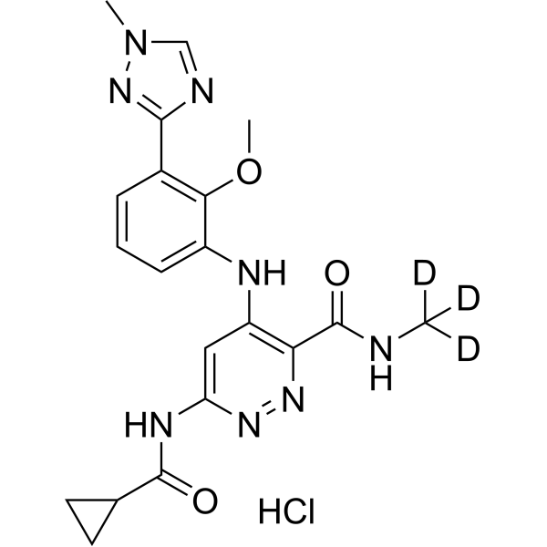
Cl.O=C(C1CC1)NC1=CC(=C(C(NC([2H])([2H])[2H])=O)N=N1)NC1C=CC=C(C2N=CN(C)N=2)C=1OC
|
| InChi Key |
LILQGPVDQZORJG-NIIDSAIPSA-N
|
| InChi Code |
InChI=1S/C20H22N8O3.ClH/c1-21-20(30)16-14(9-15(25-26-16)24-19(29)11-7-8-11)23-13-6-4-5-12(17(13)31-3)18-22-10-28(2)27-18;/h4-6,9-11H,7-8H2,1-3H3,(H,21,30)(H2,23,24,25,29);1H/i1D3;
|
| 化学名 |
6-(cyclopropanecarbonylamino)-4-[2-methoxy-3-(1-methyl-1,2,4-triazol-3-yl)anilino]-N-(trideuteriomethyl)pyridazine-3-carboxamide;hydrochloride
|
| 别名 |
BMS-986165 hydrochloride; Deucravacitinib hydrochloride; BMS-986165 hydrochloride; 95C5558CF4; UNII-95C5558CF4; 1609392-28-0; 3-Pyridazinecarboxamide, 6-((cyclopropylcarbonyl)amino)-4-((2-methoxy-3-(1-methyl-1H-1,2,4-triazol-3-yl)phenyl)amino)-N-(methyl-d3)-, hydrochloride (1:1)
|
| HS Tariff Code |
2934.99.9001
|
| 存储方式 |
Powder -20°C 3 years 4°C 2 years In solvent -80°C 6 months -20°C 1 month |
| 运输条件 |
Room temperature (This product is stable at ambient temperature for a few days during ordinary shipping and time spent in Customs)
|
| 溶解度 (体外实验) |
May dissolve in DMSO (in most cases), if not, try other solvents such as H2O, Ethanol, or DMF with a minute amount of products to avoid loss of samples
|
|---|---|
| 溶解度 (体内实验) |
注意: 如下所列的是一些常用的体内动物实验溶解配方,主要用于溶解难溶或不溶于水的产品(水溶度<1 mg/mL)。 建议您先取少量样品进行尝试,如该配方可行,再根据实验需求增加样品量。
注射用配方
注射用配方1: DMSO : Tween 80: Saline = 10 : 5 : 85 (如: 100 μL DMSO → 50 μL Tween 80 → 850 μL Saline)(IP/IV/IM/SC等) *生理盐水/Saline的制备:将0.9g氯化钠/NaCl溶解在100 mL ddH ₂ O中,得到澄清溶液。 注射用配方 2: DMSO : PEG300 :Tween 80 : Saline = 10 : 40 : 5 : 45 (如: 100 μL DMSO → 400 μL PEG300 → 50 μL Tween 80 → 450 μL Saline) 注射用配方 3: DMSO : Corn oil = 10 : 90 (如: 100 μL DMSO → 900 μL Corn oil) 示例: 以注射用配方 3 (DMSO : Corn oil = 10 : 90) 为例说明, 如果要配制 1 mL 2.5 mg/mL的工作液, 您可以取 100 μL 25 mg/mL 澄清的 DMSO 储备液,加到 900 μL Corn oil/玉米油中, 混合均匀。 View More
注射用配方 4: DMSO : 20% SBE-β-CD in Saline = 10 : 90 [如:100 μL DMSO → 900 μL (20% SBE-β-CD in Saline)] 口服配方
口服配方 1: 悬浮于0.5% CMC Na (羧甲基纤维素钠) 口服配方 2: 悬浮于0.5% Carboxymethyl cellulose (羧甲基纤维素) 示例: 以口服配方 1 (悬浮于 0.5% CMC Na)为例说明, 如果要配制 100 mL 2.5 mg/mL 的工作液, 您可以先取0.5g CMC Na并将其溶解于100mL ddH2O中,得到0.5%CMC-Na澄清溶液;然后将250 mg待测化合物加到100 mL前述 0.5%CMC Na溶液中,得到悬浮液。 View More
口服配方 3: 溶解于 PEG400 (聚乙二醇400) 请根据您的实验动物和给药方式选择适当的溶解配方/方案: 1、请先配制澄清的储备液(如:用DMSO配置50 或 100 mg/mL母液(储备液)); 2、取适量母液,按从左到右的顺序依次添加助溶剂,澄清后再加入下一助溶剂。以 下列配方为例说明 (注意此配方只用于说明,并不一定代表此产品 的实际溶解配方): 10% DMSO → 40% PEG300 → 5% Tween-80 → 45% ddH2O (或 saline); 假设最终工作液的体积为 1 mL, 浓度为5 mg/mL: 取 100 μL 50 mg/mL 的澄清 DMSO 储备液加到 400 μL PEG300 中,混合均匀/澄清;向上述体系中加入50 μL Tween-80,混合均匀/澄清;然后继续加入450 μL ddH2O (或 saline)定容至 1 mL; 3、溶剂前显示的百分比是指该溶剂在最终溶液/工作液中的体积所占比例; 4、 如产品在配制过程中出现沉淀/析出,可通过加热(≤50℃)或超声的方式助溶; 5、为保证最佳实验结果,工作液请现配现用! 6、如不确定怎么将母液配置成体内动物实验的工作液,请查看说明书或联系我们; 7、 以上所有助溶剂都可在 Invivochem.cn网站购买。 |
| 制备储备液 | 1 mg | 5 mg | 10 mg | |
| 1 mM | 2.1649 mL | 10.8244 mL | 21.6488 mL | |
| 5 mM | 0.4330 mL | 2.1649 mL | 4.3298 mL | |
| 10 mM | 0.2165 mL | 1.0824 mL | 2.1649 mL |
1、根据实验需要选择合适的溶剂配制储备液 (母液):对于大多数产品,InvivoChem推荐用DMSO配置母液 (比如:5、10、20mM或者10、20、50 mg/mL浓度),个别水溶性高的产品可直接溶于水。产品在DMSO 、水或其他溶剂中的具体溶解度详见上”溶解度 (体外)”部分;
2、如果您找不到您想要的溶解度信息,或者很难将产品溶解在溶液中,请联系我们;
3、建议使用下列计算器进行相关计算(摩尔浓度计算器、稀释计算器、分子量计算器、重组计算器等);
4、母液配好之后,将其分装到常规用量,并储存在-20°C或-80°C,尽量减少反复冻融循环。
计算结果:
工作液浓度: mg/mL;
DMSO母液配制方法: mg 药物溶于 μL DMSO溶液(母液浓度 mg/mL)。如该浓度超过该批次药物DMSO溶解度,请首先与我们联系。
体内配方配制方法:取 μL DMSO母液,加入 μL PEG300,混匀澄清后加入μL Tween 80,混匀澄清后加入 μL ddH2O,混匀澄清。
(1) 请确保溶液澄清之后,再加入下一种溶剂 (助溶剂) 。可利用涡旋、超声或水浴加热等方法助溶;
(2) 一定要按顺序加入溶剂 (助溶剂) 。
Link: https://www.clinicaltrialsregister.eu/ctr-search/search?query=2018-001925-24
Condition:Moderate-to-Severe Plaque PsoriasisLink: https://www.clinicaltrialsregister.eu/ctr-search/search?query=2017-001976-48
Condition:Crohn's DiseaseLink: https://www.clinicaltrialsregister.eu/ctr-search/search?query=2017-001203-79
Condition:Systemic Lupus Erythematosus
Title:A Phase 2, Randomized, Double-blind, Placebo-controlled Evaluation of the Safety and Efficacy of BMS-986165 with Background Treatment in Subjects with Lupus Nephritis
Status:Completed, GB - no longer in EU/EEA, Ongoing
Date:
Eudractnumber:2018-004142-42
Link: https://www.clinicaltrialsregister.eu/ctr-search/search?query=2018-004142-42
Condition:Lupus Nephritis Hydrostatin-TL1
 Madindoline B
 PIM1-IN-8
 MT204
MT204
InvivoChem的所有产品仅用于作科学研究,不面向患者销售
Copyright 2020 InvivoChem LLC | All Rights Reserved 粤ICP备20063088号-1


 463611831
