| 规格 | 价格 | 库存 | 数量 |
|---|---|---|---|
| 1mg |
|
||
| Other Sizes |
|

| 靶点 |
VEGFR2 (IC50 = 0.2 nM); VEGFR3 (IC50 = 0.7 nM); c-Kit (IC50 = 14.8 nM); c-Kit (IC50 = 14.8 nM); c-Kit (IC50 = 14.8 nM)
Anlotinib Dihydrochloride primarily targets vascular endothelial growth factor receptor-2 (VEGFR2) with an IC50 < 1 nmol/L; it also inhibits VEGF-induced signaling and proliferation in HUVEC with picomolar IC50 values [1] Anlotinib Dihydrochloride is a multi-target tyrosine kinase inhibitor that potently inhibits VEGFR2/3, FGFR1-4, PDGFR α/β, c-Kit, and Ret [2] |
|---|---|
| 体外研究 (In Vitro) |
体外活性:安罗替尼(原名 AL3818)是一种新型强效多激酶抑制剂,可抑制 VEGFR2/3、FGFR1-4、PDGFRα/β、c-Kit 和 Ret。安罗替尼作为受体酪氨酸激酶 (RTK) 抑制剂,具有潜在的抗肿瘤和抗血管生成活性。安罗替尼在体外显着减少 AN3CA 细胞数量,其特点是突变 FGFR2 蛋白的高表达。每日口服安罗替尼(5 mg/kg)使 55% 的接受治疗的动物出现完全缓解,并且在 29 次治疗后,AN3CA 肿瘤的肿瘤体积和肿瘤重量分别减少了 94% 和 96%。天治疗周期。尽管卡铂和紫杉醇未能改变肿瘤生长,但与单独使用安罗替尼治疗相比,与安罗替尼联合治疗似乎并未表现出更好的效果。 激酶检测:安罗替尼(以前称为 AL3818)是一种新型、有效的多激酶抑制剂,可抑制 VEGFR2 /3、FGFR1-4、PDGFRα/β、c-Kit 和 Ret.细胞检测:AL3818 在体外显着减少 AN3CA 细胞数量,其特征是突变的 FGFR2 蛋白高表达。每日口服 AL3818 (5 mg/kg) 可使 55% 的接受治疗动物出现完全缓解,并且在 29 次治疗后,AN3CA 肿瘤的肿瘤体积减小,肿瘤重量分别减少 94% 和 96%。天治疗周期。尽管卡铂和紫杉醇未能改变肿瘤生长,但与单独使用 AL3818 治疗相比,与 AL3818 的组合似乎并未表现出更好的效果。
盐酸安罗替尼可结合VEGFR2酪氨酸激酶的ATP结合口袋,对VEGFR2具有高选择性和抑制活性(IC50<1 nmol/L),远高于对其他酪氨酸激酶的抑制作用。它以皮摩尔级IC50抑制VEGF诱导的HUVEC信号通路激活和细胞增殖,而直接抑制肿瘤细胞增殖则需要微摩尔浓度。此外,该药物能显著抑制HUVEC的迁移和管形成,抑制大鼠主动脉外植体的微血管出芽,在体外阻断血管生成相关过程 [1] 盐酸安罗替尼在体外具有良好的膜通透性。它对大多数人细胞色素P450酶(CYPs)、尿苷二磷酸葡萄糖醛酸转移酶(UGTs)和转运体的抑制作用较弱,但对CYP3A4和CYP2C9的体外抑制IC50<1μmol/L。药物与血浆蛋白结合率较高,大鼠为97%、犬为96%、人为93% [3] |
| 体内研究 (In Vivo) |
每日口服安罗替尼(5 mg/kg)使 55% 的接受治疗的动物出现完全缓解,并且在 29 次治疗后,AN3CA 肿瘤的肿瘤体积和肿瘤重量分别减少了 94% 和 96%。天治疗周期。尽管卡铂和紫杉醇未能改变肿瘤生长,但与单独使用安罗替尼治疗相比,与安罗替尼的组合似乎并未表现出更好的效果。
安洛替尼在人异种移植物模型中的抗肿瘤疗效[1] 鉴于安洛替尼活性在体外具有令人鼓舞的抗血管生成作用,我们接下来评估了安洛替尼在人类结肠癌癌症SW620异种移植物模型中的体内抗肿瘤潜力。每日一次口服剂量的安洛替尼可引起肿瘤生长的剂量依赖性抑制(图5A,C),与对照组相比,3 mg/kg的剂量可抑制83%的肿瘤生长。相比之下,在该模型中,达到相当疗效所需的舒尼替尼剂量为50 mg/kg。此外,在所有组的实验过程中,安洛替尼对小鼠体重的影响很小(图5B)。我们通过使用内皮细胞标志物CD31的免疫组织化学分析测量提取的肿瘤中的微血管密度,进一步评估了肿瘤血管生成。安洛替尼诱导CD31阳性微血管显著减少,在0.75、1.5和3 mg/kg的剂量下,抑制率分别为48.9%、76.3%和91.2%(图5D)。相比之下,50mg/kg剂量的舒尼替尼抑制了63.6%的微血管密度。 在多种异种移植物模型中进一步研究了安洛替尼的体内抗肿瘤潜力,该模型通过接种具有不同VEGF/VEGFR2 mRNA表达水平的人癌症细胞系(图S1;数据S1)或其他RTK.35,40创建。在所有测试的肿瘤模型中,每日口服一次安洛替尼对肿瘤生长产生了剂量依赖性抑制作用(表2;图6)。在3 mg/kg的剂量下,安洛替尼在U-87MG、Caki-1、Calu-3和SK-OV-3异种移植物中分别抑制了55%、80%、91%和97%的肿瘤生长,在最后一个治疗日进行测量。此外,它在Calu-3和SK-OV-3肿瘤异种移植物模型中都导致了肿瘤消退。在后一种异种移植物模型中,用更高剂量的安洛替尼(6mg/kg)治疗可抑制95%的肿瘤生长;重要的是,在停用安洛替尼后12天内,肿瘤没有反弹。与3 mg/kg的情况一样,6 mg/kg的安洛替尼在Calu-3和SK-OV-3肿瘤异种移植物模型中均导致肿瘤消退。 裸鼠异种移植模型(SW620、U-87 MG、Caki-1、SK-OV-3、Calu-3)中,每日口服盐酸安罗替尼的体内抗肿瘤疗效比舒尼替尼更广、更强,部分模型中可诱导肿瘤消退;同时该药物通过抑制血管生成降低肿瘤组织的血管密度 [1] 晚期难治性实体瘤患者接受盐酸安罗替尼12mg每日一次、2周给药/1周停药(2/1)方案治疗后,20例可评估患者中,3例达到部分缓解,14例病情稳定(其中12例肿瘤负荷缩小),3例疾病进展 [2] 盐酸安罗替尼在大鼠中的口服生物利用度为28%-58%,犬中为41%-77%;大鼠的终末半衰期为5.1±1.6h,犬为22.8±11.0h。药物在大鼠和荷瘤小鼠各组织中的浓度显著高于血浆浓度,细胞色素P450介导的代谢是其主要消除途径 [3] |
| 酶活实验 |
使用 ELISA 可以确定安罗替尼对酪氨酸激酶的抑制活性。 ATP 和酪氨酸激酶在反应缓冲液(50 mmol/L HEPES pH 7.4、50 mmol/L MgCl2、0.5 mmol/L MnCl2、0.2 mmol/L Na 3VO4, 1 mmol/L DTT),在涂有 20 μg/mL Poly(Glu,Tyr)4:1 的 96 孔板中孵育 1 小时37°C。 PY99 抗体孵育后,将 HRP 缀合的抗小鼠 IgG 添加到板中。分析仪:与邻苯二胺溶液反应并添加2N H2SO4终止后,使用Synergy H4 Hybrid读数器测量490 nm处的吸光度。
体外代谢研究[3] 为了鉴定可以介导安洛替尼氧化的人类P450亚型,将各种用NADPH强化的cDNA表达的人类P450酶(CYP1A2、CYP2A6、CYP2B6、CYP2C8、CYP2C9、CYP2C19、CYP2D6、CYP2E1、CYP3A4和CYP3A5)在37°C下与2μmol/L安洛替尼温育60分钟。温育分两次进行,酶浓度为50 pmol P450/mL。在3000×g下离心10分钟后,通过液相色谱/质谱分析所得上清液。 为了比较P450介导的安洛替尼氧化的种间差异,在NADPH存在的情况下,将终浓度为2μmol/L的化合物与大鼠肝微粒体、犬肝微粒体制备和人肝微粒体孵育。在37°C下进行两次孵育3、7.5、15、30或60分钟。孵育条件与Hu等人所述的相同11。种间差异在代谢稳定性和代谢物形成方面具有特征。 安洛替尼对人药物代谢酶和人药物转运蛋白活性抑制作用的体外评估[3] 使用cDNA表达的CYP1A2、CYP2B6、CYP2C8、CYP2C9、CYP2C19、CYP2D6、CYP3A4和CYP3A5评估安洛替尼抑制人类P450酶活性的潜力。3-氰基-7-乙氧基香豆素、7-乙氧基-4-三氟甲基香豆素、二苄基荧光素、7-甲氧基-4-三氟甲基香豆素、3-[2-(N,N-二乙基-N-甲基氨基)乙基]-7-甲氧基-4-甲基香豆素、7-苄氧基-三氟甲基香豆素和咪达唑仑分别用作CYP1A2/CYP2C19、CYP2B6、CYP2C8、CYP2C9、CYP2D6、CYP3A4和CYP3A5的探针底物,这些探针底物在孵育混合物中的浓度分别为5.0/25.0、2.5、1.0、75.0、1.5、50.0和2.0μmol/L。最初,在孵育混合物中以100μmol/L的浓度对安洛替尼对P450酶活性的抑制作用进行了三次评估。当表现出>50%的抑制作用时,确定了安洛替尼对P450酶的半最大抑制浓度(IC50)。每种孵育混合物由安洛替尼、cDNA表达的P450酶和相关的探针底物组成。阴性对照混合物含有0.2%甲醇代替安洛替尼,而阳性对照混合物含有阳性抑制剂代替安洛替尼。CYP1A2、CYP2B6、CYP2C8、CYP2C9、CYP2C19、CYP2D6和CYP3A4/CYP3A5的阳性对照抑制剂分别为呋喃茶碱、反苯环丙胺、槲皮素、磺胺苯唑、反苯环丙丙胺、奎尼丁和酮康唑。在通过加入NADPH生成系统开始反应之前,将孵育混合物平衡10分钟,该系统包含3.3 mmol/L氯化镁、3.3 mmol/L葡萄糖-6-磷酸、0.5 U/mL葡萄糖-6-磷酸脱氢酶和1.3 mmol/L NADP。CYP3A5-、CYP1A2-、CYP2B6-/CYP2C19-/CYP2D6-/CYP3A4-、CYP2C8-和CYP2C9介导的反应的孵育时间分别为10、15、30、40和45分钟。用等体积的冰冷乙腈终止酶促反应,但CYP2C8除外,使用等体积的2 mol/L氢氧化钠溶液终止反应,然后在37°C下孵育2小时。使用SpectraMax M2酶标仪(Molecular Devices,Sunnyvale,CA,USA)在选定的激发/发射波长下测量探针底物的荧光代谢物,即410/460、409/530、485/538和390/460 nm/nm,用于3-氰基-7-羟基香豆素、7-羟基-4-三氟甲基香豆素、荧光素和7-羟基香豆素/3-[2-(N,N-二乙氨基)乙基]-7-羟基-4-甲基香豆素,分别是CYP1A2/CYP2C19、CYP2B6/CYP2C9/CYP3A4、CYP2C8和CYP2D6探针底物的代谢产物。基于液相色谱/质谱的方法用于分析CYP3A5探针底物咪达唑仑的代谢产物1'-羟基咪达唑拉姆。 使用cDNA表达的UGT1A1、UGT1A3、UGT1A4、UGT1A6、UGT1A9、UGT2B7和UGT2B15评估安洛替尼抑制人UGT酶活性的潜力。β-雌二醇、4-甲基伞形酮、三氟拉嗪和senkyunolide I分别用作UGT1A1、UGT1A3/UGT1A6/UGT1A9/UGT2B7、UGT1A4和UGT2B15的探针底物,这些探针底物在培养混合物中的浓度分别为20、1000/100/10/300、40和20μmol/L。最初,在孵育混合物中100μmol/L的安洛替尼对UGT酶活性的抑制作用进行了三次评估。当显示>50%的抑制作用时,确定了安洛替尼对UGT酶的半最大抑制浓度(IC50)。每种孵育混合物均由安洛替尼、cDNA表达的UGT酶、甲卡西星和相关探针底物组成。阴性对照混合物含有0.2%甲醇代替安洛替尼,而阳性对照混合物含有阳性抑制剂代替安洛替尼。UGT1A1/UGT1A9、UGT1A3、UGT1A4、UGT1A6和UGT2B7/UGT2B15的阳性对照抑制剂分别为硝氟酸、阿扎那韦、hecogenin、苯丁酮和双氯芬酸。在加入UDPGA引发反应之前,将孵育混合物平衡5分钟。UGT1A1-/UGT1A6-、UGT1A3-、UGT1A4-、UGT139-、UGT2B7-和UGT2B15介导的酶反应的孵育时间分别为30、75、20、15、120和20分钟。通过加入两倍体积的冰冷甲醇,然后在1000×g下离心10分钟来停止这些反应。通过液相色谱/质谱分析上清液,以确定形成的葡糖苷酸的量,即UGT1A1、UGT1A3/UGT1A6/UGT1A9/UGT2B7、UGT1A4和UGT2B15的E3G、4-MUG、TFPG和senkyunolide I-7-O-β-葡糖苷酸。 为了评估安洛替尼抑制人类转运蛋白活性的潜力,如前所述,使用Lipofectamine 2000转染试剂(Invitrogen,Carlsbad,CA,USA)将人类有机阴离子转运多肽(OATP)1B1、OATP1B3、人类有机阳离子转运蛋白(OCT)2、人类有机阴离子转运体(OAT)1和OAT3质粒以及空载体分别引入HEK-293细胞12,13。最初,评估了100μmol/L的安洛替尼在孵育混合物中对OATP1B1、OATP1B3、OCT2、OAT1和OAT3活性的抑制作用,使用的探针底物分别是雌二醇-17β-D-葡糖苷酸、对氨基马尿酸、四乙基铵和雌酮-3-硫酸酯,所有孵育混合物的浓度均为10μmol/L。当显示抑制率>50%时,测定安洛替尼对转运蛋白的IC50。所有实验均进行了三次。通过液相色谱/质谱法测定孵育后细胞中探针底物的浓度。 使用表达转运蛋白的膜囊泡评估安洛替尼对人MDR1、MRP1和BCRP活性的抑制作用,并分别使用培养混合物中10μmol/L的人参皂苷Rg1、雌二醇-17β-D-葡糖苷酸和甲氨蝶呤作为探针底物。最初,评估了100μmol/L的安洛替尼在孵育混合物中对转乘器活性的抑制作用。当显示抑制率>50%时,测定安洛替尼对转运蛋白的IC50。Jiang等人12描述了使用探针底物的囊泡运输方法的细节,所有实验都进行了三次。通过液相色谱/质谱法测定孵育后囊泡内捕获的探针底物的数量。 检测盐酸安罗替尼对VEGFR2激酶的抑制活性:将药物与VEGFR2酪氨酸激酶的ATP结合口袋结合,通过检测激酶活性确定抑制效力,结果显示其对VEGFR2的IC50<1 nmol/L。生长因子刺激的受体磷酸化实验:将血清饥饿的HUVEC用不同浓度药物处理1.5h,再用20ng/mL VEGF刺激10min,裂解细胞后通过免疫印迹检测受体磷酸化水平 [1] 评估人CYPs对盐酸安罗替尼的代谢能力:将重组人CYP酶(50pmol P450/mL)与2μmol/L安罗替尼孵育60min,定量代谢产物以确定主要代谢酶(CYP3A代谢能力最强)。检测药物对药物代谢酶的抑制作用:将安罗替尼与人CYPs、UGTs和转运体共孵育,测定酶活性以确定IC50(对CYP3A4和CYP2C9的IC50<1μmol/L) [3] |
| 细胞实验 |
将细胞接种在96孔板中,并用一系列稀释的药物处理。孵育72小时后,通过硫罗丹明B(SRB)分析评估细胞增殖。30药物抑制细胞增殖的效力表示为IC50值,使用GraphPad Prism版本5曲线拟合软件测定[1]。
将不同浓度的测试剂应用于血清饥饿的 HUVEC、Mo7e、U-87MG 和 A431 细胞 1.5 小时。然后用 VEGF (20 ng/mL)、SCF-1 (2.5 ng/mL)、PDGF-BB (10 ng/mL) 或 EGF (10 ng/mL) 刺激细胞 10 分钟。指定的抗体用于探测细胞裂解物。 1. HUVEC增殖实验:将HUVEC与不同浓度的盐酸安罗替尼及20% FBS/20ng/mL VEGF共孵育,采用磺酰罗丹明B(SRB)法检测细胞活力,评估药物对VEGF/FBS刺激的增殖抑制作用 [1] 2. 肿瘤细胞增殖实验:肿瘤细胞在含10% FBS的培养基中培养并加入盐酸安罗替尼,通过SRB法检测细胞活力,确定药物对肿瘤细胞的直接抑制作用(需微摩尔浓度) [1] 3. HUVEC迁移和管形成实验:用盐酸安罗替尼处理HUVEC,观察并定量VEGF诱导的细胞迁移和FBS刺激的管形成情况,评估药物的抗血管生成活性 [1] 4. 膜通透性实验:在Caco-2细胞中检测盐酸安罗替尼的膜通透性,结果显示其具有良好的通透性特征 [3] 5. 转运体抑制实验:利用HEK293细胞评估盐酸安罗替尼对转运体的抑制作用,发现除对特定转运体有轻微影响外,整体抑制作用有限 [3] |
| 动物实验 |
人结肠癌SW620异种移植模型(Balb/cA裸鼠,5-6周龄)
0.75、1.5、3和6 mg/kg 口服 雌性裸鼠(Balb/cA裸鼠,5-6周龄)购自中国科学院上海实验动物中心,饲养于层流罩下的无菌笼中,置于SPF级动物房内,光照/黑暗周期为12小时/12小时,自由摄取经高压灭菌的饲料和水。将人肿瘤细胞皮下接种于裸鼠左侧腋窝,建立人肿瘤异种移植模型。当肿瘤体积达到100-200 mm³时,将小鼠随机分为对照组和治疗组。对照组仅给予溶剂,治疗组每日口服安罗替尼或舒尼替尼。肿瘤体积计算公式为(长×宽²)/2。肿瘤生长抑制率的计算方法是:从治疗开始,比较对照组和治疗组肿瘤体积的变化。[1] 大鼠研究[3] 将大鼠随机分为四组(每组五只雄性大鼠和五只雌性大鼠),分别接受单次口服安罗替尼(1.5、3 或 6 mg/kg,灌胃给药)或单次静脉注射安罗替尼(1.5 mg/kg,尾静脉给药)。在异氟烷麻醉下,从大鼠眼眶静脉采集系列血样(约 0.25 mL;给药前以及给药后 5、15 和 30 分钟、1、2、4、6、8、11 和 24 小时),置于肝素化试管中,以 1300×g 离心 10 分钟,得到血浆组分。在异氟烷麻醉下,大鼠在单次口服3 mg/kg安罗替尼后1、4、8和24小时(每个时间点3只雄性大鼠和3只雌性大鼠)通过腹主动脉放血处死。[3] 荷瘤小鼠研究[3] 荷瘤雌性小鼠被随机分为三组(每组20只),分别单次灌胃给予0.75、1.5或3 mg/kg安罗替尼。在异氟烷麻醉下,小鼠在给药后2、4、8和24小时(每个时间点5只小鼠)通过眼眶静脉窦放血处死。 犬类研究[3] 将犬只随机分为四组(每组三只雄性犬和三只雌性犬),分别接受单次口服安罗替尼(0.5、1 或 2 mg/kg,灌胃给药)或单次静脉注射安罗替尼(0.5 mg/kg,左前肢静脉注射)。 1.裸鼠异种移植瘤模型:将携带 SW620、U-87 MG、Caki-1、SK-OV-3 或 Calu-3 肿瘤异种移植瘤的裸鼠每日口服一次安罗替尼二盐酸盐或舒尼替尼。SW620 模型给药持续 18 天;SK-OV-3 和 Calu-3 模型给药持续 9 天。每周两次测量肿瘤体积和小鼠体重,并使用CD31免疫组化检测肿瘤组织中的血管密度[1] 2. 大鼠主动脉环实验:将大鼠主动脉外植体进行体外培养,并用盐酸安罗替尼处理;观察并定量外植体中微血管的萌生,以评估其对血管生成的抑制作用[1] 3. 实验动物药代动力学研究:分别经口服或静脉注射途径给予大鼠和犬盐酸安罗替尼。在不同时间点采集血样,并采用液相色谱/质谱法测定血浆药物浓度,以确定药代动力学参数(生物利用度、半衰期、清除率)。对荷瘤小鼠进行口服给药,并制备组织匀浆以测定组织药物浓度[3] |
| 药代性质 (ADME/PK) |
大鼠和犬体内安罗替尼的血浆药代动力学[3]
图1显示了大鼠和犬单次服用安罗替尼后血浆中安罗替尼的平均浓度随时间的变化;表1总结了安罗替尼的血浆药代动力学参数。口服给药后,在1.5–6 mg/kg的测试剂量范围内,雌性大鼠体内安罗替尼的全身暴露水平,即血浆最大浓度(Cmax)和24小时内血浆浓度-时间曲线下面积(AUC0-24 h),往往高于雄性大鼠;而在0.5–2 mg/kg的剂量范围内,犬体内安罗替尼的血浆Cmax和AUC0-24 h的性别差异不显著。在大鼠和犬中,血浆Cmax和AUC0-24h均随安罗替尼剂量的增加呈超比例增加(表2)。安罗替尼在大鼠、犬和人血浆中的结合率很高,血浆游离分数(fu)分别为2.9%、4.0%和7.3%。这些fu值与安罗替尼的总血浆浓度无关,表明安罗替尼的总浓度可以很好地反映其在相应物种血浆中的游离浓度变化。如表3所示,安罗替尼对α1-酸性糖蛋白的结合亲和力与另一种酪氨酸激酶抑制剂伊马替尼相似。然而,安罗替尼对白蛋白的亲和力显著高于伊马替尼。与伊马替尼的 nKα1-酸性糖蛋白/nK白蛋白比值为 92.0 不同,安罗替尼的该比值为 0.9 (<7.7),表明血浆中白蛋白浓度 (600 μmol/L) 远高于 α1-酸性糖蛋白浓度 (20 μmol/L),而这种浓度差异在人体内无法被循环中的安罗替尼所补偿。值得一提的是,安罗替尼对血浆脂蛋白,特别是低密度脂蛋白和极低密度脂蛋白的亲和力显著高于对白蛋白和 α1-酸性糖蛋白的亲和力。口服给药后,安罗替尼在大鼠和犬体内均能迅速从胃肠道吸收(图 1)。犬的口服生物利用度 (F) 往往高于大鼠。大鼠和犬静脉注射安罗替尼后的终末半衰期 (t1/2) 与口服给药后的相应 t1/2 值相当。犬的平均 t1/2 值长于大鼠。这种 t1/2 差异似乎主要归因于不同物种间总血浆清除率 (CLtot,p) 的差异。大鼠体内安罗替尼的平均稳态表观分布容积 (VSS) 是大鼠体液总量的 40 倍,而犬体内的 VSS 值是犬体液总量的 12 倍15,表明该化合物广泛分布于各种体液和组织中。在接受静脉注射安罗替尼的大鼠中,仅有少量未代谢的化合物通过尿液、胆汁和粪便排出(表1),表明代谢是安罗替尼的主要清除途径。 安罗替尼的肠道吸收相关特性[3] 药物的肠道吸收是其在胃肠液中的溶解度、膜通透性和对肠上皮细胞外排系统的底物特异性的综合结果。安罗替尼的水溶性具有pH依赖性,即在pH 1.7(胃)时>1 g/mL,在pH 4.6(十二指肠)时为114 μg/mL,在pH 6.5(空肠和回肠)时为0.89 μg/mL。安罗替尼在 pH 1.7 和 4.6 时的溶解度值均高于其在 6 mg/kg 剂量下达到充分肠道吸收所需的最低溶解度,但在 pH 6.5 时的溶解度值则低于该最低溶解度。最低溶解度是根据 Lipinski 的柱状图推导出来的,该柱状图描绘了低、中、高渗透性化合物在 0.1、1 和 10 mg/kg 剂量下的最低溶解度16。安罗替尼在表达 MDR1、MRP2 和 BCRP 的 Caco-2 细胞单层膜上表现出良好的膜渗透性,平均表观渗透系数 (Papp) 为 3.5×10⁻⁶ cm/s。该化合物的平均外排比率 (EfR) 为 0.91±0.22,表明其跨细胞单层的转运不受 Caco-2 细胞顶端外排转运蛋白的影响(补充图 S1)。安罗替尼的理化性质(使用 ACD/Percepta 公司(加拿大安大略省多伦多市)预测),例如分子量(407 Da;理想值 <500 Da)、氢键能力(HBA+HBD,6+3;<12)、拓扑极性表面积(TPSA,82.4 Ų;<140 Ų)和分子柔性(NROTB,6;<10),均支持其良好的膜渗透性。在 pH 1.7 时测得的 LogD 值为 -0.89,在 pH 4.6 时为 2.10,在 pH 6.5 时为 2.38(适宜范围为 0–5)。 安罗替尼在大鼠和荷瘤小鼠体内的组织分布[3] 口服安罗替尼后,大鼠和荷瘤小鼠体内各种组织(使用相关组织匀浆样本测定)的暴露水平显著高于相关的全身暴露水平(图 2)。在大鼠中,肺的暴露水平最高,是全身暴露水平的 197 倍。同时,大鼠的肝脏、肾脏和心脏的暴露水平也较高,分别是全身暴露水平的 49 倍、54 倍和 32 倍。安罗替尼能够穿透大鼠脑组织,其脑匀浆 AUC0-24h 与相关的血浆浓度相当。在荷瘤小鼠中,肿瘤组织中化合物的暴露水平随剂量增加而增加,是全身暴露水平的13倍。 安罗替尼的代谢[3] 由于原形化合物并非主要经肝胆或肾脏排泄,因此对大鼠和犬样本中的安罗替尼代谢物进行了检测和鉴定。结果显示,给药后在大鼠的血浆、胆汁、尿液和粪便样本中共检测到12种安罗替尼代谢物(M1-M12)(表4)。其中8种代谢物,即M2、M4、M5、M6、M8、M9、M10和M11,存在于血浆中。除M7和M11仅存在于大鼠胆汁样本中外,所有这些代谢物均存在于大鼠胆汁和尿液样本中。在犬的血浆样本中共检测到5种安罗替尼代谢物;它们分别是M4、M8、M9、M10和M11。通过液相色谱/质谱联用技术对这些代谢物进行表征后,提出了安罗替尼的代谢途径(图3)。安罗替尼在大鼠体内的主要代谢途径可能是羟基化生成M10和M11,以及脱烷基化生成M8。代谢物M10和M11是安罗替尼在大鼠血浆中的两种主要代谢物,而M8进一步经葡萄糖醛酸化生成M6,M6是安罗替尼在血浆和胆汁中的主要代谢物。为了进一步表征这些代谢途径,我们对安罗替尼进行了体外代谢研究。结果发现,多种人细胞色素P450酶,即CYP1A2、CYP2A6、CYP2B6、CYP2C8、CYP2C9、CYP2C19、CYP2D6、CYP2E1、CYP3A4和CYP3A5,均能介导安罗替尼氧化生成M10、M11和M8(图4A)。其中,CYP3A4和CYP3A5的代谢能力最强。如图4B所示,在相同条件下,将安罗替尼与NADPH强化的大鼠肝微粒体、犬肝微粒体和人肝微粒体孵育后,也在样品中检测到了代谢物M10、M11和M8。不同物种肝微粒体中代谢产物的总量存在差异,表明大鼠肝微粒体的氧化速率最高,其次是犬肝微粒体,最后是人肝微粒体。 安罗替尼对药物代谢酶和转运蛋白的体外抑制活性[3] 如表5所示,安罗替尼在体外对CYP3A4和CYP2C9具有显著的抑制活性,IC50值<1 μmol/L;对CYP2C19、CYP2C8、UGT1A1、UGT1A4、UGT1A9和UGT2B15的抑制活性中等,IC50值为1–10 μmol/L。该酪氨酸激酶抑制剂在体外对人CYP2B6、CYP2D6、UGT1A6、UGT2B7、OATP1B1、OAT3、OCT2、MDR1和BCRP的抑制活性较低,IC50值大于10 μmol/L。未发现安罗替尼对人CYP1A2、CYP3A5、OATP1B3、OAT1和MRP1具有显著的抑制活性。安罗替尼并非OATP1B1、OATP1B3、OAT1、OAT3、OCT2、MDR1和BCRP的体外底物(补充表S1)。 盐酸安罗替尼在人体内具有较长的消除半衰期,多次口服给药后可发生显著蓄积。每日一次12 mg的2/1给药方案被确定为最大耐受剂量[2]。 盐酸安罗替尼吸收迅速,具有良好的膜渗透性。其在大鼠中的口服生物利用度为28%-58%,在犬中的口服生物利用度为41%-77%。在大鼠中的末端半衰期为5.1 ± 1.6 h,在犬中的末端半衰期为22.8 ± 11.0 h;在大鼠中的总血浆清除率为5.35 ± 1.31 L·h⁻¹·kg⁻¹,在犬中的总血浆清除率为0.40 ± 0.06 L·h⁻¹/kg。其表观分布容积较大(大鼠为27.6 ± 3.1 L/kg,犬为6.6 ± 2.5 L/kg)。细胞色素P450介导的代谢是主要的清除途径,其中人类CYP3A的代谢能力最强[3] |
| 毒性/毒理 (Toxicokinetics/TK) |
在4/0给药方案下,前四名患者起始剂量为5 mg/天时未观察到剂量限制性毒性(DLT)。然而,在10 mg/天剂量下,前三名接受治疗的患者中有一名出现3级高血压。随后又入组一名患者,该患者也出现3级高血压。因此,剂量递增被中止。同时,药代动力学(PK)研究显示,接受持续给药的患者体内安罗替尼持续显著蓄积(数据未显示)。基于安罗替尼的药代动力学特征以及在10 mg/天剂量下观察到的两例DLT,我们将给药方案从4/0方案修改为2/1方案。[2]
在2/1方案下,由于三名患者在10 mg/天的初始剂量下均未出现DLT,因此剂量递增至16 mg/天。16 mg剂量组的三名患者中有两名出现DLT(一名3级疲乏和一名3级高血压)。因此,最大耐受剂量(MTD)已超过,通过纳入更多患者进一步评估了下一个较低剂量12 mg/天的疗效。最初的三名患者均未出现3/4级不良事件。基于此,选择12 mg/天的剂量进行扩展研究。[2] 共有21名患者接受了12 mg/天的剂量,给药方案为2/1。在前两个疗程中,所有患者均出现任何原因导致的不良事件。所有血液学毒性均为轻度。如表2所示,最常见的非血液学不良事件包括甲状腺功能减退、甘油三酯升高、总胆固醇升高、ALT升高、腹泻和蛋白尿。在前两个疗程中,共有两名患者(10%)出现3级不良事件(一名患者出现甘油三酯升高,一名患者出现脂肪酶升高)。在所有治疗疗程中,共有六名患者(29%)出现3/4级不良事件。最常见的(>5%)非血液学3级不良事件为高血压、甘油三酯升高、手足皮肤反应和脂肪酶升高。 在4周连续(4/0)给药方案中,盐酸安罗替尼的剂量限制性毒性(DLT)为10 mg剂量下的3级高血压。在2/1给药方案中,DLT为16 mg剂量下的3级高血压和3级疲乏。患者的主要严重不良反应为高血压、甘油三酯升高、手足皮肤反应和脂肪酶升高[2]。 盐酸安罗替尼与血浆蛋白高度结合(大鼠为97%,犬为96%,人为93%),主要与人血浆中的白蛋白和脂蛋白结合(不与α₁-酸性糖蛋白或γ-球蛋白结合)。体外实验表明,该化合物对大多数人CYP、UGT和转运蛋白的抑制活性有限,但对CYP3A4和CYP2C9的抑制活性较弱(IC50 < 1 μmol/L)。其与CYP3A4和CYP2C9的药物相互作用指数分别为0.16和0.02,表明其药物相互作用的可能性较低[3]。 |
| 参考文献 | |
| 其他信息 |
盐酸卡特奎替尼是卡特奎替尼的盐酸盐形式,卡特奎替尼是一种受体酪氨酸激酶 (RTK) 抑制剂,具有潜在的抗肿瘤和抗血管生成活性。卡特奎替尼给药后可靶向多种 RTK,包括血管内皮生长因子受体 2 型 (VEGFR2) 和 3 型 (VEGFR3)。该药物既能抑制血管生成,又能阻止肿瘤细胞生长。通过抑制血管内皮生长因子受体 2 (VEGFR2) 来阻断肿瘤血管生成已被确立为一种癌症治疗策略。然而,由于选择性较低,大多数 VEGFR2 酪氨酸激酶小分子抑制剂表现出意想不到的不良反应,且抗癌疗效有限。在本研究中,我们详细阐述了安罗替尼(一种高效且选择性的 VEGFR2 抑制剂)在临床前模型中的药理学特性。安罗替尼占据了VEGFR2酪氨酸激酶的ATP结合口袋,与其他酪氨酸激酶相比,对VEGFR2表现出高度选择性和抑制效力(IC50 <1 nmol/L)。与此活性相符,安罗替尼以皮摩尔级的IC50值抑制了HUVEC中VEGF诱导的信号传导和细胞增殖。然而,在体外直接抑制肿瘤细胞增殖需要微摩尔级的安罗替尼浓度。安罗替尼显著抑制了HUVEC的迁移和管状结构形成;在体外抑制了大鼠主动脉外植体的微血管生长,并在体内降低了肿瘤组织的血管密度。与已知的酪氨酸激酶抑制剂舒尼替尼相比,每日一次口服安罗替尼显示出更广泛、更强的体内抗肿瘤疗效,并且在某些模型中,可导致裸鼠肿瘤消退。总的来说,这些结果表明安罗替尼是一种耐受性良好、口服有效的VEGFR2抑制剂,可靶向肿瘤生长中的血管生成,并支持对安罗替尼进行多种恶性肿瘤的持续临床评估。[1]
背景:安罗替尼是一种新型多靶点酪氨酸激酶抑制剂,旨在主要抑制VEGFR2/3、FGFR1-4、PDGFR α/β、c-Kit和Ret。我们旨在评估安罗替尼在晚期难治性实体瘤患者中的安全性、药代动力学和抗肿瘤活性。方法:实体瘤患者每日口服一次安罗替尼(5-16 mg),采用两种给药方案:(1)连续用药4周(4/0)或(2)用药2周/停药1周(2/1)。所有患者均进行药代动力学采样。一项扩大队列研究纳入了21例患者,以评估推荐剂量和给药方案。同时评估了初步的肿瘤反应。结果:在4/0给药方案下,剂量限制性毒性(DLT)为10 mg剂量下的3级高血压。在2/1给药方案下,DLT为16 mg剂量下的3级高血压和3级疲乏。药代动力学评估表明,安罗替尼的消除半衰期较长,多次口服给药后药物蓄积显著。因此,选择2/1给药方案,并将每日一次12 mg作为扩大研究的最大耐受剂量。21例患者中有20例(患有结肠腺癌、非小细胞肺癌、肾透明细胞癌、甲状腺髓样癌和软组织肉瘤)可评估安罗替尼的抗肿瘤活性:3例患者达到部分缓解,14例患者病情稳定(其中12例肿瘤负荷缩小),3例患者病情进展。主要严重不良反应包括高血压、甘油三酯升高、手足皮肤反应和脂肪酶升高。结论:每日一次,每次12 mg,按2/1给药方案给药,安罗替尼显示出可控的毒性、较长的循环时间和广谱抗肿瘤潜力,值得开展进一步研究。[2]安罗替尼是一种新型口服酪氨酸激酶抑制剂;本研究旨在表征其药代动力学和体内分布。在大鼠、荷瘤小鼠和犬中评估了安罗替尼,并在体外评估了其药代动力学、体内分布和药物相互作用潜力。采用液相色谱/质谱法分析样品。安罗替尼具有良好的膜渗透性,吸收迅速,大鼠口服生物利用度为28%-58%,犬口服生物利用度为41%-77%。安罗替尼在犬体内的终末半衰期(22.8±11.0 h)长于大鼠(5.1±1.6 h)。这种差异似乎主要与种间总血浆清除率的差异有关(大鼠:5.35±1.31 L·h⁻¹·kg⁻¹;犬:0.40±0.06 L·h⁻¹/kg⁻¹)。细胞色素P450介导的代谢可能是主要的清除途径。人CYP3A的代谢能力最强,其他人P450的作用较小。安罗替尼在大鼠(27.6±3.1 L/kg)和犬(6.6±2.5 L/kg)体内均表现出较大的表观分布容积,并且在大鼠(97%)、犬(96%)和人(93%)血浆中的血药结合率均较高。在人血浆中,安罗替尼主要与白蛋白和脂蛋白结合,而非与α1-酸性糖蛋白或γ-球蛋白结合。大鼠各种组织匀浆和荷瘤小鼠组织匀浆中的安罗替尼浓度显著高于相应的血浆浓度。安罗替尼对多种人P450酶、UDP-葡萄糖醛酸转移酶和转运蛋白的体外抑制作用有限,但对CYP3A4和CYP2C9有抑制作用(体外半数抑制浓度<1 μmol/L)。根据早期报道的人体药代动力学数据,安罗替尼与CYP3A4和CYP2C9的药物相互作用指数分别为0.16和0.02,表明安罗替尼与这些酶发生药物相互作用的可能性较低。安罗替尼的药代动力学特征与其他酪氨酸激酶抑制剂相似,但末端半衰期、与药物代谢酶和转运蛋白的相互作用以及血浆蛋白结合率有所不同。[3] 盐酸安罗替尼是一种高效且选择性的VEGFR2抑制剂,靶向肿瘤血管生成。它是一种耐受性良好的口服活性药物,在临床前模型中,其体内抗肿瘤疗效强于舒尼替尼,且作用范围更广,支持对多种恶性肿瘤进行临床评估。[1] 盐酸安罗替尼是一种新型口服多靶点酪氨酸激酶抑制剂。在2/1给药方案下,每日一次12 mg的剂量,安罗替尼在晚期难治性实体瘤患者中显示出可控的毒性、较长的循环时间和广谱抗肿瘤潜力[2] 盐酸安罗替尼的药代动力学特征与其他酪氨酸激酶抑制剂相似,但在末端半衰期、与药物代谢酶/转运蛋白的相互作用以及血浆蛋白结合方面存在差异。尽管体外实验表明其可抑制CYP3A4和CYP2C9,但其引发药物相互作用的倾向较低[3] |
| 分子式 |
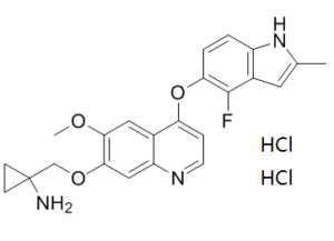
C23H24CL2FN3O3
|
|---|---|
| 分子量 |
480.36
|
| 精确质量 |
479.117
|
| 元素分析 |
C, 57.51; H, 5.04; Cl, 14.76; F, 3.96; N, 8.75; O, 9.99
|
| CAS号 |
1360460-82-7
|
| 相关CAS号 |
1058156-90-3;1360460-82-7 (HCl);
|
| PubChem CID |
57380530
|
| 外观&性状 |
Solid powder
|
| tPSA |
82.4Ų
|
| 氢键供体(HBD)数目 |
4
|
| 氢键受体(HBA)数目 |
6
|
| 可旋转键数目(RBC) |
6
|
| 重原子数目 |
32
|
| 分子复杂度/Complexity |
606
|
| 定义原子立体中心数目 |
0
|
| SMILES |
CC1=CC2=C(N1)C=CC(=C2F)OC3=C4C=C(C(=CC4=NC=C3)OCC5(CC5)N)OC.Cl.Cl
|
| InChi Key |
UUAKQNIPIXQZFN-UHFFFAOYSA-N
|
| InChi Code |
InChI=1S/C23H22FN3O3.2ClH/c1-13-9-15-16(27-13)3-4-19(22(15)24)30-18-5-8-26-17-11-21(20(28-2)10-14(17)18)29-12-23(25)6-7-23;;/h3-5,8-11,27H,6-7,12,25H2,1-2H3;2*1H
|
| 化学名 |
1-[[4-[(4-fluoro-2-methyl-1H-indol-5-yl)oxy]-6-methoxyquinolin-7-yl]oxymethyl]cyclopropan-1-amine;dihydrochloride
|
| 别名 |
AL3818 dihydrochloride; AL-3818 dihydrochloride; AL 3818 dihydrochloride; Anlotinib HCl; AL3818; Anlotinib dihydrochloride; Anlotinib HCl; 1360460-82-7; Anlotinib hydrochloride; AL3818 dihydrochloride; Catequentinib Hydrochloride; CATEQUENTINIB DIHYDROCHLORIDE; A3749M6582;AL 3818; AL-3818; Anlotinib; Catequentinib
|
| HS Tariff Code |
2934.99.9001
|
| 存储方式 |
Powder -20°C 3 years 4°C 2 years In solvent -80°C 6 months -20°C 1 month |
| 运输条件 |
Room temperature (This product is stable at ambient temperature for a few days during ordinary shipping and time spent in Customs)
|
| 溶解度 (体外实验) |
DMSO:≥ 60 mg/mL
Water: Ethanol: |
|---|---|
| 溶解度 (体内实验) |
注意: 如下所列的是一些常用的体内动物实验溶解配方,主要用于溶解难溶或不溶于水的产品(水溶度<1 mg/mL)。 建议您先取少量样品进行尝试,如该配方可行,再根据实验需求增加样品量。
注射用配方
注射用配方1: DMSO : Tween 80: Saline = 10 : 5 : 85 (如: 100 μL DMSO → 50 μL Tween 80 → 850 μL Saline)(IP/IV/IM/SC等) *生理盐水/Saline的制备:将0.9g氯化钠/NaCl溶解在100 mL ddH ₂ O中,得到澄清溶液。 注射用配方 2: DMSO : PEG300 :Tween 80 : Saline = 10 : 40 : 5 : 45 (如: 100 μL DMSO → 400 μL PEG300 → 50 μL Tween 80 → 450 μL Saline) 注射用配方 3: DMSO : Corn oil = 10 : 90 (如: 100 μL DMSO → 900 μL Corn oil) 示例: 以注射用配方 3 (DMSO : Corn oil = 10 : 90) 为例说明, 如果要配制 1 mL 2.5 mg/mL的工作液, 您可以取 100 μL 25 mg/mL 澄清的 DMSO 储备液,加到 900 μL Corn oil/玉米油中, 混合均匀。 View More
注射用配方 4: DMSO : 20% SBE-β-CD in Saline = 10 : 90 [如:100 μL DMSO → 900 μL (20% SBE-β-CD in Saline)] 口服配方
口服配方 1: 悬浮于0.5% CMC Na (羧甲基纤维素钠) 口服配方 2: 悬浮于0.5% Carboxymethyl cellulose (羧甲基纤维素) 示例: 以口服配方 1 (悬浮于 0.5% CMC Na)为例说明, 如果要配制 100 mL 2.5 mg/mL 的工作液, 您可以先取0.5g CMC Na并将其溶解于100mL ddH2O中,得到0.5%CMC-Na澄清溶液;然后将250 mg待测化合物加到100 mL前述 0.5%CMC Na溶液中,得到悬浮液。 View More
口服配方 3: 溶解于 PEG400 (聚乙二醇400) 请根据您的实验动物和给药方式选择适当的溶解配方/方案: 1、请先配制澄清的储备液(如:用DMSO配置50 或 100 mg/mL母液(储备液)); 2、取适量母液,按从左到右的顺序依次添加助溶剂,澄清后再加入下一助溶剂。以 下列配方为例说明 (注意此配方只用于说明,并不一定代表此产品 的实际溶解配方): 10% DMSO → 40% PEG300 → 5% Tween-80 → 45% ddH2O (或 saline); 假设最终工作液的体积为 1 mL, 浓度为5 mg/mL: 取 100 μL 50 mg/mL 的澄清 DMSO 储备液加到 400 μL PEG300 中,混合均匀/澄清;向上述体系中加入50 μL Tween-80,混合均匀/澄清;然后继续加入450 μL ddH2O (或 saline)定容至 1 mL; 3、溶剂前显示的百分比是指该溶剂在最终溶液/工作液中的体积所占比例; 4、 如产品在配制过程中出现沉淀/析出,可通过加热(≤50℃)或超声的方式助溶; 5、为保证最佳实验结果,工作液请现配现用! 6、如不确定怎么将母液配置成体内动物实验的工作液,请查看说明书或联系我们; 7、 以上所有助溶剂都可在 Invivochem.cn网站购买。 |
| 制备储备液 | 1 mg | 5 mg | 10 mg | |
| 1 mM | 2.0818 mL | 10.4089 mL | 20.8177 mL | |
| 5 mM | 0.4164 mL | 2.0818 mL | 4.1635 mL | |
| 10 mM | 0.2082 mL | 1.0409 mL | 2.0818 mL |
1、根据实验需要选择合适的溶剂配制储备液 (母液):对于大多数产品,InvivoChem推荐用DMSO配置母液 (比如:5、10、20mM或者10、20、50 mg/mL浓度),个别水溶性高的产品可直接溶于水。产品在DMSO 、水或其他溶剂中的具体溶解度详见上”溶解度 (体外)”部分;
2、如果您找不到您想要的溶解度信息,或者很难将产品溶解在溶液中,请联系我们;
3、建议使用下列计算器进行相关计算(摩尔浓度计算器、稀释计算器、分子量计算器、重组计算器等);
4、母液配好之后,将其分装到常规用量,并储存在-20°C或-80°C,尽量减少反复冻融循环。
计算结果:
工作液浓度: mg/mL;
DMSO母液配制方法: mg 药物溶于 μL DMSO溶液(母液浓度 mg/mL)。如该浓度超过该批次药物DMSO溶解度,请首先与我们联系。
体内配方配制方法:取 μL DMSO母液,加入 μL PEG300,混匀澄清后加入μL Tween 80,混匀澄清后加入 μL ddH2O,混匀澄清。
(1) 请确保溶液澄清之后,再加入下一种溶剂 (助溶剂) 。可利用涡旋、超声或水浴加热等方法助溶;
(2) 一定要按顺序加入溶剂 (助溶剂) 。
| NCT Number | Recruitment | interventions | Conditions | Sponsor/Collaborators | Start Date | Phases |
| NCT05481645 | Recruiting | Drug: TQB2450 injection Drug: Carboplatin Injection |
Advanced Endometrial Cancer Sarcoma of Uterus |
Chia Tai Tianqing Pharmaceutical Group Co., Ltd. |
August 22, 2022 | Phase 2 |
The lung metastasis changes in patients of alveolar soft tissue sarcoma with lung metastasis during treatment.J Hematol Oncol.2016 Oct 4;9(1):105. |
Duration of treatment and tumor size changes of 20 patients who received 12mg QD at the 2/1 schedule. J Hematol Oncol.2016 Oct 4;9(1):105. |
Plasma concentrations of anlotinib over time after a single oral dose of anlotinib capsules at 5 (green line), 10 (purple line), 12 (blue line), or 16mg anlotinib/person (red line) in male (solid circles) and female cancer patients (open circles) (a).bCorrelation of dose with plasma AUC0–120h.cCorrelation of dose with plasmaCmax.dCorrelation of dose witht1/2.ePlasma concentrations of anlotinib (24h after daily dosing) over time during multiple oral doses of anlotinib capsules at 12mg anlotinib/person/day in female cancer patients.fPlasma concentrations of anlotinib (24h after daily dosing) over time during multiple oral doses of anlotinib capsules at 12mg anlotinib/person/day in male cancer patients.J Hematol Oncol.2016 Oct 4;9(1):105. |
InvivoChem的所有产品仅用于作科学研究,不面向患者销售
Copyright 2020 InvivoChem LLC | All Rights Reserved 粤ICP备20063088号-1


 COA
COA


 463611831
