| 规格 | 价格 | 库存 | 数量 |
|---|---|---|---|
| 5mg |
|
||
| 10mg |
|
||
| 25mg |
|
||
| 50mg |
|
||
| 100mg |
|
||
| 250mg |
|
||
| Other Sizes |
|

| 靶点 |
DDP-38003 dihydrochloride targets Histone Lysine-Specific Demethylase 1A (KDM1A/LSD1, amine oxidase family enzyme); the IC₅₀ value for KDM1A inhibition was 16 nM (in vitro enzymatic assay) [1]
|
|---|---|
| 体外研究 (In Vitro) |
KDM1A 被 DDP-38003 抑制,IC50 为 84 nM。与1R、2S对应物相比,DDP-38003在抑制THP-1细胞形成集落的能力和促进其分化方面更有效[1]。
1. DDP-38003二盐酸盐 在体外脱甲基酶试验中以剂量依赖性方式有效抑制重组人KDM1A酶的活性(IC₅₀ = 16 nM)(通过单因素方差分析,p<0.001)。该试验使用H3K4me2肽作为底物[1] 2. 选择性试验显示,DDP-38003二盐酸盐(10 μM)对其他胺氧化酶(MAO-A、MAO-B)或组蛋白脱甲基酶(KDM2A、KDM3A、KDM4A、KDM6A)没有显著的抑制作用(剩余酶活性>85%,与对照组相比,p>0.05,经学生t检验得出)[1] 3. DDP-38003二盐酸盐 (0.1/1/10 μM, 72小时)以浓度依赖性的方式降低人类癌细胞系(HL-60、MV4-11、MOLM-13、A549、H1299)的存活率(CCK-8试验,通过单因素方差分析p<0.01/p<0.001),其IC₅₀值范围从0.32 μM(HL-60)到1.85 μM(H1299)[1] 4. 在HL-60急性髓系白血病(AML)细胞中,DDP-38003二盐酸盐(0.5/1/2 μM,48小时)诱导G0/G1细胞周期停滞(流式细胞仪检测,经学生t检验p<0.01)和凋亡(Annexin V-FITC/PI染色,经学生t检验p< 0.001)[1] 5. Western blot分析显示,DDP-38003二盐酸盐(0.5/1/2 μM,24/48小时)在HL-60细胞中增加了H3K4me2水平(KDM1A的底物)(p<0.01,采用学生t检验),下调了c-Myc和Bcl-2(促癌蛋白),并上调了p21和Bax(肿瘤抑制/凋亡相关蛋白)(p<0.01/p<0.001,采用学生t检验)[1] 6. qRT-PCR分析显示,DDP-38003二盐酸盐(1 μM,24小时)上调HL-60细胞中分化相关基因(CD11b、CD14)的表达(通过学生t检验,p<0.01),并下调KDM1A靶标肿瘤基因(HOXA9、MEIS1)(通过学生t检验,p< 0.01)[1] |
| 体内研究 (In Vivo) |
口服给药后,DDP-38003 具有体内功效,小鼠白血病模型的存活率提高了 62%,并有 KDM1A 抑制的证据。 DDP-38003 的半衰期为八小时。 DDP-38003 治疗可显着提高小鼠的存活率,且呈剂量依赖性。剂量分别为 11.25 和 22.50 mg/kg 时,存活率分别提高 35% 和 62%。一种可能的口服抗癌药物是 DDP-38003[1]。
1. 皮下AML异种移植模型:HL-60细胞(1×10⁷)被皮下注射到BALB/c裸小鼠的侧腹(6-8周龄);当肿瘤体积达到约100毫米³时,DDP-38003二盐酸盐 被口服给药(PO),剂量为10/20/40毫克/千克,每日一次,持续21天(溶剂:0.5%羧甲基纤维素钠(CMC-Na)+ 0.1%吐温80溶于水中);肿瘤体积每3天测量一次(体积=长度×宽度² / 2),并在处死时记录肿瘤重量(每组8只小鼠);DDP-38003 二盐酸盐 在20/40毫克每千克剂量下显著降低了肿瘤体积(通过双向ANOVA分析,p<0.01/p<0.001)和肿瘤重量(通过学生t检验,p<0.01 / p<0.001),且不影响小鼠体重[1] 2. 异种移植肿瘤的免疫组织化学(IHC)结果显示,DDP-38003二盐酸盐(40 mg/kg)增加了H3K4me2水平(H-评分定量分析,经学生t检验p<0.01),降低了Ki-67(增殖标志物,经学生t检验p< 0.01),并增加了裂解型caspase-3(凋亡标志物,经学生t检验 p<0.01)[1] 3. 原位AML模型:MV4-11细胞(5×10⁶)通过尾静脉注射到NOD/SCID小鼠(6-8周龄);口服给予DDP-38003二盐酸盐,剂量为20毫克/千克,每日一次,持续28天;生存分析显示DDP-38003 二盐酸盐显著延长了小鼠的中位生存时间(从对照组的32天延长至治疗组的48天,通过对数秩和检验p<0.01)[1] |
| 酶活实验 |
1. KDM1A脱甲基酶活性测定:重组人KDM1A蛋白与H3K4me2肽底物(50 μM)和不同浓度的DDP-38003二盐酸盐(0.001至10 μM)在测定缓冲液中在37°C下孵育1小时;反应通过添加终止缓冲液终止,然后使用比色法试剂盒定量甲醛(脱甲基反应产物);IC₅₀值由剂量响应曲线计算得出(每个组n=3重复,采用单因素方差分析进行统计分析)[1]
2. 胺氧化酶/组蛋白脱甲基酶的选择性测定:重组的MAO-A、MAO-B、KDM2A、KDM3A、KDM4A、KDM6A蛋白分别与各自的底物和DDP-38003二盐酸盐(10 μM)在37°C下孵育1小时;酶活性使用针对底物的检测试剂盒进行测量,剩余活性被计算为与载体对照的百分比(每个组n=3重复,采用学生t检验进行统计分析)[1] |
| 细胞实验 |
1. 癌细胞存活率测定: HL-60、MV4-11、MOLM-13、A549、H1299 细胞被接种在96孔板中(每孔5×10³细胞),并过夜培养;细胞分别被处理于DDP-38003二盐酸盐(0/0.1/1/10 μM)或溶剂(DMSO)72小时;加入CCK-8试剂,在450 nm处测量吸光度,细胞存活率被计算为与溶剂对照组的百分比(每个组n=6重复,采用单因素方差分析进行统计分析)[1]
2. 细胞周期分析: HL-60细胞(1×10⁵个细胞/孔)被接种在6孔板中,分别处理于DDP-38003二盐酸盐(0/0.5/1/2 μM)48小时;随后收集细胞,在4°C下用70%乙醇固定过夜,用含RNase A的碘化丙啶(PI)染色,并使用流式细胞仪进行分析(每个样本10,000个细胞,进行统计分析时采用学生t检验)[1] 3. 凋亡检测:HL-60细胞(1×10⁵个细胞/孔)被接种在6孔板中,分别处理于DDP-38003二盐酸盐(0/0.5/1/2 μM)48小时;收集细胞,用Annexin V-FITC和PI染色,并使用流式细胞仪进行分析(每个样本10,000个细胞,进行统计分析时采用学生t检验)[1] 4. Western印迹分析: HL-60细胞分别处理于DDP-38003二盐酸盐(0/0.5/1/2 μM)中,时间为24/48小时;随后提取总蛋白,进行定量,使用SDS-PAGE进行分离,转移至PVDF膜上,并使用针对H3K4me2、c-Myc、Bcl-2、p21、Bax和β-actin(内参)的抗体进行探针检测;进行条带密度分析,计算相对蛋白水平(n=3个独立实验,使用学生t检验进行统计分析)[1] 5. qRT-PCR检测:HL-60细胞被处理于DDP-38003二盐酸盐(1 μM)中24小时;随后提取总RNA,反转录成cDNA,并使用针对CD11b、CD14、HOXA9、MEIS1和GAPDH(参照基因)的引物进行qRT-PCR;相对基因表达值使用2⁻ΔΔCt法计算(n=3个独立实验,使用Student's t-test进行统计分析)[1] |
| 动物实验 |
溶于 40% PEG 400 的 5% 葡萄糖溶液中;11.25 和 22.50 mg/kg;口服给药。
小鼠白血病模型 1.皮下 AML 异种移植模型:6-8 周龄 BALB/c 裸鼠饲养于 SPF 条件下;将 HL-60 细胞(1×10⁷ 溶于 0.1 mL PBS + Matrigel (1:1 v/v))皮下注射到每只小鼠的右侧腹部;每 3 天用游标卡尺测量肿瘤体积(体积 = 长 × 宽² / 2);当肿瘤达到约 100 mm³ 时,将小鼠随机分为 4 组(载体组、10/20/40 mg/kg DDP-38003 二盐酸盐 组,每组 n=8);将DDP-38003二盐酸盐溶于0.5% CMC-Na + 0.1% Tween 80水溶液中,每日一次口服(PO)给药,持续21天;每3天记录一次小鼠体重;在研究终点(第21天),处死小鼠,切除肿瘤,称重,并用4%多聚甲醛固定,用于免疫组化分析[1] 2. 原位AML模型:将6-8周龄的NOD/SCID小鼠饲养于SPF条件下;通过尾静脉注射MV4-11细胞(5×10⁶个细胞溶于0.1 mL PBS);细胞注射24小时后,将小鼠随机分为2组(载体组,20 mg/kg DDP-38003二盐酸盐组,每组n=10);将DDP-38003二盐酸盐溶于0.5% CMC-Na + 0.1% Tween 80水溶液中,每日口服一次,连续28天;每日监测小鼠存活情况,并使用Kaplan-Meier法计算中位生存时间(统计分析采用log-rank检验)[1] 3. 异种移植瘤的IHC染色:将固定的肿瘤组织包埋于石蜡中,切片(4 μm),脱蜡,并进行抗原修复;将切片与针对H3K4me2、Ki-67和cleaved caspase-3的一抗在4°C下孵育过夜,然后与二抗孵育;进行DAB染色,并用苏木精复染切片;由两位独立的病理学家(对治疗分组不知情)对H评分(H3K4me2)或阳性细胞百分比(Ki-67/cleaved caspase-3)进行定量分析[1] |
| 药代性质 (ADME/PK) |
1. 口服生物利用度:在SD大鼠中,DDP-38003二盐酸盐的口服生物利用度为42%(单次口服10 mg/kg剂量与静脉注射2 mg/kg剂量比较,血浆浓度采用LC-MS/MS法测定)[1]
2. 血浆半衰期:DDP-38003二盐酸盐在SD大鼠中的终末血浆半衰期(t₁/₂)分别为6.8 h(口服,10 mg/kg)和5.2 h(静脉注射,2 mg/kg)[1] 3. 组织分布:SD大鼠口服10 mg/kg DDP-38003二盐酸盐后,药物广泛分布于组织中,其中肝脏(12.5 μg/g)和肾脏中的浓度最高。给药后2小时,DDP-38003在肝脏(8.7 μg/g)和脾脏(7.2 μg/g)中的浓度较高;脑组织浓度较低(0.8 μg/g),表明其血脑屏障穿透性较差[1] 4. 代谢和排泄:DDP-38003二盐酸盐主要在大鼠肝微粒体中通过氧化和葡萄糖醛酸化代谢;口服给药72小时内,35%的剂量经尿液排出,52%经粪便排出(放射性标记法测定)[1] |
| 毒性/毒理 (Toxicokinetics/TK) |
1. 急性毒性:ICR 小鼠中 DDP-38003 二盐酸盐的半数致死剂量 (LD₅₀) >200 mg/kg(口服),剂量高达 100 mg/kg 时未观察到急性毒性(无死亡或明显的体重减轻)[1]
2. 亚慢性毒性:SD 大鼠每日一次口服 10/30/100 mg/kg 的 DDP-38003 二盐酸盐,持续 28 天;在 10/30 mg/kg 剂量下未观察到体重、食物摄入量或器官重量的显著变化;在 100 mg/kg 剂量下,观察到轻度肝毒性(ALT/AST 升高,p<0.05,Student's t 检验)和肾毒性(BUN/肌酐升高,p<0.05,Student's t 检验),其他器官未见组织病理学改变 [1] 3. 血浆蛋白结合率:DDP-38003 二盐酸盐在大鼠血浆中的血浆蛋白结合率为 89%(超滤法,n=3 次重复)[1] 4. 药物相互作用:DDP-38003 二盐酸盐 (10 μM) 对人肝微粒体中的 CYP450 酶(CYP1A2、CYP2C9、CYP2C19、CYP2D6、CYP3A4)无显著抑制作用(残余酶活性)与车辆对照组相比,>80%(p>0.05,采用学生t检验)[1] |
| 参考文献 | |
| 其他信息 |
1. DDP-38003 二盐酸盐是一种新型、高效且选择性的小分子 KDM1A/LSD1 抑制剂。KDM1A/LSD1 是一种组蛋白去甲基化酶,它通过去除 H3K4me1/me2 上的甲基来调控基因表达 [1]。
2. KDM1A 在急性髓系白血病 (AML) 和实体瘤中过度表达,DDP-38003 二盐酸盐抑制 KDM1A 可导致 H3K4me2 积累、肿瘤抑制基因重新激活以及致癌通路(c-Myc/Bcl-2)抑制,最终导致细胞周期阻滞、细胞凋亡和癌细胞分化 [1]。 3. DDP-38003 二盐酸盐是首个具有良好药代动力学特性(口服生物利用度 42%,半衰期长)且低毒性的口服活性 KDM1A 抑制剂。毒性较低,使其成为治疗急性髓系白血病 (AML) 和实体瘤的潜在先导化合物 [1] 4. 临床前研究表明,DDP-38003 二盐酸盐 在皮下和原位 AML 模型中均表现出显著的抗肿瘤活性,可延长原位 AML 小鼠的生存期,且无严重的全身毒性 [1] |
| 分子式 |
C21H28CL2N4O
|
|
|---|---|---|
| 分子量 |
423.38
|
|
| 精确质量 |
422.164
|
|
| CAS号 |
1831167-98-6
|
|
| 相关CAS号 |
DDP-38003 trihydrochloride
|
|
| PubChem CID |
102004295
|
|
| 外观&性状 |
Brown to reddish brown solid powder
|
|
| tPSA |
61.6
|
|
| 氢键供体(HBD)数目 |
4
|
|
| 氢键受体(HBA)数目 |
4
|
|
| 可旋转键数目(RBC) |
4
|
|
| 重原子数目 |
28
|
|
| 分子复杂度/Complexity |
474
|
|
| 定义原子立体中心数目 |
2
|
|
| SMILES |
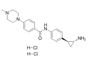
CN1CCN(CC1)C2=CC=C(C=C2)C(=O)NC3=CC=C(C=C3)[C@@H]4C[C@H]4N.Cl.Cl
|
|
| InChi Key |
HFSFENIQYFFMGC-HUQPAIQZSA-N
|
|
| InChi Code |
InChI=1S/C21H26N4O.2ClH/c1-24-10-12-25(13-11-24)18-8-4-16(5-9-18)21(26)23-17-6-2-15(3-7-17)19-14-20(19)22;;/h2-9,19-20H,10-14,22H2,1H3,(H,23,26);2*1H/t19-,20+;;/m0../s1
|
|
| 化学名 |
N-[4-[(1S,2R)-2-aminocyclopropyl]phenyl]-4-(4-methylpiperazin-1-yl)benzamide;dihydrochloride
|
|
| 别名 |
|
|
| HS Tariff Code |
2934.99.9001
|
|
| 存储方式 |
Powder -20°C 3 years 4°C 2 years In solvent -80°C 6 months -20°C 1 month |
|
| 运输条件 |
Room temperature (This product is stable at ambient temperature for a few days during ordinary shipping and time spent in Customs)
|
| 溶解度 (体外实验) |
|
|||
|---|---|---|---|---|
| 溶解度 (体内实验) |
注意: 如下所列的是一些常用的体内动物实验溶解配方,主要用于溶解难溶或不溶于水的产品(水溶度<1 mg/mL)。 建议您先取少量样品进行尝试,如该配方可行,再根据实验需求增加样品量。
注射用配方
注射用配方1: DMSO : Tween 80: Saline = 10 : 5 : 85 (如: 100 μL DMSO → 50 μL Tween 80 → 850 μL Saline)(IP/IV/IM/SC等) *生理盐水/Saline的制备:将0.9g氯化钠/NaCl溶解在100 mL ddH ₂ O中,得到澄清溶液。 注射用配方 2: DMSO : PEG300 :Tween 80 : Saline = 10 : 40 : 5 : 45 (如: 100 μL DMSO → 400 μL PEG300 → 50 μL Tween 80 → 450 μL Saline) 注射用配方 3: DMSO : Corn oil = 10 : 90 (如: 100 μL DMSO → 900 μL Corn oil) 示例: 以注射用配方 3 (DMSO : Corn oil = 10 : 90) 为例说明, 如果要配制 1 mL 2.5 mg/mL的工作液, 您可以取 100 μL 25 mg/mL 澄清的 DMSO 储备液,加到 900 μL Corn oil/玉米油中, 混合均匀。 View More
注射用配方 4: DMSO : 20% SBE-β-CD in Saline = 10 : 90 [如:100 μL DMSO → 900 μL (20% SBE-β-CD in Saline)] 口服配方
口服配方 1: 悬浮于0.5% CMC Na (羧甲基纤维素钠) 口服配方 2: 悬浮于0.5% Carboxymethyl cellulose (羧甲基纤维素) 示例: 以口服配方 1 (悬浮于 0.5% CMC Na)为例说明, 如果要配制 100 mL 2.5 mg/mL 的工作液, 您可以先取0.5g CMC Na并将其溶解于100mL ddH2O中,得到0.5%CMC-Na澄清溶液;然后将250 mg待测化合物加到100 mL前述 0.5%CMC Na溶液中,得到悬浮液。 View More
口服配方 3: 溶解于 PEG400 (聚乙二醇400) 请根据您的实验动物和给药方式选择适当的溶解配方/方案: 1、请先配制澄清的储备液(如:用DMSO配置50 或 100 mg/mL母液(储备液)); 2、取适量母液,按从左到右的顺序依次添加助溶剂,澄清后再加入下一助溶剂。以 下列配方为例说明 (注意此配方只用于说明,并不一定代表此产品 的实际溶解配方): 10% DMSO → 40% PEG300 → 5% Tween-80 → 45% ddH2O (或 saline); 假设最终工作液的体积为 1 mL, 浓度为5 mg/mL: 取 100 μL 50 mg/mL 的澄清 DMSO 储备液加到 400 μL PEG300 中,混合均匀/澄清;向上述体系中加入50 μL Tween-80,混合均匀/澄清;然后继续加入450 μL ddH2O (或 saline)定容至 1 mL; 3、溶剂前显示的百分比是指该溶剂在最终溶液/工作液中的体积所占比例; 4、 如产品在配制过程中出现沉淀/析出,可通过加热(≤50℃)或超声的方式助溶; 5、为保证最佳实验结果,工作液请现配现用! 6、如不确定怎么将母液配置成体内动物实验的工作液,请查看说明书或联系我们; 7、 以上所有助溶剂都可在 Invivochem.cn网站购买。 |
| 制备储备液 | 1 mg | 5 mg | 10 mg | |
| 1 mM | 2.3619 mL | 11.8097 mL | 23.6194 mL | |
| 5 mM | 0.4724 mL | 2.3619 mL | 4.7239 mL | |
| 10 mM | 0.2362 mL | 1.1810 mL | 2.3619 mL |
1、根据实验需要选择合适的溶剂配制储备液 (母液):对于大多数产品,InvivoChem推荐用DMSO配置母液 (比如:5、10、20mM或者10、20、50 mg/mL浓度),个别水溶性高的产品可直接溶于水。产品在DMSO 、水或其他溶剂中的具体溶解度详见上”溶解度 (体外)”部分;
2、如果您找不到您想要的溶解度信息,或者很难将产品溶解在溶液中,请联系我们;
3、建议使用下列计算器进行相关计算(摩尔浓度计算器、稀释计算器、分子量计算器、重组计算器等);
4、母液配好之后,将其分装到常规用量,并储存在-20°C或-80°C,尽量减少反复冻融循环。
计算结果:
工作液浓度: mg/mL;
DMSO母液配制方法: mg 药物溶于 μL DMSO溶液(母液浓度 mg/mL)。如该浓度超过该批次药物DMSO溶解度,请首先与我们联系。
体内配方配制方法:取 μL DMSO母液,加入 μL PEG300,混匀澄清后加入μL Tween 80,混匀澄清后加入 μL ddH2O,混匀澄清。
(1) 请确保溶液澄清之后,再加入下一种溶剂 (助溶剂) 。可利用涡旋、超声或水浴加热等方法助溶;
(2) 一定要按顺序加入溶剂 (助溶剂) 。
InvivoChem的所有产品仅用于作科学研究,不面向患者销售
Copyright 2020 InvivoChem LLC | All Rights Reserved 粤ICP备20063088号-1


 COA
COA
 463611831
