| 规格 | 价格 | 库存 | 数量 |
|---|---|---|---|
| 5mg |
|
||
| 10mg |
|
||
| 25mg |
|
||
| 50mg |
|
||
| Other Sizes |
|
| 靶点 |
GLP-1 receptor
|
|---|---|
| 体外研究 (In Vitro) |
Dulaglutide(50 nM 和 100 nM;24 小时)可改善 ox-LDL 诱导的氧化应激并抑制 ox-LDL 诱导的人主动脉内皮细胞 (HAEC) 线粒体功能障碍[1]。细胞活力测定[1] 细胞系:人主动脉内皮细胞 (HAEC) 浓度:50 nM、100 nM 孵育时间:24 小时 结果:抑制 ox-LDL 诱导的细胞活力降低和乳酸脱氢酶 (LDH) 释放。
杜拉鲁肽改善了ox-LDL诱导的氧化应激和线粒体功能障碍。[1] 杜拉鲁肽抑制ox-LDL诱导的IL-1β、IL-6、MCP-1和HMG-1的分泌。[1] 杜拉鲁肽抑制ox-LDL诱导的细胞存活率降低和LDH释放。[1] 杜拉鲁肽通过抑制VCAM-1、E-选择素抑制THP-1与HAEC的附着。[1] 杜拉鲁肽通过抑制p53的激活来促进KLF2的表达。[1] |
| 体内研究 (In Vivo) |
在大鼠致癌性研究中,杜拉鲁肽(0、0.05、0.5、1.5 或 5 mg/kg;皮下注射;每周两次,持续 93 周)会增加甲状腺 C 细胞增生和肿瘤的发生率[2]。动物模型:大鼠和转基因小鼠[1] 剂量:0、0.05、0.5、1.5或5 mg/kg; 0、0.3、1 或 3 mg/kg 给药:皮下注射,每周两次,持续 93 周; SC,每周两次,持续 26 周 结果:弥漫性 C 细胞增生和腺瘤在统计学上增加,>0.5 mg/kg。随着时间的推移,小鼠的全身暴露量减少。
在大鼠和转基因小鼠中评估杜拉鲁肽的致瘤潜力。大鼠每周皮下注射两次杜拉鲁肽0、0.05、0.5、1.5或5mg/kg,持续93周,分别相当于基于血浆曲线下面积的最大推荐人体剂量的0、0.5、7、20和58倍。转基因小鼠每周两次皮下注射杜拉鲁肽0、0.3、1或3mg/kg,持续26周。杜拉鲁肽的作用仅限于甲状腺C细胞。在大鼠中,0.5 mg/kg或更高剂量的弥漫性C细胞增生和腺瘤在统计学上增加(5 mg/kg时P≤.01),5 mg/kg时C细胞癌在数量上增加。与对照组相比,0.5、1.5和5 mg/kg的雌性大鼠局灶性C细胞增殖更高。在转基因小鼠中,在任何剂量下都没有观察到杜拉鲁肽相关的C细胞增生或肿瘤;然而,在所有杜拉鲁肽组中都观察到C细胞的最小细胞质肥大。随着时间的推移,小鼠的全身暴露量逐渐减少,这可能是由于抗药物抗体反应。在一项为期52周的研究中,旨在定量C细胞质量和血浆降钙素反应,大鼠每周两次皮下注射杜拉鲁肽0或5mg/kg。杜拉鲁肽增加了局灶性C细胞增生;C细胞质量没有发生定量增加。与C细胞质量缺乏形态计量学变化一致,杜拉鲁肽不影响弥漫性C细胞增生或基础或钙刺激的血浆降钙素的发生率,这表明在大鼠致癌性研究的最初52周内没有发生C细胞质量的弥漫性增加[3]。 |
| 酶活实验 |
活性氧(ROS)的评估[1]
使用2′,7′-二氯二氢荧光素二乙酸酯(DCFH-DA)染色测量HAEC中的细胞内ROS。在有或没有浓度为50和100 nM的dulaglutide/杜拉鲁肽的情况下,用ox-LDL(100μg/ml)刺激HAEC 24小时,并用PBS洗涤3次。然后在37°C的黑暗中用5μM DCFH-DA加载细胞15分钟。使用蔡司荧光显微镜观察荧光信号。使用Image J软件计算细胞内ROS。简而言之,在荧光图像中定义了感兴趣区域(ROI),并计算了所定义ROI中存在的细胞的平均数量。计算ROI中的积分密度值(IDV),并除以平均细胞数。结果用于表示细胞内ROS的平均水平。 还原型谷胱甘肽(GSH)测定[1] 使用荧光测定法测定HAEC中还原型谷胱甘肽(GSH)的细胞内水平。在有或没有浓度为50和100 nM的dulaglutide/杜拉鲁肽的情况下,用ox-LDL(100μg/ml)刺激HAEC 24小时。然后将细胞收集在冰冷的5%偏磷酸(MPA)中。然后对细胞进行超声处理,并在14000×g下离心5分钟。上清液与等体积的OPAME在甲醇和硼酸盐缓冲液中孵育,并在室温下孵育15分钟。在350 nm激发和420 nm发射下记录荧光信号。 线粒体膜电位(MMP)的测定[1] 使用四甲基罗丹明甲酯(TMRM)染色测定HAEC中MMP的细胞内水平。在存在或不存在浓度为50和100 nM的dulaglutide的情况下,用ox-LDL(100μg/ml)刺激HAEC 24小时。然后用PBS洗涤细胞3次,用20 nmol/L TMRM探测。在37°C下孵育1小时后,将细胞洗涤3次,并使用蔡司荧光显微镜观察荧光信号。 |
| 细胞实验 |
细胞系:人主动脉内皮细胞 (HAEC)
浓度:50 nM、100 nM 孵育时间:24 小时 结果:抑制 ox-LDL 诱导的细胞活力降低和乳酸脱氢酶 (LDH) 释放)。 细胞粘附试验[1] 将HAEC培养至80%融合。在存在或不存在浓度为50和100 nM的杜拉鲁肽的情况下,用ox-LDL(100μg/ml)刺激细胞24小时。共有2×105个THP-1单核细胞用钙黄绿素乙酰氧基甲酯染色30分钟,并与HAEC孵育2小时。将未附着的THP-1细胞洗掉,并用荧光显微镜观察附着的THP-1细胞。 细胞活力评估[1] 将HAEC接种到6孔板中,在有或没有浓度为50和100 nM的<强>杜拉鲁肽的情况下,用ox-LDL(100μg/ml)刺激24小时。经过3次温和洗涤后,加入终浓度为5 mg/ml的3-(4,5-二甲基噻唑-2-基)-2,5-二苯基溴化四氮唑(MTT)的无酚红培养基,在37°C的黑暗中孵育4小时,产物用二甲亚砜(DMSO)溶解。测量570 nm处的OD值以反映存活率百分比。 乳酸脱氢酶(LDH)释放的测定[1] 将HAEC接种到6孔板中,在有或没有浓度为50和100 nM的杜拉鲁肽的情况下,用ox-LDL(100μg/ml)刺激24小时。收集50μl上清液,并在新鲜的96孔板中与50μl LDH测定试剂混合。在黑暗中孵育30分钟后,用50μl停止缓冲液停止反应。记录490 nm处的OD值以评估LDH释放。 |
| 动物实验 |
大鼠和转基因小鼠
0、0.05、0.5、1.5 或 5 mg/kg;0、0.3、1 或 3 mg/kg 皮下注射,每周两次,持续 93 周;皮下注射,每周两次,持续 26 周 血浆度拉糖肽毒代动力学[3] 为期 26 周的小鼠研究[3] 在给药阶段的第 1、85 和 176 天采集血样。每个时间点,每个性别、每个组别均采集三只动物的血液样本。分别于第1天和第176天给药前以及给药后4、12、24、48和96小时,以及第85天给药前和给药后24小时采集血样。采用经验证的ELISA方法分析血浆样本中的度拉糖肽浓度。 微孔板包被小鼠抗人IgG(Fc)抗体。度拉糖肽标准品、对照品和样本均用小鼠血浆配制。配制完成后,将样本滴加到包被板上,室温孵育约1.5小时。板上的度拉糖肽复合物与豚鼠抗GLP-1活性抗血清结合,然后使用山羊抗豚鼠IgG-辣根过氧化物酶和四甲基联苯胺底物进行检测。标准曲线范围为 0.25 至 125 ng/mL,定量下限为 2.0 ng/mL,定量上限为 30 ng/mL。 93 周致癌性研究(基于 52 周大鼠甲状腺 C 细胞研究)[3] 在 93 周致癌性研究中,分别于第 1 天和第 24 天给药前以及给药后 4、12、24、48 和 96 小时采集血液样本(每组每性别每时间点各取三只大鼠),并在第 13、26、78 和 93 周给药前以及给药后 24 小时采集血液样本。在 52 周形态计量学研究中,分别于第 94、185、276 和 374 天处死动物时(给药后约 6 天)采集血液样本(每组每性别每时间点各取三只大鼠)。两项研究的血浆样本均采用经验证的 ELISA 方法分析度拉糖肽浓度。 微量滴定板包被单克隆抗GLP-1抗体。度拉糖肽标准品、对照品和样品均用大鼠血浆配制。配制完成后,将样品滴加到包被板上,室温孵育约1.5小时。使用小鼠抗人IgG4-辣根过氧化物酶抗体(Southern Biotech)和四甲基联苯胺底物检测板上的度拉糖肽复合物。标准曲线范围为0.39至50 ng/mL,定量下限和上限分别为0.80 ng/mL和40 ng/mL。 52周大鼠甲状腺C细胞研究[3] 微量滴定板包被小鼠抗人IgG(Fc)抗体。度拉糖肽标准品、对照品和样品均用大鼠血浆配制。配制完成后,将样品滴加到包被板上,室温孵育约1小时。将板上的度拉糖肽复合物与小鼠IgG2a kappa抗GLP-1抗体结合,然后用山羊抗小鼠IgG2a-辣根过氧化物酶和四甲基联苯胺底物进行检测。标准曲线范围为0.40至100 ng/mL,定量下限和上限分别为0.80 ng/mL和40 ng/mL。 |
| 毒性/毒理 (Toxicokinetics/TK) |
为期26周的小鼠研究[3]
生命阶段[3] 度拉糖肽的给药对存活率没有影响。未观察到与度拉糖肽相关的临床症状。与对照组相比,度拉糖肽治疗组雄性小鼠的平均食物消耗量普遍下降,导致平均体重相应降低(补充图1-4)。治疗组雌性小鼠的食物消耗量也观察到类似但通常不太显著的变化,且未导致生长减缓。 血浆度拉糖肽毒代动力学[3] 给药后4至12小时观察到度拉糖肽血浆浓度达峰。根据曲线下面积 (AUC) 和血浆峰浓度 (Cmax) 值评估,度拉糖肽的暴露量随剂量增加而增加,但在第 176 天,其与剂量增加的比例通常低于剂量增加的比例。男性和女性的度拉糖肽全身暴露量相似(表 1)。第 85 天的度拉糖肽血浆浓度(未显示)以及第 176 天的 Cmax 和 AUC 值通常比第 1 天的相应值低 0.5 倍或更少(表 1)。研究期间暴露量的下降可能是由于度拉糖肽抗药抗体 (ADA) 的形成所致;然而,并未进行度拉糖肽ADA的特异性测定。 解剖病理学[3] 在对照组和治疗组中,使用常规苏木精-伊红染色甲状腺切片,未检测到与度拉糖肽相关的效应,也未发现甲状腺C细胞增生或肿瘤的证据。在降钙素染色的甲状腺切片中,所有接受试验药物治疗的组均检测到C细胞胞质体积增大,并记录为C细胞胞质肥大/降钙素染色增强(表2)。程度较轻,度拉糖肽治疗小鼠的甲状腺C细胞数量未见明显增加。在MNU阳性对照动物中观察到死亡率增加,以及细支气管肺泡腺瘤和癌、鳞状细胞乳头状瘤和癌、血管瘤和血管肉瘤的发生率增加,这反映了该品系小鼠在接受MNU给药后的典型反应。 93周大鼠致癌性研究[3] 体内阶段[3] 由于对照动物存活率低(每性别<20只动物),经FDA致癌性评估执行委员会同意,该研究在第93周终止。然而,有足够数量的动物存活至第80周,足以充分评估致癌性。未发现与度拉糖肽相关的特定死亡原因。在所有度拉糖肽治疗组中,无论男女,生存率均有所提高,且在0.5 mg/kg和5 mg/kg雄性组以及0.05 mg/kg、0.5 mg/kg和1.5 mg/kg雌性组中达到统计学意义(P ≤ 0.05)(表3)。与对照组相比,药物引起的平均体重和平均食物消耗量下降通常呈剂量依赖性(补充图5-8)。 血浆度拉糖肽毒代动力学[3] 在第1天给药后12小时观察到度拉糖肽血浆峰浓度的平均时间,并在第52周时介于12至48小时之间。根据AUC和Cmax值评估,在所有评估日期,度拉糖肽的暴露量均随剂量从0.05 mg/kg增加到5 mg/kg而增加(表1)。第1天和第52周的峰浓度(Cmax)和AUC0-96h值与剂量呈近似正比关系。在所有剂量组和评估日期,雄性和雌性大鼠的药物暴露量相似。稳态似乎在第13周达到,并且药物暴露量维持至第93周。第52周的Cmax和AUC0-96h值高于第1天,表明在92周多次给药后,度拉糖肽可能在大鼠血浆中蓄积。此外,在第93周研究结束时,各毒性组的平均血浆度拉糖肽浓度与相应毒代动力学组的平均浓度基本相似。 解剖病理学[3] 在度拉糖肽剂量为0.5、1.5和5 mg/kg时,男性和女性甲状腺C细胞腺瘤的发生率均显著高于对照组(P ≤ 0.05)(表4)。5 mg/kg剂量组的甲状腺C细胞癌发生率在数值上较高,但未达到统计学意义。在接受度拉糖肽1.5 mg/kg和5 mg/kg治疗的男性以及接受度拉糖肽5 mg/kg治疗的雌性中,弥漫性C细胞增生的发生率均显著高于对照组(P ≤ 0.05)。与对照组相比,接受度拉糖肽0.5、1.5和5 mg/kg治疗的雌性大鼠局灶性C细胞增生的发生率更高。接受度拉糖肽0.5、1.5或5 mg/kg治疗的雄性大鼠局灶性C细胞增生的发生率较低,且在甲状腺C细胞腺瘤发生率呈上升趋势后,局灶性C细胞增生的发生率也呈下降趋势(补充图9和10)。 为期52周的大鼠甲状腺C细胞研究[3] 体内研究阶段[3] 未观察到与度拉糖肽相关的死亡或临床症状。3只对照组大鼠在给药阶段第365或372天接受氯化钙(CaCl2)后数分钟内死亡。此外,3只对照组大鼠和2只接受5 mg/kg度拉糖肽治疗的大鼠在非计划时间间隔内死亡。一只接受 5 mg/kg 剂量的大鼠死于造血系统肿瘤;其余大鼠的死因不明。 血浆度拉糖肽毒代动力学[3] 在动物处死的中间时间点(末次给药后 6 天,即第 94、185、276 和 374 天),血浆度拉糖肽浓度通常随着研究的进行而降低,且第 185 天的度拉糖肽浓度存在相当大的变异性。在末次给药后 13 天(第 374 天),19 只接受 5 mg/kg 剂量的动物中有 18 只的血浆度拉糖肽浓度低于定量限。其中一只动物的血浆浓度为 0.91 ng/mL。研究期间度拉糖肽暴露量的降低可能是由于抗体的形成;然而,并未进行度拉糖肽抗药抗体(ADA)的特异性测定。尽管度拉糖肽血浆浓度在研究期间总体呈下降趋势,但在整个给药阶段均观察到与度拉糖肽药理学相关的效应[食物摄入量减少和体重增加减少(补充图11和12)],表明在整个研究期间,活性度拉糖肽始终存在于受试动物体内。 解剖病理学[3] 在所有尸检中,受试动物的平均终末体重均有所下降(为对照组平均值的83%–88%)。受试动物的平均甲状腺/甲状旁腺绝对重量(为对照组平均值的81%)和甲状腺/甲状旁腺与脑重量比(为对照组平均值的84%)的下降被认为是继发于体重下降。唯一被认为与度拉糖肽相关的显微镜检查结果是,在给药52周后,治疗组大鼠甲状腺中局灶性或多灶性C细胞肥大/增生的发生率和严重程度增加(表5)。在给药26周或39周后,一两只治疗组动物出现局灶性或多灶性C细胞肥大/增生,这被认为与度拉糖肽无关;这种病变是大鼠的自发性背景变化,正如一只对照动物在给药39周后出现该病变所证实的那样。局灶性和多灶性C细胞肥大/增生的特征是大小不一的分化良好的C细胞结节,这些结节通常具有增大的胞质体积(肥大)。这些病灶中的C细胞通常比周围的C细胞的降钙素免疫阳性强度低。仅在接受 52 周给药后检查的动物中发现了 C 细胞肿瘤,在对照组和治疗组动物中发生率同样很低,并且被认为与度拉糖肽无关。 |
| 参考文献 |
|
| 其他信息 |
动脉粥样硬化是II型糖尿病的常见合并症,也是全球主要的死亡原因之一。氧化低密度脂蛋白(ox-LDL)通过诱导氧化应激、线粒体功能障碍、促炎细胞因子和趋化因子(包括白细胞介素(IL)-1β、IL-6和单核细胞趋化蛋白1(MCP-1))、黏附分子(包括血管细胞黏附分子1(VCAM-1)和E-选择素)的表达以及下调Krüppel样因子2(KLF2)转录因子的表达,促进动脉粥样硬化的发生发展。值得注意的是,ox-LDL可诱导THP-1单核细胞黏附于内皮细胞。在本研究中,我们首次证实,特异性胰高血糖素样肽-1受体(GLP-1R)激动剂度拉糖肽可通过阻止p53蛋白对人主动脉内皮细胞中KLF2的抑制,从而预防ox-LDL的动脉粥样硬化作用。KLF2已被证实对保护血管内皮细胞免受ox-LDL和振荡剪切力损伤起着重要作用,因此,能够调节KLF2信号通路的疗法可能是一种有吸引力且可用于预防动脉粥样硬化发生和发展的有效治疗方案。[1]
背景:三种不同的胰高血糖素样肽-1(GLP-1)受体激动剂可降低糖化血红蛋白A1c(HbA1c)浓度高且心血管风险高的2型糖尿病患者的心血管事件发生率。我们评估了在现有降糖方案中加入GLP-1受体激动剂度拉糖肽对2型糖尿病患者(无论既往是否有心血管疾病史,且血糖控制水平差异较大)主要不良心血管事件的影响。方法:这项多中心、随机、双盲、安慰剂对照试验在24个国家的371个研究中心进行。年龄至少50岁、患有2型糖尿病且既往有心血管事件或存在心血管危险因素的男性和女性,按1:1的比例随机分配至度拉糖肽(1.5 mg)皮下注射组(每周一次)或安慰剂组。随机化采用计算机生成的随机代码,并按研究中心进行分层。所有研究者和受试者均对治疗分配不知情。受试者至少每6个月随访一次,以评估新发心血管事件和其他严重临床结局。主要终点为首次发生非致死性心肌梗死、非致死性卒中或心血管原因死亡(包括不明原因死亡)的复合终点,该终点在符合意向治疗原则的人群中进行评估。本研究已在ClinicalTrials.gov注册,注册号为NCT01394952。研究结果:2011年8月18日至2013年8月14日期间,共纳入9901名受试者(平均年龄66.2岁[标准差6.5],糖化血红蛋白中位数为7.2%[四分位距6.6-8.1],其中女性4589名[46.3%]),并随机分配至度拉糖肽组(n=4949)或安慰剂组(n=4952)。中位随访时间为 5.4 年(四分位距 5.1-5.9 年),度拉糖肽组有 594 例(12.0%)受试者发生主要复合终点事件,发生率为每 100 人年 2.4 例;安慰剂组有 663 例(13.4%)受试者发生主要复合终点事件,发生率为每 100 人年 2.7 例(风险比 [HR] 0.88,95% 置信区间 [CI] 0.79-0.99;p=0.026)。两组的全因死亡率无显著差异(度拉糖肽组 536 例 [10.8%] vs 安慰剂组 592 例 [12.0%];HR 0.90,95% CI 0.80-1.01;p=0.067)。在随访期间,接受度拉糖肽治疗的受试者中有 2347 例(47.4%)报告了胃肠道不良事件,而接受安慰剂治疗的受试者中有 1687 例(34.1%)报告了胃肠道不良事件(p<0.0001)。结论:度拉糖肽可用于治疗既往有心血管疾病或心血管危险因素的中老年 2 型糖尿病患者的血糖控制。[2] |
| 精确质量 |
3313.597
|
|---|---|
| CAS号 |
923950-08-7
|
| 相关CAS号 |
GLP-1 moiety from Dulaglutide; 1197810-60-8
|
| PubChem CID |
171042928
|
| 序列 |
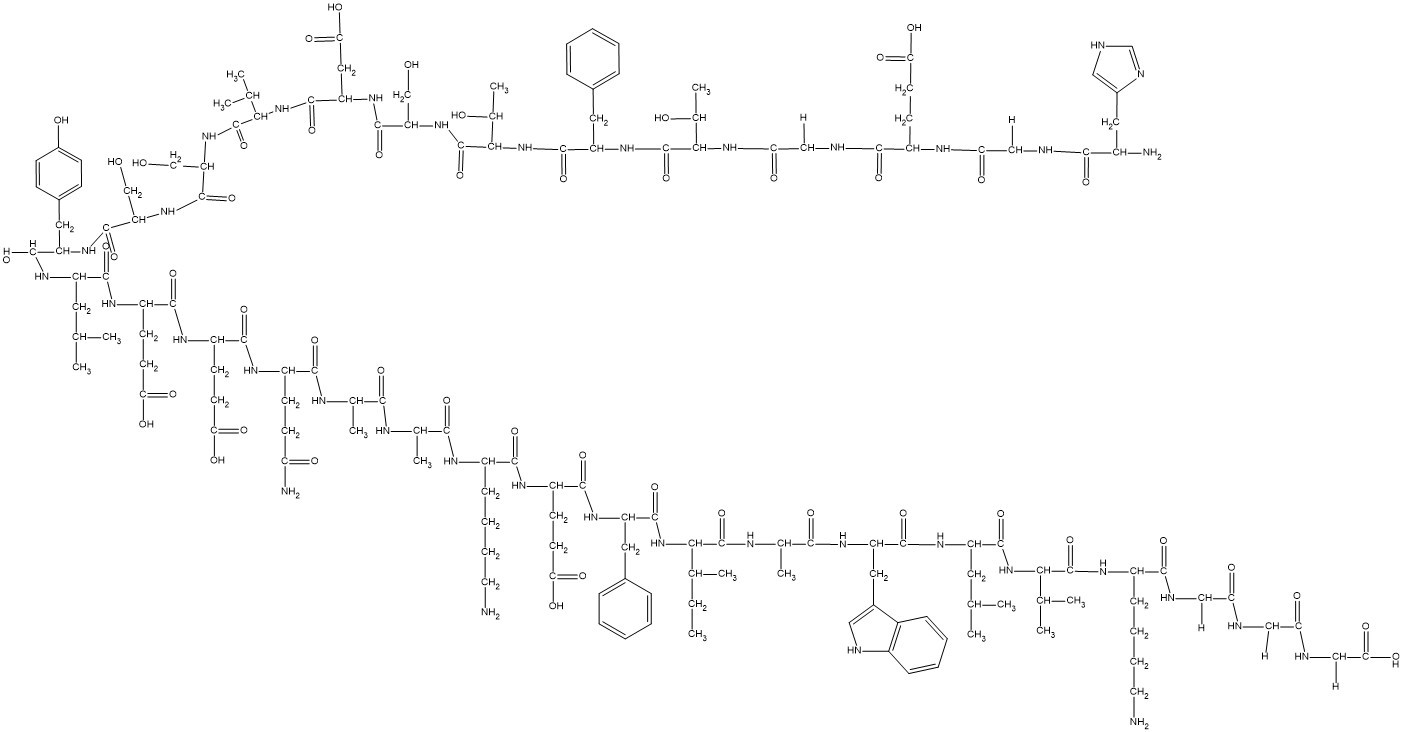
HGEGTFTSDVSSYLEEQAAKEFIAWLVKGGG
|
| 外观&性状 |
Typically exists as solid at room temperature
|
| 密度 |
1.4±0.1 g/cm3
|
| 折射率 |
1.706
|
| LogP |
3.81
|
| tPSA |
1380
|
| 氢键供体(HBD)数目 |
48
|
| 氢键受体(HBA)数目 |
53
|
| 可旋转键数目(RBC) |
109
|
| 重原子数目 |
235
|
| 分子复杂度/Complexity |
7740
|
| 定义原子立体中心数目 |
29
|
| SMILES |
CC[C@H](C)[C@@H](C(=O)N[C@@H](C)C(=O)N[C@@H](CC1=CNC2=CC=CC=C21)C(=O)N[C@@H](CC(C)C)C(=O)N[C@@H](C(C)C)C(=O)N[C@@H](CCCCN)C(=O)NCC(=O)NCC(=O)NCC(=O)O)NC(=O)[C@H](CC3=CC=CC=C3)NC(=O)[C@H](CCC(=O)O)NC(=O)[C@H](CCCCN)NC(=O)[C@H](C)NC(=O)[C@H](C)NC(=O)[C@H](CCC(=O)N)NC(=O)[C@H](CCC(=O)O)NC(=O)[C@H](CCC(=O)O)NC(=O)[C@H](CC(C)C)NC(=O)[C@H](CC4=CC=C(C=C4)O)NC(=O)[C@H](CO)NC(=O)[C@H](CO)NC(=O)[C@H](C(C)C)NC(=O)[C@H](CC(=O)O)NC(=O)[C@H](CO)NC(=O)[C@H]([C@@H](C)O)NC(=O)[C@H](CC5=CC=CC=C5)NC(=O)[C@H]([C@@H](C)O)NC(=O)CNC(=O)[C@H](CCC(=O)O)NC(=O)CNC(=O)[C@H](CC6=CNC=N6)N
|
| InChi Key |
HPNPLWNTQBSMAJ-FBXRENMFSA-N
|
| InChi Code |
InChI=1S/C149H221N37O49/c1-16-76(10)121(147(233)164-79(13)126(212)172-103(58-85-61-155-90-34-24-23-33-88(85)90)137(223)174-99(54-73(4)5)138(224)183-119(74(6)7)145(231)171-91(35-25-27-51-150)128(214)159-64-110(195)156-63-109(194)157-67-118(208)209)185-139(225)101(55-82-29-19-17-20-30-82)175-134(220)97(45-50-116(204)205)168-131(217)92(36-26-28-52-151)166-125(211)78(12)162-124(210)77(11)163-130(216)94(41-46-108(153)193)167-132(218)95(43-48-114(200)201)169-133(219)96(44-49-115(202)203)170-135(221)98(53-72(2)3)173-136(222)100(57-84-37-39-87(192)40-38-84)176-142(228)105(68-187)179-144(230)107(70-189)180-146(232)120(75(8)9)184-141(227)104(60-117(206)207)177-143(229)106(69-188)181-149(235)123(81(15)191)186-140(226)102(56-83-31-21-18-22-32-83)178-148(234)122(80(14)190)182-112(197)66-160-129(215)93(42-47-113(198)199)165-111(196)65-158-127(213)89(152)59-86-62-154-71-161-86/h17-24,29-34,37-40,61-62,71-81,89,91-107,119-123,155,187-192H,16,25-28,35-36,41-60,63-70,150-152H2,1-15H3,(H2,153,193)(H,154,161)(H,156,195)(H,157,194)(H,158,213)(H,159,214)(H,160,215)(H,162,210)(H,163,216)(H,164,233)(H,165,196)(H,166,211)(H,167,218)(H,168,217)(H,169,219)(H,170,221)(H,171,231)(H,172,212)(H,173,222)(H,174,223)(H,175,220)(H,176,228)(H,177,229)(H,178,234)(H,179,230)(H,180,232)(H,181,235)(H,182,197)(H,183,224)(H,184,227)(H,185,225)(H,186,226)(H,198,199)(H,200,201)(H,202,203)(H,204,205)(H,206,207)(H,208,209)/t76-,77-,78-,79-,80+,81+,89-,91-,92-,93-,94-,95-,96-,97-,98-,99-,100-,101-,102-,103-,104-,105-,106-,107-,119-,120-,121-,122-,123-/m0/s1
|
| 化学名 |
(4S)-5-[[2-[[(2S,3R)-1-[[(2S)-1-[[(2S,3R)-1-[[(2S)-1-[[(2S)-1-[[(2S)-1-[[(2S)-1-[[(2S)-1-[[(2S)-1-[[(2S)-1-[[(2S)-1-[[(2S)-1-[[(2S)-5-amino-1-[[(2S)-1-[[(2S)-1-[[(2S)-6-amino-1-[[(2S)-1-[[(2S)-1-[[(2S,3S)-1-[[(2S)-1-[[(2S)-1-[[(2S)-1-[[(2S)-1-[[(2S)-6-amino-1-[[2-[[2-(carboxymethylamino)-2-oxoethyl]amino]-2-oxoethyl]amino]-1-oxohexan-2-yl]amino]-3-methyl-1-oxobutan-2-yl]amino]-4-methyl-1-oxopentan-2-yl]amino]-3-(1H-indol-3-yl)-1-oxopropan-2-yl]amino]-1-oxopropan-2-yl]amino]-3-methyl-1-oxopentan-2-yl]amino]-1-oxo-3-phenylpropan-2-yl]amino]-4-carboxy-1-oxobutan-2-yl]amino]-1-oxohexan-2-yl]amino]-1-oxopropan-2-yl]amino]-1-oxopropan-2-yl]amino]-1,5-dioxopentan-2-yl]amino]-4-carboxy-1-oxobutan-2-yl]amino]-4-carboxy-1-oxobutan-2-yl]amino]-4-methyl-1-oxopentan-2-yl]amino]-3-(4-hydroxyphenyl)-1-oxopropan-2-yl]amino]-3-hydroxy-1-oxopropan-2-yl]amino]-3-hydroxy-1-oxopropan-2-yl]amino]-3-methyl-1-oxobutan-2-yl]amino]-3-carboxy-1-oxopropan-2-yl]amino]-3-hydroxy-1-oxopropan-2-yl]amino]-3-hydroxy-1-oxobutan-2-yl]amino]-1-oxo-3-phenylpropan-2-yl]amino]-3-hydroxy-1-oxobutan-2-yl]amino]-2-oxoethyl]amino]-4-[[2-[[(2S)-2-amino-3-(1H-imidazol-4-yl)propanoyl]amino]acetyl]amino]-5-oxopentanoic acid
|
| 别名 |
Dulaglutide; GLP-1 moiety from Dulaglutide; 923950-08-7; Dulaglutide; 1197810-60-8; HPNPLWNTQBSMAJ-FBXRENMFSA-N; LY2189265
|
| HS Tariff Code |
2934.99.9001
|
| 存储方式 |
Powder -20°C 3 years 4°C 2 years In solvent -80°C 6 months -20°C 1 month |
| 运输条件 |
Room temperature (This product is stable at ambient temperature for a few days during ordinary shipping and time spent in Customs)
|
| 溶解度 (体外实验) |
May dissolve in DMSO (in most cases), if not, try other solvents such as H2O, Ethanol, or DMF with a minute amount of products to avoid loss of samples
|
|---|---|
| 溶解度 (体内实验) |
Note: 如何溶解多肽产品?请参考本产品网页右上角“产品说明书”文件,第4页。 注意: 如下所列的是一些常用的体内动物实验溶解配方,主要用于溶解难溶或不溶于水的产品(水溶度<1 mg/mL)。 建议您先取少量样品进行尝试,如该配方可行,再根据实验需求增加样品量。 注射用配方
注射用配方1: DMSO : Tween 80: Saline = 10 : 5 : 85 (如: 100 μL DMSO → 50 μL Tween 80 → 850 μL Saline)(IP/IV/IM/SC等) *生理盐水/Saline的制备:将0.9g氯化钠/NaCl溶解在100 mL ddH ₂ O中,得到澄清溶液。 注射用配方 2: DMSO : PEG300 :Tween 80 : Saline = 10 : 40 : 5 : 45 (如: 100 μL DMSO → 400 μL PEG300 → 50 μL Tween 80 → 450 μL Saline) 注射用配方 3: DMSO : Corn oil = 10 : 90 (如: 100 μL DMSO → 900 μL Corn oil) 示例: 以注射用配方 3 (DMSO : Corn oil = 10 : 90) 为例说明, 如果要配制 1 mL 2.5 mg/mL的工作液, 您可以取 100 μL 25 mg/mL 澄清的 DMSO 储备液,加到 900 μL Corn oil/玉米油中, 混合均匀。 View More
注射用配方 4: DMSO : 20% SBE-β-CD in Saline = 10 : 90 [如:100 μL DMSO → 900 μL (20% SBE-β-CD in Saline)] 口服配方
口服配方 1: 悬浮于0.5% CMC Na (羧甲基纤维素钠) 口服配方 2: 悬浮于0.5% Carboxymethyl cellulose (羧甲基纤维素) 示例: 以口服配方 1 (悬浮于 0.5% CMC Na)为例说明, 如果要配制 100 mL 2.5 mg/mL 的工作液, 您可以先取0.5g CMC Na并将其溶解于100mL ddH2O中,得到0.5%CMC-Na澄清溶液;然后将250 mg待测化合物加到100 mL前述 0.5%CMC Na溶液中,得到悬浮液。 View More
口服配方 3: 溶解于 PEG400 (聚乙二醇400) 请根据您的实验动物和给药方式选择适当的溶解配方/方案: 1、请先配制澄清的储备液(如:用DMSO配置50 或 100 mg/mL母液(储备液)); 2、取适量母液,按从左到右的顺序依次添加助溶剂,澄清后再加入下一助溶剂。以 下列配方为例说明 (注意此配方只用于说明,并不一定代表此产品 的实际溶解配方): 10% DMSO → 40% PEG300 → 5% Tween-80 → 45% ddH2O (或 saline); 假设最终工作液的体积为 1 mL, 浓度为5 mg/mL: 取 100 μL 50 mg/mL 的澄清 DMSO 储备液加到 400 μL PEG300 中,混合均匀/澄清;向上述体系中加入50 μL Tween-80,混合均匀/澄清;然后继续加入450 μL ddH2O (或 saline)定容至 1 mL; 3、溶剂前显示的百分比是指该溶剂在最终溶液/工作液中的体积所占比例; 4、 如产品在配制过程中出现沉淀/析出,可通过加热(≤50℃)或超声的方式助溶; 5、为保证最佳实验结果,工作液请现配现用! 6、如不确定怎么将母液配置成体内动物实验的工作液,请查看说明书或联系我们; 7、 以上所有助溶剂都可在 Invivochem.cn网站购买。 |
计算结果:
工作液浓度: mg/mL;
DMSO母液配制方法: mg 药物溶于 μL DMSO溶液(母液浓度 mg/mL)。如该浓度超过该批次药物DMSO溶解度,请首先与我们联系。
体内配方配制方法:取 μL DMSO母液,加入 μL PEG300,混匀澄清后加入μL Tween 80,混匀澄清后加入 μL ddH2O,混匀澄清。
(1) 请确保溶液澄清之后,再加入下一种溶剂 (助溶剂) 。可利用涡旋、超声或水浴加热等方法助溶;
(2) 一定要按顺序加入溶剂 (助溶剂) 。
Perioperative Stress Hyperglycemia in General and Vascular Surgery Patients
CTID: NCT04862234
Phase: Phase 4 Status: Recruiting
Date: 2024-09-20
InvivoChem的所有产品仅用于作科学研究,不面向患者销售
Copyright 2020 InvivoChem LLC | All Rights Reserved 粤ICP备20063088号-1


 COA
COA
 463611831
