| 规格 | 价格 | 库存 | 数量 |
|---|---|---|---|
| 2g |
|
||
| 5g |
|
||
| 10g |
|
||
| 50g |
|
||
| 100g |
|
||
| 200g |
|
||
| Other Sizes |
|

| 靶点 |
Surfactant
|
|---|---|
| 体外研究 (In Vitro) |
分别采用膜水合法和反溶剂法制备了负载厚朴酚的混合胶束(MMs)和厚朴酚纳米混悬剂(MNs)。最佳的MMs和MNs制剂分别以1:12:5和2:1:1的比例使用厚朴酚、Soluplus®和泊洛沙姆188制备。MMs的平均粒径为111.8±14.6,MNs的平均粒度为78.53±5.4nm。MMs的包封率和载药率分别为89.58±2.54%和5.46±0.65%。MNs的载药效率为42.50±1.57%。在体外释放研究中,MMs表现出缓慢的药物释放,而MNs表现出快速的药物释放。Caco-2跨细胞转运研究的结果表明,MMs和MNs都增加了厚朴酚的渗透。如药代动力学研究所示,MMs和MNs分别显著促进胃肠道药物吸收2.85倍和2.27倍 [1]。
|
| 体内研究 (In Vivo) |
创伤性脑损伤(TBI)是全球发病率和死亡率的主要原因,可破坏血管内皮系统的细胞膜完整性,危及血脑屏障功能并威胁细胞生存。对血管内皮系统的保护可能会增强TBI后的临床结果。泊洛沙姆188(P188)已被证明可改善缺血/再灌注(I/R)损伤后和TBI后神经元功能。我们旨在建立一种体外压缩型TBI模型,比较轻度、中度和重度损伤,以观察P188对小鼠脑微血管内皮细胞(MBEC)的直接影响。将汇合的MBEC暴露于常氧或缺氧条件下5或15小时(小时)。加入压缩1小时,并在2小时复氧期间给予P188。通过评估细胞数量/活力、细胞毒性/膜损伤、代谢活性和总一氧化氮生成(tNOp)来测试P188对MBEC的直接影响。虽然P188增强了细胞数量/活力、代谢活性和tOp,但阻止了轻度至中度损伤后细胞毒性/膜损伤的增加。在严重损伤的MBEC中,P188仅改善代谢活性。P188在复氧过程中存在,直接影响模拟I/R和压缩型TBI中的MBEC功能[2]。
|
| 酶活实验 |
代谢活性[2]
使用CellTiter 96 AQueous单溶液细胞增殖测定法测定代谢活性。四唑化合物(3-(4,5-二甲基噻唑-2-基)-5-(3-羧甲氧基苯基)-2-(4-磺基苯基)-2H-四唑鎓,内盐;MTS)被代谢活性细胞生物还原,形成可溶于培养基中的有色甲zan产物。在20–25°C下解冻90分钟后,将20µL CellTiter 96 AQueous One Solution Reagent添加到每个含有100µL培养基的孔中(达到约16.67%的浓度)。将平板在37°C的细胞培养箱中,在含5%CO2的加湿空气中培养1-4小时。在板读数器中在490nm处读取abs。最后,从总腹肌中减去空白腹肌。[2] NO总生成量[2] tOp使用开曼硝酸盐/亚硝酸盐比色测定试剂盒进行测定。一氧化氮(NO)是一种高活性、短命的自由基,在生物系统中由NO合酶(NOS)家族的酶合成,包括内皮型NOS(eNOS)、神经元型NOS(nNOS)和诱导型NOS(iNOS)。不同的细胞类型可以产生NO,包括内皮细胞。NO的主要作用是鸟苷酸环化酶的旁分泌激活,这导致细胞内环状鸟苷单磷酸的增加,导致平滑肌细胞放松。该测定通过添加亚硝酸盐(NO2−)和硝酸盐(NO3−)来测量NO的总产量,这两种物质都代表NO的最终产物。首先,NO3−暴露于NO3−还原酶,将其转化为NO2−。在第二步中,Griess试剂用于将NO2−转化为偶氮发色团。测量所得abs可以确定NO2−浓度。制备试剂和样品,并按照测定方案中的指示进行测定。简言之,将10µL的酶辅因子混合物和硝酸还原酶混合物添加到多达80µL的样品中(所得浓度:10%的酶辅剂混合物、10%的硝酸还原酶混合物、80%的样品)。在所需的孵育时间后,补充50µL Griess试剂R1和50µL Griess试剂R2(每个结果的浓度为25%)。使用平板读数器在540nm处测量abs。每当进行测定时,按照测定方案中的说明制备标准曲线。 |
| 细胞实验 |
皮尔斯乳酸脱氢酶(LDH)细胞毒性测定试剂盒(Thermo Fisher Scientific;Waltham,MA,USA)用于测定细胞毒性/膜损伤。在健康细胞中,LDH是一种胞浆酶。在质膜损伤之后,LDH被释放到培养基中。我们使用比色法,通过使用偶联酶反应测量细胞外LDH来量化细胞毒性。LDH催化乳酸盐到丙酮酸盐的反应,导致烟酰胺腺嘌呤二核苷酸从其氧化态(NAD+)还原为其还原态(NADH)。四唑盐碘硝基四唑鎓通过黄递酶使用NADH还原为红色甲酰胺产物。甲酰胺的形成与LDH的释放量成正比。这可用于指示细胞毒性/膜破坏的水平。按照制造商的说明制备和储存试剂。简言之,将含有基质混合物(冻干物)的小瓶用11.4mL超纯水稀释。将测定缓冲液解冻,同时遮光。反应混合物由0.6 mL测定缓冲液(5%)和11.4 mL底物混合物(95%)组成。首先,将每个孔的50µL介质转移到未预涂的透明96孔板上。之后,加入50µL反应混合物(达到50%反应混合物的浓度),轻轻拍打板,并使用铝箔保护板不受光照。将平板在室温下孵育。30分钟后,加入50µL停止溶液(浓度约33%)。然后,10分钟后,使用平板读数器(Synergy H1,BioTek Instruments股份有限公司)在490nm处测量培养基内的吸光度。在第二步中,将5µL裂解缓冲液(10X)加入到含有细胞和50µL残余培养基的原始平板中。将平板在37°C下孵育60分钟,并如上所述进行测定。计算吸光度(abs):
abs(培养基)/(abs[培养基]+abs[裂解的细胞])[2]。
|
| 动物实验 |
动物/疾病模型: 大鼠止血带诱导缺血再灌注损伤 [2]
剂量: 150 mg/kg 给药途径: 静脉注射/iv 实验结果: 显著降低了升高的 TBARS 水平,但未降至对照水平,SOD 活性与对照水平相当。 雄性 Sprague-Dawley 大鼠接受 180 分钟的止血带诱导缺血。在解除止血带前 5 分钟,大鼠通过颈静脉导管接受 (1) 泊洛沙姆-188 (P-188)(150 mg/kg;P-188 组)或 (2) 赋形剂(赋形剂组)的推注(每组 n=10)。再灌注240分钟后,两组大鼠均通过尾静脉导管接受第二次推注,分别注射泊洛沙姆-188 (P-188)或载体(Vehicle)。16小时后,处死大鼠;测定肌肉重量,梗死面积(2,3,5-氯化三苯基四氮唑法),并对腓肠肌和胫前肌进行盲法组织学分析(苏木精-伊红染色),以及抗氧化状态指标。[3] 实验步骤和监测[3] 大鼠被随机分为泊洛沙姆-188 (P-188)组(P-188 + 载体)或载体组(仅载体)(每组n = 10)。称量大鼠体重后,使用1.5%至2.5%的异氟烷进行麻醉,并腹腔注射丁丙诺啡(0.1 mg/kg)进行镇痛。剃除双侧后肢毛发,并在直肠括约肌远端5 cm处插入润滑的直肠温度探头。随后将动物仰卧放置于温水循环温控床上,并将核心体温维持在37°C ± 1°C。在止血带充气前,将实验腿抬高至高于心脏水平5分钟,以引流血液。然后将气动止血带套于抬高后肢的近端,并充气至250 mmHg。所有操作步骤均已在之前的文献中详细描述16。止血带保留180分钟。图 1 详细描述了实验过程,包括麻醉、恢复、导尿和药物给药等所有步骤。在两次麻醉之间,大鼠被放回笼中,并可自由摄取水和食物。实验期间未给予复苏液。 给予泊洛沙姆-188 (P-188) [3] 无菌 P-188 溶液包含 150 mg/mL 高纯度 P-188、3.08 mg/mL 氯化钠、2.38 mg/mL 柠檬酸钠和 0.366 mg/mL 柠檬酸。安慰剂溶液包含相同的成分,但不含泊洛沙姆-188 (P-188)。剂量为 1.0 mL/kg 体重的 P-188 溶液或赋形剂。通过尾静脉注射P-188或载体。分别在(1)止血带松开前5分钟和(2)止血带松开后240分钟进行两次注射。第二次注射前,大鼠接受轻度麻醉(1.5%异氟烷)。此给药方案旨在最大程度地提高P-188的生物利用度。 |
| 参考文献 |
|
| 其他信息 |
泊洛沙姆是一种环氧化物。
泊洛沙姆是一种非离子型三嵌段共聚物,由疏水性聚氧丙烯核心和两侧的亲水性聚氧乙烯侧链组成,可用作药物递送系统中的固定剂、增溶剂、乳化剂和稳定剂。 另见:泊洛沙伦(注释已移至);泊洛沙姆188(注释已移至);泊洛沙姆331(注释已移至)…… 创伤性脑损伤(TBI)是全球发病率和死亡率的主要原因,它会破坏血管内皮系统的细胞膜完整性,危及血脑屏障功能并威胁细胞存活。保护血管内皮系统可能改善TBI后的临床预后。泊洛沙姆188 (P188) 已被证实能够改善缺血/再灌注 (I/R) 损伤以及创伤性脑损伤 (TBI) 后的神经元功能。本研究旨在建立一种体外压迫型TBI模型,比较轻度至中度损伤和重度损伤,以观察P188对小鼠脑微血管内皮细胞 (MBEC) 的直接作用。将汇合的MBEC暴露于常氧或低氧条件下5小时或15小时。随后进行1小时的压迫处理,并在2小时的复氧期间给予P188。通过评估细胞数量/活力、细胞毒性/膜损伤、代谢活性和总一氧化氮生成量 (tNOp) 来检测P188对MBEC的直接作用。结果显示,P188能够提高细胞数量/活力、代谢活性和tNOp,同时抑制轻度至中度损伤后细胞毒性/膜损伤的增加。在严重受损的MBEC中,P188仅改善了代谢活性。在复氧过程中存在的P188可直接影响模拟缺血/再灌注损伤和压迫型TBI中的MBEC功能。[1] 背景:骨骼肌损伤可导致显著水肿,进而可能导致急性肢体筋膜室综合征(CS)的发生。泊洛沙姆-188(P-188)是一种多嵌段共聚物表面活性剂,已被证明可通过封闭多种组织在各种损伤方式后的受损膜来减轻水肿。本研究旨在确定给予P-188是否能显著减轻与缺血/再灌注损伤(IR)相关的骨骼肌水肿。 方法:雄性Sprague-Dawley大鼠接受180分钟的止血带诱导缺血。在止血带松开前5分钟,大鼠经颈静脉导管分别接受(1)P-188(150 mg/kg;P-188组)或(2)载体(载体组)的推注(每组n=10)。再灌注240分钟后,两组大鼠均经尾静脉导管再次接受P-188(P-188组)或载体(载体组)的推注。16小时后,处死大鼠;测定肌肉重量,采用2,3,5-氯化三苯基四氮唑法测定梗死面积,并对腓肠肌和胫前肌进行盲法组织学分析(苏木精-伊红染色),同时检测抗氧化状态指标。 结果:与载体组相比,P-188组显著减轻了水肿(湿重),并降低了脂质过氧化指数(p<0.05)。 P-188组的湿重/干重比值较低(表明水肿程度较轻)。通过2,3,5-氯化三苯基四氮唑染色或常规组织学检查评估的肌肉活力在各组间无统计学差异。 结论:P-188显著降低了缺血再灌注相关的肌肉水肿和脂质过氧化,但对肌肉活力没有影响。过度水肿可导致急性肢体缺血综合征(CS),而CS与显著的发病率和死亡率相关。P-188可能为减轻CS提供一种潜在的辅助治疗方法。[2] 本研究旨在探讨三嵌段共聚物泊洛沙姆188对纳米颗粒形态、粒径、癌细胞摄取和细胞毒性的影响。采用油包水乳液/溶剂蒸发法,以可生物降解的聚乳酸-羟基乙酸共聚物(PLGA)为基底,分别添加或不添加泊洛沙姆188,制备了载有多西他赛的纳米颗粒。所得纳米颗粒呈球形,表面粗糙多孔。纳米颗粒的平均粒径约为200 nm,粒径分布较窄。体外药物释放曲线显示,两种纳米颗粒制剂均呈现双相释放模式。与PLGA纳米颗粒相比,PLGA/泊洛沙姆188纳米颗粒在多西他赛耐药的MCF-7 TAX30人乳腺癌细胞系中的摄取量显著增加。此外,载多西他赛的PLGA/泊洛沙姆188纳米颗粒的细胞毒性显著高于载多西他赛的PLGA纳米颗粒和泰索帝(P < .05)。总之,结果表明,与未包封泊洛沙姆188的纳米颗粒相比,包封泊洛沙姆188的载多西他赛PLGA纳米颗粒在乳腺癌化疗中具有更佳的药物缓释性和疗效。[3] |
| 分子量 |
8800 (Average)
|
|---|---|
| 精确质量 |
304.115
|
| CAS号 |
9003-11-6
|
| 相关CAS号 |
691397-13-4;126925-06-2;697765-47-2;9003-11-6;106392-12-5
|
| PubChem CID |
24751
|
| 外观&性状 |
White to yellow solid
|
| 密度 |
1.2±0.1 g/cm3
|
| 沸点 |
370.7±37.0 °C at 760 mmHg
|
| 熔点 |
60-50ºC
|
| 闪点 |
160.5±26.5 °C
|
| 蒸汽压 |
0.0±0.8 mmHg at 25°C
|
| 折射率 |
1.452
|
| LogP |
0
|
| tPSA |
25.06
|
| 氢键供体(HBD)数目 |
0
|
| 氢键受体(HBA)数目 |
2
|
| 可旋转键数目(RBC) |
0
|
| 重原子数目 |
7
|
| 分子复杂度/Complexity |
36.7
|
| 定义原子立体中心数目 |
0
|
| SMILES |
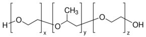
CC(COCCO)OCCO
|
| InChi Key |
OQNWUUGFAWNUME-UHFFFAOYSA-N
|
| InChi Code |
InChI=1S/C7H16O4/c1-7(11-5-3-9)6-10-4-2-8/h7-9H,2-6H2,1H3
|
| 化学名 |
2-[2-(2-hydroxyethoxy)propoxy]ethanol
|
| 别名 |
Pluronic F-68; Pluronic F-127; 2-[2-(2-hydroxyethoxy)propoxy]ethanol; 721929-01-7; Therabloat (TN); ...; CAS-9003-11-6;
|
| HS Tariff Code |
2934.99.9001
|
| 存储方式 |
Powder -20°C 3 years 4°C 2 years In solvent -80°C 6 months -20°C 1 month |
| 运输条件 |
Room temperature (This product is stable at ambient temperature for a few days during ordinary shipping and time spent in Customs)
|
| 溶解度 (体外实验) |
DMSO: ~100 mg/mL
Water: ~100 mg/mL Ethanol: ~100 mg/mL |
|---|---|
| 溶解度 (体内实验) |
注意: 如下所列的是一些常用的体内动物实验溶解配方,主要用于溶解难溶或不溶于水的产品(水溶度<1 mg/mL)。 建议您先取少量样品进行尝试,如该配方可行,再根据实验需求增加样品量。
注射用配方
注射用配方1: DMSO : Tween 80: Saline = 10 : 5 : 85 (如: 100 μL DMSO → 50 μL Tween 80 → 850 μL Saline)(IP/IV/IM/SC等) *生理盐水/Saline的制备:将0.9g氯化钠/NaCl溶解在100 mL ddH ₂ O中,得到澄清溶液。 注射用配方 2: DMSO : PEG300 :Tween 80 : Saline = 10 : 40 : 5 : 45 (如: 100 μL DMSO → 400 μL PEG300 → 50 μL Tween 80 → 450 μL Saline) 注射用配方 3: DMSO : Corn oil = 10 : 90 (如: 100 μL DMSO → 900 μL Corn oil) 示例: 以注射用配方 3 (DMSO : Corn oil = 10 : 90) 为例说明, 如果要配制 1 mL 2.5 mg/mL的工作液, 您可以取 100 μL 25 mg/mL 澄清的 DMSO 储备液,加到 900 μL Corn oil/玉米油中, 混合均匀。 View More
注射用配方 4: DMSO : 20% SBE-β-CD in Saline = 10 : 90 [如:100 μL DMSO → 900 μL (20% SBE-β-CD in Saline)] 口服配方
口服配方 1: 悬浮于0.5% CMC Na (羧甲基纤维素钠) 口服配方 2: 悬浮于0.5% Carboxymethyl cellulose (羧甲基纤维素) 示例: 以口服配方 1 (悬浮于 0.5% CMC Na)为例说明, 如果要配制 100 mL 2.5 mg/mL 的工作液, 您可以先取0.5g CMC Na并将其溶解于100mL ddH2O中,得到0.5%CMC-Na澄清溶液;然后将250 mg待测化合物加到100 mL前述 0.5%CMC Na溶液中,得到悬浮液。 View More
口服配方 3: 溶解于 PEG400 (聚乙二醇400) 请根据您的实验动物和给药方式选择适当的溶解配方/方案: 1、请先配制澄清的储备液(如:用DMSO配置50 或 100 mg/mL母液(储备液)); 2、取适量母液,按从左到右的顺序依次添加助溶剂,澄清后再加入下一助溶剂。以 下列配方为例说明 (注意此配方只用于说明,并不一定代表此产品 的实际溶解配方): 10% DMSO → 40% PEG300 → 5% Tween-80 → 45% ddH2O (或 saline); 假设最终工作液的体积为 1 mL, 浓度为5 mg/mL: 取 100 μL 50 mg/mL 的澄清 DMSO 储备液加到 400 μL PEG300 中,混合均匀/澄清;向上述体系中加入50 μL Tween-80,混合均匀/澄清;然后继续加入450 μL ddH2O (或 saline)定容至 1 mL; 3、溶剂前显示的百分比是指该溶剂在最终溶液/工作液中的体积所占比例; 4、 如产品在配制过程中出现沉淀/析出,可通过加热(≤50℃)或超声的方式助溶; 5、为保证最佳实验结果,工作液请现配现用! 6、如不确定怎么将母液配置成体内动物实验的工作液,请查看说明书或联系我们; 7、 以上所有助溶剂都可在 Invivochem.cn网站购买。 |
计算结果:
工作液浓度: mg/mL;
DMSO母液配制方法: mg 药物溶于 μL DMSO溶液(母液浓度 mg/mL)。如该浓度超过该批次药物DMSO溶解度,请首先与我们联系。
体内配方配制方法:取 μL DMSO母液,加入 μL PEG300,混匀澄清后加入μL Tween 80,混匀澄清后加入 μL ddH2O,混匀澄清。
(1) 请确保溶液澄清之后,再加入下一种溶剂 (助溶剂) 。可利用涡旋、超声或水浴加热等方法助溶;
(2) 一定要按顺序加入溶剂 (助溶剂) 。
InvivoChem的所有产品仅用于作科学研究,不面向患者销售
Copyright 2020 InvivoChem LLC | All Rights Reserved 粤ICP备20063088号-1


 COA
COA
 463611831
