| 规格 | 价格 | 库存 | 数量 |
|---|---|---|---|
| 500μg |
|
||
| 1mg |
|
||
| Other Sizes |
|
| 靶点 |
Aflibercept inhibits vascular endothelial growth factor (VEGF) signaling by acting as a soluble decoy receptor for VEGF-A, VEGF-B, and placental growth factor (PlGF) [1]
|
|---|---|
| 体外研究 (In Vitro) |
在台盼蓝排除实验或 MTT 测定中,阿柏西普(500 μg/mL;24 小时和 7 d)对 RPE 细胞均未表现出任何毒性[1]。在具有三个伤口的汇合 RPE 细胞层中,与对照相比,阿柏西普(500 μg/mL;24 小时)对伤口愈合表现出统计学上显着的影响[1]。与未处理的对照相比,阿柏西普(500 μg/mL;7 d)表现出对调理乳胶珠的吞噬作用明显降低[1]。阿柏西普(1 和 10 μg/mL;10 小时)可降低血管生成和通透性(这两个 VEGF 调节过程),进而抑制 VEGF 信号传导[2]。
Aflibercept (500 µg/mL) 对原代猪RPE细胞无细胞毒性:MTT实验中细胞存活率为97.8±3.0%(24小时)和102.4±0.9%(7天);台盼蓝排除实验中为97.9±7.3%(24小时)和97.2±17.4%(7天)。[1] 临床相关浓度(500 µg/mL)下,Aflibercept 显著抑制RPE伤口愈合(划痕实验),伤口闭合率降至70.3±10.3%(对照组87.3±8.4%,p<0.01)。125 µg/mL时无影响(72.2±6.1% vs. 对照组74.2±5.5%)。[1] Aflibercept (500 µg/mL) 降低RPE细胞对调理化乳胶珠的吞噬能力(28.3±2.9 珠/细胞 vs. 对照组42.8±4.3 珠/细胞,p<0.001),效果与贝伐单抗相当(30.2±5.2 珠/细胞)。[1] Aflibercept (1–10 µg/mL) 降低rVEGF-A诱导的人脐静脉内皮细胞(HUVECs)通透性18.8–27.1%(p<0.01–0.0001),经FITC-葡聚糖跨孔实验测定。管腔形成呈剂量依赖性抑制,证实抗血管生成活性。[2] 在原代猪RPE细胞中,Aflibercept (500 µg/mL) 损害伤口愈合(闭合率70.3±10.3% vs. 对照组87.3±8.4%,p<0.01),并降低对调理化珠粒的吞噬能力(28.3±2.9 珠/细胞 vs. 对照组42.8±4.3,p<0.001)。[2] HUVECs经Aflibercept(50 nM,24小时)处理后,细胞内NO水平显著降低(DAF-FM荧光检测),ROS积累增加(DHE染色显示),与NOX1/NOX4蛋白表达上调相关[3] Western blot分析表明,Aflibercept(50 nM,24小时)降低Akt(Ser473)和eNOS(Ser1177)磷酸化,抑制CAT-1转运蛋白表达,并减少细胞内L-精氨酸浓度[3] |
| 体内研究 (In Vivo) |
在肥胖小鼠中,阿柏西普(10 mg/kg;大脑中动脉闭塞后 3 小时;MCAO))通过减少中风诱导的 VEGF-A 和 VEGFR2 表达、脑水肿和 BBB 破坏来增加中风后存活率 [2]。阿柏西普(18.2 mg/kg 和 36.4 mg/kg;腹腔注射一次)影响小鼠的血压、ROS 的产生和 eNOS[3]。
在饮食诱导的肥胖卒中小鼠中,Aflibercept (10 mg/kg 静脉注射,大脑中动脉闭塞(MCAO)后3h单次给药) 降低死亡率(17% vs. IgG对照组40%)、出血性转化(27% vs. 43%)和脑水肿(18% vs. 28%,p<0.01)。血脑屏障破坏(IgG/葡聚糖外渗)显著减弱(p<0.05)。[2] 神经行为评分在卒中后1/3天改善(p<0.05)。在非肥胖恶性梗死小鼠中无获益,证实疗效具共病选择性。[2] C57BL/6小鼠单次静脉注射Aflibercept(18.2或36.4 mg/kg)诱发急性剂量依赖性高血压(收缩压峰值出现在第2天),损害乙酰胆碱介导的内皮依赖性舒张(EDR),并降低主动脉NO生物利用度[3] 长期给药(14天内5次注射)导致持续高血压,增加主动脉ET-1水平,升高ROS(通过NOX1/NOX4介导),并持续抑制p-Akt/p-eNOS信号及CAT-1表达[3] |
| 细胞实验 |
伤口愈合(划痕实验): 融合的原代猪RPE细胞用牙签划伤创口,洗涤后加入含Aflibercept (125 µg/mL或500 µg/mL)的无酚红DMEM(含10% FBS)。24小时后通过显微镜定量伤口闭合率(AxioVision软件)。[1]
吞噬功能实验: RPE细胞经Aflibercept (500 µg/mL)处理7天后,与猪光感受器外节调理化的FITC标记乳胶珠共孵育4小时。通过荧光显微镜计数每个细胞的内化珠粒量化吞噬能力。[1] 细胞摄取: RPE细胞经Aflibercept (125 µg/mL或500 µg/mL)处理1小时至7天后,固定、透化,并用山羊抗人AlexaFluor555抗体染色。荧光显微镜显示囊泡状摄取(≥82.5%细胞阳性),持续7天。[1] |
| 动物实验 |
动物/疾病模型:雄性C57BL/6小鼠[3]
剂量:18.2 mg/kg和36.4 mg/kg 给药途径:静脉注射;18.2 mg/kg和36.4 mg/kg单次注射 实验结果:小鼠血压迅速且呈剂量依赖性升高,内皮依赖性舒张(EDR)显著受损,导致NADPH氧化酶1(NOX1)和NADPH氧化酶4(NOX4)介导的活性氧(ROS)生成减少,蛋白激酶B(Akt)和内皮型一氧化氮合酶(eNOS)的活化降低,同时一氧化氮(NO)生成减少,小鼠主动脉内皮素-1(ET-1)水平升高。 肥胖的C57BL/6小鼠(高脂饮食喂养8-12周)接受30分钟的短暂性大脑中动脉闭塞(MCAO)。闭塞后3小时,静脉注射阿柏西普(10 mg/kg)或IgG对照。72小时后评估结局(梗死体积、肿胀、血脑屏障完整性)。[2] 对于瘦小鼠的严重卒中:40分钟的MCAO,随后给予相同的阿柏西普剂量(10 mg/kg,静脉注射,闭塞后3小时)。[2] 雄性C57BL/6小鼠(8-10周龄)接受静脉注射阿柏西普(18.2或36.4 mg/kg),单次给药(短期)或每隔2天给药5次(长期)。剂量根据人鼠剂量转换计算(人剂量当量:4 mg/kg)[3] 在 L-精氨酸联合给药研究中,小鼠在注射阿柏西普前 3 天经口预先给予 L-精氨酸(0.5 或 1.0 g/kg,每日两次),并持续给药直至研究结束[3] |
| 药代性质 (ADME/PK) |
吸收、分布和排泄
在新生血管性(湿性)年龄相关性黄斑变性患者中,玻璃体内注射阿柏西普(每眼2 mg)后,游离阿柏西普的平均血浆峰浓度在1-3天内达到;据估计,该浓度比达到全身性VEGF半数最大结合所需的阿柏西普浓度低100倍以上。玻璃体内注射后2周,血浆中游离阿柏西普的浓度无法检测到。重复玻璃体内注射(即每4周一次)后,未观察到阿柏西普在血浆中的蓄积。 吸收 单眼玻璃体内注射8 mg阿柏西普后,血浆中游离阿柏西普的平均(标准差)Cmax为0.30 (0.27) mg/L,达到血浆最大浓度的中位时间为2.9天。最初三个月每月一次玻璃体内注射阿柏西普后,血浆中游离阿柏西普的蓄积量极低(平均蓄积比为1.2);随后未观察到进一步蓄积。在湿性年龄相关性黄斑变性(AMD)、早产儿视网膜病变(RVO)和糖尿病性黄斑水肿(DME)患者中,血浆中游离阿柏西普的平均Cmax分别为0.02 mcg/mL(范围:0至0.054 mcg/mL)、0.05 mcg/mL(范围:0至0.081 mcg/mL)和0.03 mcg/mL(范围:0至0.076 mcg/mL),在每眼玻璃体内注射2 mg后1至3天内达到。所有患者在给药两周后,血浆中均检测不到游离阿柏西普。 分布容积 静脉注射阿柏西普后,游离阿柏西普的分布容积约为 7 升。 清除率 在癌症患者中,每 2 或 3 周静脉输注 2 至 9 mg/kg 阿柏西普 1 小时后,游离阿柏西普和结合阿柏西普的清除率分别估计为 0.88 升/天和 0.14 升/天。健康受试者的游离阿柏西普清除率与上述情况相似,但结合阿柏西普的清除率略快(0.19 升/天)。白蛋白水平低或碱性磷酸酶水平高的患者通常也表现出游离阿柏西普清除率加快。 代谢/代谢物 阿柏西普是一种治疗性蛋白质,尚未进行药物代谢研究。预计阿柏西普的消除途径包括:通过与游离内源性 VEGF 结合进行靶向介导的清除,以及通过蛋白水解进行代谢。 生物半衰期 对于玻璃体内注射制剂,半衰期估计为 7.13 天。对于静脉注射制剂,每两周静脉注射 4 mg/kg 剂量后,游离 ziv-aflibercept 的消除半衰期约为 6 天(范围 4-7 天)。 |
| 毒性/毒理 (Toxicokinetics/TK) |
单次静脉注射(10 mg/kg)未在小鼠中引起高血压或白细胞减少症。出血性转化减少(27% vs. 对照组的 43%),表明出血风险未增加。[2]
在视网膜色素上皮细胞中,500 µg/mL 的阿柏西普未显示细胞毒性(MTT/台盼蓝检测显示,24 小时/7 天时细胞活力 >97%)。[1] 阿柏西普在 42.4% 的患者中诱发高血压,在 17.4% 的患者中诱发高血压危象(临床数据)。在小鼠中,它通过抑制 eNOS/NO 信号通路、ROS 过度产生和 ET-1 升高引起血管功能障碍。[3] 毒性总结 识别和用途:玻璃体内注射阿柏西普用于治疗新生血管性(湿性)年龄相关性黄斑变性。人体暴露和毒性:在初始视力较差的情况下,阿柏西普在改善视力方面更有效。接受阿柏西普治疗的患者的无进展生存期高于安慰剂组。观察到的最常见不良反应是贫血、腹泻和中性粒细胞减少症。对于接受含奥沙利铂治疗后病情进展的转移性结直肠癌患者,阿柏西普联合FOLFIRI方案是一种安全有效的治疗选择。尚未证实其优于其他抗血管生成治疗。对于既往接受过贝伐珠单抗和/或雷珠单抗注射治疗的患者,阿柏西普是一种有价值的替代治疗方案。在既往接受过每4-6周一次雷珠单抗和/或贝伐珠单抗注射治疗的患者中,开始阿柏西普治疗后观察到视力稳定,且光谱域光学相干断层扫描(SD-OCT)显示眼部解剖结构有所改善。 52 周时,接受每 8 周一次 2 mg 阿柏西普、每 4 周一次 2 mg 阿柏西普或每 4 周一次 0.5 mg 雷珠单抗治疗的患者,其视力平均变化(定义为与基线相比视力增加或下降的字母数)分别为 7.9-8.9 个字母、7.6-10.9 个字母和 8.1-9.4 个字母。动物研究:在接受玻璃体内注射阿柏西普治疗的猴子中,每只眼睛玻璃体内注射 2 mg 或 4 mg 阿柏西普时,观察到鼻甲呼吸上皮出现糜烂和溃疡。在猴子中评估的最低剂量(3 mg/kg)的阿柏西普静脉注射后,其全身暴露量(AUC)约为人类玻璃体内注射 2 mg 阿柏西普后全身暴露量的 1500 倍。治疗停止后20周内,所有变化均可逆转。在兔模型中,所有评估剂量的阿柏西普均导致胎儿畸形。在兔模型中,给予最低评估剂量(0.1 mg/kg)后,其全身暴露量(AUC)约为人体玻璃体内注射2 mg后全身暴露量的10倍。 阿柏西普旨在通过玻璃体内注射发挥眼部局部作用。玻璃体内注射可使药物渗透所有视网膜层,从而最大限度地减少全身作用。在将培养的角膜内皮细胞暴露于不同浓度阿柏西普的研究中,未观察到细胞毒性作用。 然而,由于预充式注射器活塞的轻微错位,人们对玻璃体内注射阿柏西普治疗可能导致药物过量产生了兴趣。这些微小的测量误差可能导致注射的药物体积是预期体积的两倍。自阿柏西普上市以来,部分眼压升高病例是由过量用药引起的。遗憾的是,目前尚无已知的阿柏西普过量解毒剂。严格按照使用说明进行操作可以最大限度地减少剂量错误,并预防玻璃体内注射阿柏西普的毒性。 妊娠和哺乳期影响 ◉ 哺乳期用药概述 本记录涉及玻璃体内注射阿柏西普的使用。阿柏西普可抑制血管内皮生长因子 (VEGF)。阿柏西普是一种分子量为 115,000 的大分子蛋白质,由于其可能在婴儿的胃肠道内部分被破坏且口服吸收不良,因此不太可能被吸收,预计不会对婴儿产生全身性影响。在一位母亲接受玻璃体内注射后,四天内仅有一天在母乳中检测到少量阿柏西普。阿柏西普已获准用于治疗早产儿视网膜病变的玻璃体内注射。对哺乳婴儿的风险似乎非常低。由于人乳中含有血管内皮生长因子(VEGF),且VEGF被认为有助于婴儿胃肠道的成熟,因此人们对哺乳期母亲使用VEGF抑制剂表示担忧。然而,母乳的典型替代品是婴儿配方奶粉,其中不含VEGF。 ◉ 对母乳喂养婴儿的影响 截至修订日期,未找到相关的已发表信息。 ◉ 对泌乳和母乳的影响 一名患有糖尿病性黄斑水肿的妇女在产后一周接受了2毫克阿柏西普玻璃体内注射。她没有进行母乳喂养。在注射前和注射后第1至4天采集了乳汁样本。 VEGF 水平从基线时的 10.6 mcg/L 降至第 1 天的 4.9 mcg/L,并在接下来的 3 天内保持在该水平。 不良反应 阿柏西普的使用相关不良反应包括眼刺激、玻璃体脱离、暂时性视力模糊、眼睑肿胀和结膜出血。与阿柏西普给药和注射操作相关的严重不良反应包括视网膜脱离、外伤性白内障、血栓栓塞事件和眼压升高。这些并发症在阿柏西普玻璃体内注射中的发生率低于 0.1%。 预计给药后 60 分钟内会出现暂时性眼压升高,并在注射后数分钟内恢复至基线水平。青光眼和其他眼部合并症患者的眼压可能需要更长时间才能恢复正常。患者应了解常见和严重不良反应,并知道何时应告知医生。 人体毒性摘录 /人体暴露研究/ 玻璃体内注射阿柏西普、贝伐珠单抗和雷珠单抗治疗糖尿病性黄斑水肿的相对疗效和安全性尚不明确。方法:我们在89个临床中心随机分配了660名患有累及黄斑中心的糖尿病性黄斑水肿的成年患者(平均年龄61±10岁),分别接受玻璃体内注射阿柏西普2.0 mg(224名受试者)、贝伐珠单抗1.25 mg(218名受试者)或雷珠单抗0.3 mg(218名受试者)。根据方案规定的算法,研究药物最快每4周给药一次。主要终点是1年时视力的平均变化。结果:从基线到1年,平均视力字母评分(范围0至100,分数越高表示视力越好;85分约等于20/20)在阿柏西普组提高了13.3分,在贝伐珠单抗组提高了9.7分,在雷珠单抗组提高了11.2分。虽然阿柏西普组的改善幅度大于其他两种药物(阿柏西普组与贝伐珠单抗组比较,P<0.001;阿柏西普组与雷珠单抗组比较,P=0.03),但这种差异不具有临床意义,因为这种差异主要来自基线视力较差的眼睛(交互作用P<0.001)。当初始视力字母评分在 78 至 69 之间(相当于约 20/32 至 20/40)(占受试者的 51%)时,阿柏西普组平均改善 8.0 分,贝伐单抗组为 7.5 分,雷珠单抗组为 8.3 分(各组间两两比较的 P 值均 > 0.50)。当初始视力字母评分低于 69 分(约 20/50 或更差)时,阿柏西普组平均改善 18.9 分,贝伐单抗组为 11.8 分,雷珠单抗组为 14.2 分(阿柏西普组与贝伐单抗组比较的 P 值 < 0.001,阿柏西普组与雷珠单抗组比较的 P 值为 0.003,雷珠单抗组与贝伐单抗组比较的 P 值为 0.21)。研究组间严重不良事件发生率(P=0.40)、住院率(P=0.51)、死亡率(P=0.72)或主要心血管事件发生率(P=0.56)均无显著差异。结论:玻璃体内注射阿柏西普、贝伐珠单抗或雷珠单抗可改善中心型糖尿病性黄斑水肿患者的视力,但相对疗效取决于基线视力。当初始视力下降较轻时,各研究组间平均疗效无明显差异。在初始视力较差的情况下,阿柏西普在改善视力方面更有效。PMID:25692915 /人体暴露研究/ 综述了阿柏西普联合FOLFIRI方案治疗对含奥沙利铂方案耐药或进展的转移性结直肠癌(mCRC)的药理学、药代动力学、疗效和安全性……阿柏西普是一种选择性血管内皮生长因子拮抗剂,在一项II期研究中作为单药治疗mCRC进行了评估,并在一项III期试验中将其与FOLFIRI方案联合使用。患者对阿柏西普单药治疗的反应未达到统计学意义。结果表明,既往贝伐珠单抗治疗不影响患者对阿柏西普治疗的反应。一项3期临床试验比较了ziv-aflibercept联合FOLFIRI方案与安慰剂联合FOLFIRI方案在接受含奥沙利铂方案治疗后出现疾病进展的转移性结直肠癌(mCRC)患者中的安全性和有效性。ziv-aflibercept组患者的中位总生存期为13.5个月,而安慰剂组为12.06个月(风险比[HR] = 0.817,95%置信区间[CI] = 0.713至0.937)。ziv-aflibercept组患者的无进展生存期也高于安慰剂组(HR = 0.758;95% CI = 0.661至0.869)。最常见的不良反应为贫血、腹泻和中性粒细胞减少症。对于接受含奥沙利铂方案治疗后出现疾病进展的mCRC患者,ziv-aflibercept联合FOLFIRI方案是一种安全有效的治疗选择。尚未证实其优于其他抗血管生成疗法。PMID:24259608 /人体暴露研究/ 玻璃体内注射阿柏西普是一种对血管内皮生长因子具有高亲和力的融合蛋白,可为渗出性年龄相关性黄斑变性提供一种替代疗法。临床前研究以及早期和晚期临床试验表明,与雷珠单抗或贝伐珠单抗相比,阿柏西普的高结合亲和力可能赋予其更持久的活性和更高的疗效。本回顾性研究纳入了249例渗出性年龄相关性黄斑变性患者的266只眼,这些患者在接受贝伐珠单抗和/或雷珠单抗治疗后接受了阿柏西普治疗。在首次注射阿柏西普后1、3、6和12个月,计算了光谱域光学相干断层扫描(SD-OCT)测得的平均中心凹下视网膜厚度和平均最小分辨角对数(logMAR)视力。对在阿柏西普治疗前6个月内接受过至少5次贝伐珠单抗和/或雷珠单抗注射的眼睛,以及在阿柏西普治疗前12个月内接受过至少10次注射的眼睛进行了亚组分析。在开始阿柏西普治疗前,这些眼睛平均接受了14.7次(范围1-43次)雷珠单抗和/或贝伐珠单抗治疗。平均中心凹下视网膜厚度在1个月时从300 μm下降至275 μm(p<0.001),并在6个月时维持稳定。平均logMAR视力在1个月时从0.60(相当于Snellen视力表20/80)提高到0.54(相当于20/70,p = 0.01),并在6个月时稳定在0.55(相当于Snellen视力表20/70,p = 0.11,n = 251)。在接受阿柏西普治疗前6个月内至少接受过5次注射的82只眼(平均共注射18.1次)中,中心凹下视网膜厚度在1个月时从296 μm改善到279 μm(p<0.0001),并在6个月时维持稳定(p<0.0001)。视力未发生变化(1 个月时为 0.48 [20/61],基线值为 0.49 [20/62],p = 0.634;6 个月时为 0.51 [20/65],p = 0.601)。在接受阿柏西普治疗前 12 个月内至少接受过 10 次注射的 50 只眼(平均总共注射 21.8 次)中,中心凹下视网膜厚度平均值在 1 个月时下降了 17 μm(p = 0.0007),并在 6 个月时保持不变(p = 0.013)。同样,视力未发生变化(1 个月时为 0.46 [20/56],基线值为 0.44 [20/56],p = 0.547;6 个月时为 0.50 [20/63],p = 0.2445)。对于既往接受过贝伐单抗和/或雷珠单抗注射治疗的患者,阿柏西普是一种有价值的治疗选择。在既往每 4-6 周接受一次雷珠单抗和/或贝伐单抗注射治疗的患者中,开始阿柏西普治疗后,观察到视力稳定,且光谱域光学相干断层扫描(SD-OCT)显示眼部解剖结构有所改善。PMID:24706352 /人体暴露研究/ 旨在评估既往接受过玻璃体内注射贝伐单抗或雷珠单抗治疗的新生血管性年龄相关性黄斑变性患者改用阿柏西普治疗后眼压(IOP)的变化。本研究回顾性分析了截至2013年3月6日接受至少两次2 mg阿柏西普(溶于0.05 mL溶液中)治疗的前53例患者(53只眼)的病历。这些患者此前至少接受过两次0.5 mg雷珠单抗(溶于0.05 mL溶液中)注射,部分患者此前接受过1.25 mg贝伐珠单抗(溶于0.05 mL溶液中)注射。分析仅限于每位患者的首次阿柏西普治疗。所有纳入研究的病例在改用阿柏西普前最后一次抗血管内皮生长因子(VEGF)注射均为雷珠单抗。每位患者均作为自身对照。阿柏西普治疗前(贝伐珠单抗或雷珠单抗治疗前)的眼压值是指首次阿柏西普注射就诊时散瞳前的眼压。统计分析使用Microsoft Excel软件进行。共有41例患者先接受雷珠单抗治疗,随后接受阿柏西普治疗;另有12例患者先接受雷珠单抗和贝伐珠单抗治疗,随后接受阿柏西普治疗。对于每种治疗顺序,在阿柏西普治疗期间(治疗后状态),采用三种不同的方法计算受试眼的眼压:首次测量眼压、末次测量眼压以及阿柏西普治疗期间的平均眼压。汇总数据显示,阿柏西普治疗前(治疗前状态)的平均眼压为14.87 mmHg,治疗期间首次测量眼压降至14.57 mmHg,末次测量眼压降至13.79 mmHg,平均眼压降至14.14 mmHg。该结论基于汇总分析。首次、末次和平均眼压的差异(治疗后减去治疗前)的95%置信区间分别为-0.30(-1.12至0.52)、-1.08(-1.83至-0.32)和-0.73(-1.30至-0.17)。在阿柏西普治疗期间,首次、末次和平均眼压的相应P值分别为0.46、0.006和0.01。既往接受过雷珠单抗和/或贝伐珠单抗治疗的患者,改用阿柏西普治疗后,眼压显著降低。对于先前接受过其他抗血管内皮生长因子治疗的患者,阿柏西普可能具有更佳的眼压安全性。PMID:25072648 非人类毒性摘录 /实验动物:急性暴露/在接受玻璃体内注射阿柏西普治疗的猴子中,每眼玻璃体内注射剂量为 2 或 4 mg 时,观察到鼻甲呼吸上皮的糜烂和溃疡。在猴子眼内注射0.5 mg的无观察到不良反应剂量(NOAEL)下,全身暴露量(AUC)比人类玻璃体内注射2 mg后观察到的暴露量高56倍。 /实验动物:发育或生殖毒性/ 在一项为期6个月的猴子研究中,评估了每周静脉注射3至30 mg/kg剂量的阿柏西普对雄性和雌性生育能力的影响。在所有剂量水平下均观察到闭经或月经不调,并伴有雌性生殖激素水平改变以及精子形态和活力变化。此外,雌性动物表现出卵巢和子宫重量下降,伴有黄体发育受损和成熟卵泡数量减少。这些变化与子宫和阴道萎缩相关。未确定未观察到不良反应剂量(NOAEL)。在猴子中评估的最低剂量阿柏西普(3 mg/kg)静脉注射后,其全身暴露量(AUC)约为人类玻璃体内注射2 mg后观察到的全身暴露量的1500倍。所有变化在停止治疗后20周内均可逆转。 /实验动物:发育或生殖毒性/ 在器官形成期,每三天向妊娠兔静脉注射≥3 mg/kg剂量,或每六天皮下注射≥0.1 mg/kg剂量,均可产生胚胎-胎儿毒性。对胚胎-胎儿的不良影响包括着床后胚胎丢失发生率增加和胎儿畸形,例如全身水肿、脐疝、膈疝、腹裂、腭裂、缺指(趾)畸形、肠闭锁、脊柱裂、脑膜膨出、心脏和主要血管缺陷以及骨骼畸形(椎骨、胸骨和肋骨融合;椎弓和肋骨增生;以及骨化不全)。这些研究中,母体未观察到不良反应剂量(NOAEL)为3 mg/kg。在兔中,所有评估剂量的阿柏西普均导致胎儿畸形,胎儿NOAEL低于0.1 mg/kg。在兔子身上评估的最低剂量(0.1 mg/kg)的给药导致全身暴露量(AUC)约为人类玻璃体内注射 2 mg 剂量后观察到的全身暴露量的 10 倍。 |
| 参考文献 |
|
| 其他信息 |
阿柏西普可在1小时内被RPE细胞内化,并在细胞内以囊泡状和网状结构的形式滞留≥7天。这种摄取与吞噬功能降低相关,且与VEGF抑制无关。[1]
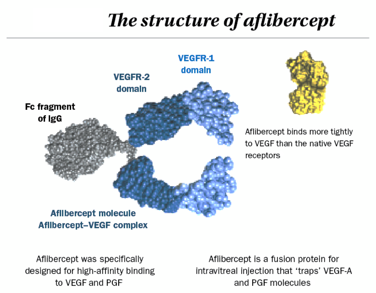
临床浓度(500 µg/mL)的阿柏西普会损害RPE伤口愈合和吞噬作用,提示长期治疗可能产生潜在的副作用,包括视网膜修复延迟和感光细胞变性。[1] 阿柏西普被RPE细胞内化(以囊泡状/网状结构),并滞留≥7天,这与吞噬作用受损相关。在肥胖性卒中中,它下调同侧 VEGF-A/VEGFR2/NRP-1 的表达 (p<0.05)。[2] FDA 批准用于治疗眼部新生血管疾病;由于其选择性降低血管通透性而不改变梗死灶,因此具有治疗肥胖加重卒中水肿的潜力。[2] 阿柏西普 是一种融合蛋白,由 VEGFR1/R2 的 Ig 结构域和人 IgG1 的 Fc 区组成,用作癌症的一线单药治疗。它抑制 VEGF/PlGF 驱动的血管生成,但通过 CAT-1/AKT/eNOS/NO 通路破坏引发高血压[3] 从机制上讲,它降低 NO 的生物利用度,增加 ET-1 和氧化应激(NOX1/NOX4 介导),并下调阳离子氨基酸转运蛋白 CAT-1[3] 药物适应症 该眼科药物用于治疗新生血管性(湿性)年龄相关性黄斑变性 (AMD)、视网膜静脉阻塞 (RVO) 后的黄斑水肿、糖尿病性黄斑水肿 (DME)、糖尿病视网膜病变 (DR) 和早产儿视网膜病变 (ROP)。全身注射剂 ziv-aflibercept 与 5-氟尿嘧啶、亚叶酸钙、伊立替康(FOLFIRI 方案)联合使用,用于治疗对奥沙利铂耐药或奥沙利铂治疗后进展的转移性结直肠癌。 Yesafili 适用于成人,用于治疗新生血管性(湿性)年龄相关性黄斑变性 (AMD)(参见 5.1 节)、视网膜静脉阻塞(分支 RVO 或中央 RVO)继发的黄斑水肿引起的视力障碍(参见 5.1 节)、糖尿病性黄斑水肿 (DME) 引起的视力障碍(参见 5.1 节)以及近视性脉络膜新生血管 (CNV) 引起的视力障碍(参见 5.1 节)。 Eylea 适用于成人,用于治疗:新生血管性(湿性)年龄相关性黄斑变性 (AMD);视网膜静脉阻塞(分支型视网膜静脉阻塞或中央型视网膜静脉阻塞)继发的黄斑水肿引起的视力障碍;糖尿病性黄斑水肿(DME)引起的视力障碍;近视性脉络膜新生血管(近视性CNV)引起的视力障碍。 治疗用途 综述ziv-aflibercept联合FOLFIRI方案治疗对含奥沙利铂方案耐药或进展的转移性结直肠癌(mCRC)的药理学、药代动力学、疗效和安全性……Ziv-aflibercept是一种选择性血管内皮生长因子拮抗剂,在一项II期研究中作为单药治疗mCRC进行了评估,并在一项III期试验中将其与FOLFIRI方案联合使用。患者对ziv-aflibercept单药治疗的反应未达到统计学意义。结果表明,对ziv-aflibercept治疗的反应不受既往贝伐珠单抗治疗的影响。一项3期临床试验比较了ziv-aflibercept联合FOLFIRI方案与安慰剂联合FOLFIRI方案在接受含奥沙利铂方案治疗后出现疾病进展的转移性结直肠癌(mCRC)患者中的安全性和有效性。ziv-aflibercept组患者的中位总生存期为13.5个月,而安慰剂组为12.06个月(风险比[HR] = 0.817,95%置信区间[CI] = 0.713至0.937)。ziv-aflibercept组患者的无进展生存期也高于安慰剂组(HR = 0.758;95% CI = 0.661至0.869)。最常见的不良反应为贫血、腹泻和中性粒细胞减少症。对于接受含奥沙利铂方案治疗后出现疾病进展的mCRC患者,ziv-aflibercept联合FOLFIRI方案是一种安全有效的治疗选择。尚未证实其优于其他抗血管生成疗法。PMID:24259608 玻璃体内注射阿柏西普是一种对血管内皮生长因子具有高亲和力的融合蛋白,为渗出性年龄相关性黄斑变性提供了一种新的治疗选择。临床前研究以及早期和晚期临床试验表明,与雷珠单抗或贝伐珠单抗相比,阿柏西普的高结合亲和力可能赋予其更持久的活性和更高的疗效。本回顾性研究纳入了249例渗出性年龄相关性黄斑变性患者的266只眼,这些患者在接受贝伐珠单抗和/或雷珠单抗治疗后接受了阿柏西普治疗。在首次注射阿柏西普后1、3、6和12个月,计算了光谱域光学相干断层扫描(SD-OCT)测得的平均中心凹下视网膜厚度和平均最小分辨角对数(logMAR)视力。对接受阿柏西普治疗前6个月内至少接受过5次贝伐单抗和/或雷珠单抗注射的眼以及接受阿柏西普治疗前12个月内至少接受过10次注射的眼进行了亚组分析。在开始阿柏西普治疗前,这些眼平均接受了14.7次(范围1-43次)雷珠单抗和/或贝伐单抗治疗。中心凹下视网膜厚度平均值在1个月时从300 μm下降至275 μm(p<0.001),并在6个月时保持稳定。平均logMAR视力在1个月时从0.60(相当于Snellen视力表20/80)提高至0.54(相当于Snellen视力表20/70,p=0.01),并在6个月时稳定在0.55(相当于Snellen视力表20/70,p=0.11,n=251)。在接受阿柏西普治疗前 6 个月内至少接受过 5 次注射的 82 只眼(平均总共注射 18.1 次)中,中心凹下视网膜厚度在 1 个月时从 296 微米改善至 279 微米(p<0.0001),并在 6 个月时维持稳定(p<0.0001)。视力没有变化(1 个月时为 0.48 [20/61],与基线相比为 0.49 [20/62],p = 0.634;6 个月时为 0.51 [20/65],p = 0.601)。在接受阿柏西普治疗前12个月内至少接受过10次注射(平均共注射21.8次)的50只眼中,中心凹下视网膜厚度平均值在治疗1个月时下降了17 μm(p = 0.0007),并在治疗6个月时维持稳定(p = 0.013)。此外,视力未发生变化(治疗1个月时为0.46 [20/56],基线值为0.44 [20/56],p = 0.547;治疗6个月时为0.50 [20/63],p = 0.2445)。对于既往接受过贝伐珠单抗和/或雷珠单抗注射治疗的患者,阿柏西普是一种有价值的替代治疗方案。在既往接受过每4-6周一次雷珠单抗和/或贝伐珠单抗注射治疗的患者中,开始使用阿柏西普治疗后,观察到视力稳定,且光谱域光学相干断层扫描(SD-OCT)显示眼部解剖结构有所改善。PMID:24706352 本研究旨在评估既往接受过玻璃体内注射贝伐珠单抗或雷珠单抗治疗的新生血管性年龄相关性黄斑变性(NVA)患者改用阿柏西普治疗后眼压(IOP)的变化。这是一项回顾性病例分析,纳入了截至2013年3月6日接受过至少2次2 mg/0.05 mL阿柏西普注射治疗的患者,这些患者此前至少接受过2次0.5 mg/0.05 mL雷珠单抗注射治疗,部分患者此前接受过1.25 mg/0.05 mL贝伐珠单抗注射治疗。分析仅限于每位患者的首次此类治疗序列。所有纳入研究的病例在改用阿柏西普之前最后一次抗血管内皮生长因子注射均为雷珠单抗。每位患者均作为自身对照。阿柏西普治疗前(贝伐珠单抗或雷珠单抗治疗前)的眼压为首次注射阿柏西普时散瞳前的眼压值。统计分析使用 Microsoft Excel 进行。共有 41 例患者先接受雷珠单抗治疗后改用阿柏西普,另有 12 例患者先接受雷珠单抗和贝伐珠单抗治疗后改用阿柏西普。对于每种治疗序列,在阿柏西普治疗期间(治疗后)治疗眼的眼压以三种不同的方式计算:首次眼压、最后一次眼压以及阿柏西普治疗期间的平均眼压。汇总数据显示,阿柏西普治疗前(治疗前状态)平均眼压为14.87 mmHg,治疗期间首次平均眼压降至14.57 mmHg,末次平均眼压降至13.79 mmHg,平均眼压降至14.14 mmHg。该推断基于汇总分析。首次、末次和平均眼压差值(治疗后减去治疗前)的95%置信区间分别为-0.30(-1.12至0.52)、-1.08(-1.83至-0.32)和-0.73(-1.30至-0.17)。阿柏西普治疗期间,首次、末次和平均眼压的相应P值分别为0.46、0.006和0.01。研究发现,既往接受过雷珠单抗和/或贝伐珠单抗治疗的患者,在改用阿柏西普治疗后,眼压显著降低。对于既往接受过其他抗血管内皮生长因子治疗的患者,阿柏西普可能具有更佳的眼压安全性。PMID:25072648 玻璃体内注射阿柏西普用于治疗新生血管性(湿性)年龄相关性黄斑变性。 药效学 阿柏西普与各种人受体的平衡解离常数(KD)如下:VEGF-A165为0.5 pM,VEGF-A121为0.36 pM,VEGF-B为1.92 pM,PlGF-2为39 pM。在一项随机、安慰剂对照研究中,研究人员评估了每三周静脉注射6 mg/kg阿柏西普对87例实体瘤患者QTc间期的影响。研究结果显示,根据Fridericia校正法,平均QT间期较基线未发生显著变化(即,校正安慰剂后变化大于20 ms)。然而,由于研究设计的局限性,不能排除平均QTc间期出现小幅增加(即小于10 ms)的可能性。 作用机制 阿柏西普是一种重组人源化融合蛋白,是血管内皮生长因子A (VEGF-A) 和胎盘生长因子 (PlGF) 的拮抗剂。该药物由人血管内皮生长因子受体 (VEGFR) 1和2的胞外结构域与人免疫球蛋白G1 (IgG1) 的Fc段融合而成。阿柏西普是一种可溶性诱饵受体,它能与 VEGF-A 和 PlGF 结合并抑制其生物活性。VEGF-A 和 PlGF 是血管生成因子,可作为内皮细胞的促有丝分裂因子、趋化因子和血管通透性因子。VEGF-A 可诱导新生血管形成(血管生成)并增加血管通透性,这似乎在新生血管性(湿性)年龄相关性黄斑变性的发病机制和进展中发挥作用,而年龄相关性黄斑变性是发达国家老年人失明的主要原因之一。阿柏西普与 VEGF-A 和 PlGF 的结合可阻止这些因子与内源性 VEGF 受体(即 VEGFR-1、VEGFR-2)结合,从而减少新生血管形成和血管通透性。阿柏西普对 VEGF-A 亚型的结合亲和力高于对内源性受体的结合亲和力。阿柏西普即使在低浓度下也能阻断 VEGF 与 VEGFR-1 和 VEGFR-2 的结合和激活。 阿柏西普是一种重组融合蛋白,可作为血管内皮生长因子 A (VEGF-A) 和胎盘生长因子 (PIGF) 配体的诱饵受体。它能阻止这些配体与内皮细胞受体 VEGFR-1 和 VEGFR-2 结合,从而抑制新生血管形成并降低血管通透性。最终,这将延缓视力丧失或转移性结直肠癌的进展。 |
| 分子量 |
97kD
|
|---|---|
| CAS号 |
862111-32-8
|
| 相关CAS号 |
Aflibercept (VEGF Trap); 862111-32-8; 845771-78-0
|
| 外观&性状 |
Colorless to light yellow liquid
|
| HS Tariff Code |
2934.99.9001
|
| 存储方式 |
Powder -20°C 3 years 4°C 2 years In solvent -80°C 6 months -20°C 1 month |
| 运输条件 |
Room temperature (This product is stable at ambient temperature for a few days during ordinary shipping and time spent in Customs)
|
| 溶解度 (体外实验) |
May dissolve in DMSO (in most cases), if not, try other solvents such as H2O, Ethanol, or DMF with a minute amount of products to avoid loss of samples
|
|---|---|
| 溶解度 (体内实验) |
注意: 如下所列的是一些常用的体内动物实验溶解配方,主要用于溶解难溶或不溶于水的产品(水溶度<1 mg/mL)。 建议您先取少量样品进行尝试,如该配方可行,再根据实验需求增加样品量。
注射用配方
注射用配方1: DMSO : Tween 80: Saline = 10 : 5 : 85 (如: 100 μL DMSO → 50 μL Tween 80 → 850 μL Saline)(IP/IV/IM/SC等) *生理盐水/Saline的制备:将0.9g氯化钠/NaCl溶解在100 mL ddH ₂ O中,得到澄清溶液。 注射用配方 2: DMSO : PEG300 :Tween 80 : Saline = 10 : 40 : 5 : 45 (如: 100 μL DMSO → 400 μL PEG300 → 50 μL Tween 80 → 450 μL Saline) 注射用配方 3: DMSO : Corn oil = 10 : 90 (如: 100 μL DMSO → 900 μL Corn oil) 示例: 以注射用配方 3 (DMSO : Corn oil = 10 : 90) 为例说明, 如果要配制 1 mL 2.5 mg/mL的工作液, 您可以取 100 μL 25 mg/mL 澄清的 DMSO 储备液,加到 900 μL Corn oil/玉米油中, 混合均匀。 View More
注射用配方 4: DMSO : 20% SBE-β-CD in Saline = 10 : 90 [如:100 μL DMSO → 900 μL (20% SBE-β-CD in Saline)] 口服配方
口服配方 1: 悬浮于0.5% CMC Na (羧甲基纤维素钠) 口服配方 2: 悬浮于0.5% Carboxymethyl cellulose (羧甲基纤维素) 示例: 以口服配方 1 (悬浮于 0.5% CMC Na)为例说明, 如果要配制 100 mL 2.5 mg/mL 的工作液, 您可以先取0.5g CMC Na并将其溶解于100mL ddH2O中,得到0.5%CMC-Na澄清溶液;然后将250 mg待测化合物加到100 mL前述 0.5%CMC Na溶液中,得到悬浮液。 View More
口服配方 3: 溶解于 PEG400 (聚乙二醇400) 请根据您的实验动物和给药方式选择适当的溶解配方/方案: 1、请先配制澄清的储备液(如:用DMSO配置50 或 100 mg/mL母液(储备液)); 2、取适量母液,按从左到右的顺序依次添加助溶剂,澄清后再加入下一助溶剂。以 下列配方为例说明 (注意此配方只用于说明,并不一定代表此产品 的实际溶解配方): 10% DMSO → 40% PEG300 → 5% Tween-80 → 45% ddH2O (或 saline); 假设最终工作液的体积为 1 mL, 浓度为5 mg/mL: 取 100 μL 50 mg/mL 的澄清 DMSO 储备液加到 400 μL PEG300 中,混合均匀/澄清;向上述体系中加入50 μL Tween-80,混合均匀/澄清;然后继续加入450 μL ddH2O (或 saline)定容至 1 mL; 3、溶剂前显示的百分比是指该溶剂在最终溶液/工作液中的体积所占比例; 4、 如产品在配制过程中出现沉淀/析出,可通过加热(≤50℃)或超声的方式助溶; 5、为保证最佳实验结果,工作液请现配现用! 6、如不确定怎么将母液配置成体内动物实验的工作液,请查看说明书或联系我们; 7、 以上所有助溶剂都可在 Invivochem.cn网站购买。 |
计算结果:
工作液浓度: mg/mL;
DMSO母液配制方法: mg 药物溶于 μL DMSO溶液(母液浓度 mg/mL)。如该浓度超过该批次药物DMSO溶解度,请首先与我们联系。
体内配方配制方法:取 μL DMSO母液,加入 μL PEG300,混匀澄清后加入μL Tween 80,混匀澄清后加入 μL ddH2O,混匀澄清。
(1) 请确保溶液澄清之后,再加入下一种溶剂 (助溶剂) 。可利用涡旋、超声或水浴加热等方法助溶;
(2) 一定要按顺序加入溶剂 (助溶剂) 。
InvivoChem的所有产品仅用于作科学研究,不面向患者销售
Copyright 2020 InvivoChem LLC | All Rights Reserved 粤ICP备20063088号-1


 463611831
