| 规格 | 价格 | |
|---|---|---|
| 500mg | ||
| 1g | ||
| Other Sizes |

| 靶点 |
ziv-aflibercept binds VEGF and placental growth factor (PIGF) as a soluble decoy receptor (no IC50/Ki/EC50 data provided)[1]
|
|---|---|
| 体外研究 (In Vitro) |
Ziv-aflibercept(0.25、0.5、1.0 和 5 mg/mL;24 小时)以剂量依赖性方式降低 ARPE-19 细胞中的线粒体膜电位 [1]。
ziv-aflibercept 在10×临床浓度(2 mg/4 mL玻璃体等效)下显著降低ARPE-19人视网膜色素上皮细胞活力至77.25% ± 2.1%(p < 0.0001),但在1/2×、1×或2×剂量下与对照组相比无统计学显著变化[1] ziv-aflibercept 诱导线粒体毒性,在1×(73.50% ± 2.93%,p < 0.0001)、2×(64.83% ± 2.7%,p < 0.0001)和10×(49.65% ± 4.22%,p = 0.0002)浓度下降低线粒体膜电位(ΔΨm),表明早期凋亡;1/2×剂量(91.57% ± 2.54%)未观察到显著ΔΨm变化[1] ziv-aflibercept 在10×浓度下增加渗透压至418 mOsm/kg(对照组为324 mOsm/kg),但1/2×、1×和2×剂量下的渗透压(分别为324、330和342 mOsm/kg)与对照组无显著差异[1] Ziv-aflibercept 在临床剂量(1×)下对人视网膜色素上皮细胞(ARPE-19系)暴露24小时后未降低细胞活力(基于Malik等研究)。[2] 在临床剂量两倍(2×)浓度下,其在Müller细胞(MIO-M1系)中显著降低活力,而雷珠单抗、阿柏西普或贝伐珠单抗无此效应,表明高浓度下存在细胞类型特异性毒性。[2] |
| 体内研究 (In Vivo) |
在兔子的右眼中,ziv-aflibercept(25 mg/mL;眼内注射;单剂量)基本上不会诱发白内障、视网膜脱离或任何相关问题[2]。
兔模型中,玻璃体内注射ziv-aflibercept(0.05 ml, 25 mg/ml)后1天和7天,眼底检查、光学相干断层扫描(OCT)和全视野视网膜电图(ERG)均未显示毒性;组织学和透射电镜证实无解剖损伤。[2] 人类案例报告(如新生血管性年龄相关性黄斑变性、糖尿病性黄斑水肿)显示,玻璃体内注射(1.25 mg/剂)后视力改善,视网膜内/下液减少,随访期间未见炎症、白内障进展或视网膜毒性。[2] |
| 细胞实验 |
细胞活力测定[1]
细胞类型: ARPE-19 细胞。 测试浓度:0.25、0.5、1.0 和 5 mg/mL。 孵化持续时间:24小时。 实验结果:严重影响细胞活力(低于 1 mg/mL)。 细胞活力实验中,ARPE-19细胞以5.0 × 10⁵细胞/孔接种于六孔板,加入2 mL培养基,标准条件(37°C、5% CO₂、95%湿度)孵育24小时,后用ziv-aflibercept在1/2×、1×、2×或10×临床浓度或对照(无药物)处理24小时。细胞经胰蛋白酶消化、离心(1000 rpm,5分钟),重悬于1 mL培养基,通过自动化台盼蓝染色排除法分析活细胞/死细胞;实验重复三次并独立重复两次[1] 线粒体膜电位(ΔΨm)实验中,ARPE-19细胞以1.0 × 10⁵细胞/孔接种于24孔板,过夜孵育,后用ziv-aflibercept处理24小时。JC-1染料检测ΔΨm:健康细胞(红色荧光,590 nm)对比凋亡/坏死细胞(绿色荧光,529 nm)。使用荧光成像扫描仪测量红绿荧光比值,实验重复四次并独立重复两次[1] 含ziv-aflibercept培养基的渗透压通过自动渗透压仪(基于冰点降低法)测量;所有浓度(1/2×至10×)样本均被测试[1] 活力测试中,ARPE-19细胞暴露于ziv-aflibercept的1/2×、1×、2×和10×临床浓度(基于玻璃体分布)24小时,通过自动化台盼蓝排除法评估活力,结果归一化至对照组并进行统计分析。[2] Müller细胞(MIO-M1系)实验采用低剂量(1/2×)和临床等效剂量;2×浓度下的活力降低通过标准化方法量化。[2] |
| 动物实验 |
九只兔子接受了单侧玻璃体内注射0.05 ml的ziv-aflibercept(25 mg/ml)。评估内容包括基线/注射后血清/玻璃体/房水渗透压、眼底镜检查、OCT、ERG(第1天和第7天)以及组织病理学分析。未设置假手术对照组;主要观察指标为无并发症(例如白内障、视网膜脱离)。[2]
|
| 药代性质 (ADME/PK) |
ziv-aflibercept的半衰期为7.1天,与aflibercept相同,这是基于其结构相似性。[2]
|
| 毒性/毒理 (Toxicokinetics/TK) |
在ARPE-19细胞中,ziv-aflibercept在临床相关浓度(1×、2×和10×)下表现出线粒体毒性,ΔΨm降低提示早期细胞凋亡;细胞活力仅在10×剂量下下降[1]
与aflibercept相比,ziv-aflibercept的细胞毒性增加可能归因于其较低的pH值和较高的渗透压,但尚未评估全身毒代动力学数据(例如,血浆蛋白结合率、器官毒性)[1] 在兔或人中,玻璃体内注射ziv-aflibercept后未报告眼部或全身毒性。注射后血清和眼内渗透压保持不变。[2] 体外超临床剂量下观察到线粒体毒性(例如,ARPE-19细胞中ΔΨm降低),但体内未发现器官毒性或药物相互作用的证据。[2] 妊娠和哺乳期影响 ◉哺乳期用药概述 本记录涉及使用ziv-aflibercept治疗癌症。目前尚无关于哺乳期使用ziv-aflibercept的信息。由于ziv-aflibercept是一种分子量为115,000的大分子蛋白质,其在乳汁中的含量可能非常低,且由于可能在婴儿的胃肠道中被破坏,因此不太可能被吸收。然而,阿柏西普(ziv-aflibercept)通常与其他潜在毒性化疗药物联合使用,大多数资料认为,在母亲接受抗肿瘤药物治疗期间,母乳喂养是禁忌的。一名女性在接受玻璃体内注射阿柏西普后,乳汁中的血管内皮生长因子(VEGF)水平降低。由于VEGF存在于母乳中,且被认为有助于婴儿胃肠道的成熟,因此人们对哺乳期母亲使用VEGF抑制剂表示担忧。需要注意的是,母乳的典型替代品是婴儿配方奶粉,其中不含VEGF。 ◉ 对母乳喂养婴儿的影响 截至修订日期,未找到相关的已发表信息。 ◉ 对泌乳和母乳的影响 截至修订日期,未找到相关的已发表信息。 |
| 参考文献 |
|
| 其他信息 |
目的:比较抗血管内皮生长因子(VEGF)药物雷珠单抗、贝伐珠单抗、阿柏西普和ziv-阿柏西普在体外培养的视网膜色素上皮细胞中的安全性。方法:将人视网膜色素上皮细胞(ARPE-19)暴露于四种抗VEGF药物中,浓度分别为临床浓度的1/2倍、1倍、2倍和10倍,培养24小时。通过细胞活力和线粒体膜电位检测评估早期细胞凋亡变化和总体细胞死亡率。结果:在10倍浓度下,贝伐珠单抗(82.38%,p=0.0001)、阿柏西普(82.68%,p=0.0002)和ziv-阿柏西普(77.25%,p<0.0001)均导致细胞活力下降,但在较低浓度下未观察到此现象。然而,在所有浓度(包括10倍浓度)下,雷珠单抗处理的细胞活力均未发生变化。10倍浓度雷珠单抗处理的细胞线粒体膜电位略有下降(89.61%,p=0.0006),2倍和10倍浓度阿柏西普处理的细胞线粒体膜电位也略有下降(分别为88.76%和81.46%;p<0.01)。贝伐珠单抗(1倍、2倍和10倍浓度)和阿柏西普(73.50%、64.83%和49.65%;p<0.01)均导致线粒体膜电位更大幅度的下降,提示在较低剂量(包括临床剂量)下即可诱导早期细胞凋亡。结论:在临床剂量下,雷珠单抗和阿柏西普均未显示出线粒体毒性或细胞死亡的证据。然而,贝伐珠单抗和阿柏西普(ziv-aflibercept)在临床相关剂量下显示出轻微的线粒体毒性。[1]
近年来,单克隆抗体彻底改变了视网膜新生血管疾病的治疗。最近,另一类药物——融合蛋白——提供了一种具有不同药理学特性的替代治疗策略。除了市售的阿柏西普外,另外两种药物——阿柏西普(ziv-aflibercept)和康柏西普(conbercept)——也已在眼科疾病的抗血管生成治疗中进行了研究。在此背景下,对目前关于融合蛋白在眼科疾病中的应用数据进行批判性回顾可能是一项及时且重要的贡献。阿柏西普,曾用名VEGF Trap Eye,是一种VEGF受体1和2的融合蛋白,用于治疗多种与血管生成相关的视网膜疾病。它已与雷珠单抗和贝伐珠单抗一起,成为治疗新生血管性年龄相关性黄斑变性(AMD)、糖尿病性黄斑水肿(DME)和视网膜静脉阻塞(RVO)相关性黄斑水肿的重要治疗选择。Ziv-aflibercept是一种全身化疗药物,已获批用于治疗转移性结直肠癌,由于其玻璃体内注射给药的潜力,近期备受关注。一项实验研究和人体病例报告显示,Ziv-aflibercept未出现与视网膜电图(ERG)相关的毒性反应。Conbercept是一种可溶性受体诱饵,可阻断所有VEGF-A、VEGF-B、VEGF-C和PlGF亚型,对VEGF具有高亲和力,且在玻璃体内半衰期长。Conbercept已完成三期临床试验,并显示出疗效和安全性。本综述讨论了三种在眼科领域研究过的融合蛋白:阿柏西普 (aflibercept)、齐夫-阿柏西普 (ziv-aflibercept) 和康柏西普 (conbercept),重点关注它们在治疗视网膜疾病方面的临床应用。[2] 齐夫-阿柏西普 的结构与阿柏西普相同,但赋形剂和渗透压不同(1000 mOsm/L 对比阿柏西普的 286 mOsm/L)。齐夫-阿柏西普于 2012 年获得 FDA 批准用于治疗转移性结直肠癌,由于价格较低,它也被用于治疗视网膜疾病(属于超适应症用药)。临床玻璃体内注射剂量为 1.25 mg(0.05 ml 浓度为 25 mg/ml 的溶液)。[2] 初步数据显示,齐夫-阿柏西普可有效减轻年龄相关性黄斑变性/糖尿病性黄斑水肿 (AMD/DME) 患者的黄斑水肿并改善视力,但仍需进行大规模试验来验证其安全性和剂量。[2] |
| CAS号 |
1609655-49-3
|
|---|---|
| 外观&性状 |
Colorless to light yellow liquid
|
| HS Tariff Code |
2934.99.9001
|
| 存储方式 |
Powder -20°C 3 years 4°C 2 years In solvent -80°C 6 months -20°C 1 month |
| 运输条件 |
Room temperature (This product is stable at ambient temperature for a few days during ordinary shipping and time spent in Customs)
|
| 溶解度 (体外实验) |
May dissolve in DMSO (in most cases), if not, try other solvents such as H2O, Ethanol, or DMF with a minute amount of products to avoid loss of samples
|
|---|---|
| 溶解度 (体内实验) |
注意: 如下所列的是一些常用的体内动物实验溶解配方,主要用于溶解难溶或不溶于水的产品(水溶度<1 mg/mL)。 建议您先取少量样品进行尝试,如该配方可行,再根据实验需求增加样品量。
注射用配方
注射用配方1: DMSO : Tween 80: Saline = 10 : 5 : 85 (如: 100 μL DMSO → 50 μL Tween 80 → 850 μL Saline)(IP/IV/IM/SC等) *生理盐水/Saline的制备:将0.9g氯化钠/NaCl溶解在100 mL ddH ₂ O中,得到澄清溶液。 注射用配方 2: DMSO : PEG300 :Tween 80 : Saline = 10 : 40 : 5 : 45 (如: 100 μL DMSO → 400 μL PEG300 → 50 μL Tween 80 → 450 μL Saline) 注射用配方 3: DMSO : Corn oil = 10 : 90 (如: 100 μL DMSO → 900 μL Corn oil) 示例: 以注射用配方 3 (DMSO : Corn oil = 10 : 90) 为例说明, 如果要配制 1 mL 2.5 mg/mL的工作液, 您可以取 100 μL 25 mg/mL 澄清的 DMSO 储备液,加到 900 μL Corn oil/玉米油中, 混合均匀。 View More
注射用配方 4: DMSO : 20% SBE-β-CD in Saline = 10 : 90 [如:100 μL DMSO → 900 μL (20% SBE-β-CD in Saline)] 口服配方
口服配方 1: 悬浮于0.5% CMC Na (羧甲基纤维素钠) 口服配方 2: 悬浮于0.5% Carboxymethyl cellulose (羧甲基纤维素) 示例: 以口服配方 1 (悬浮于 0.5% CMC Na)为例说明, 如果要配制 100 mL 2.5 mg/mL 的工作液, 您可以先取0.5g CMC Na并将其溶解于100mL ddH2O中,得到0.5%CMC-Na澄清溶液;然后将250 mg待测化合物加到100 mL前述 0.5%CMC Na溶液中,得到悬浮液。 View More
口服配方 3: 溶解于 PEG400 (聚乙二醇400) 请根据您的实验动物和给药方式选择适当的溶解配方/方案: 1、请先配制澄清的储备液(如:用DMSO配置50 或 100 mg/mL母液(储备液)); 2、取适量母液,按从左到右的顺序依次添加助溶剂,澄清后再加入下一助溶剂。以 下列配方为例说明 (注意此配方只用于说明,并不一定代表此产品 的实际溶解配方): 10% DMSO → 40% PEG300 → 5% Tween-80 → 45% ddH2O (或 saline); 假设最终工作液的体积为 1 mL, 浓度为5 mg/mL: 取 100 μL 50 mg/mL 的澄清 DMSO 储备液加到 400 μL PEG300 中,混合均匀/澄清;向上述体系中加入50 μL Tween-80,混合均匀/澄清;然后继续加入450 μL ddH2O (或 saline)定容至 1 mL; 3、溶剂前显示的百分比是指该溶剂在最终溶液/工作液中的体积所占比例; 4、 如产品在配制过程中出现沉淀/析出,可通过加热(≤50℃)或超声的方式助溶; 5、为保证最佳实验结果,工作液请现配现用! 6、如不确定怎么将母液配置成体内动物实验的工作液,请查看说明书或联系我们; 7、 以上所有助溶剂都可在 Invivochem.cn网站购买。 |
计算结果:
工作液浓度: mg/mL;
DMSO母液配制方法: mg 药物溶于 μL DMSO溶液(母液浓度 mg/mL)。如该浓度超过该批次药物DMSO溶解度,请首先与我们联系。
体内配方配制方法:取 μL DMSO母液,加入 μL PEG300,混匀澄清后加入μL Tween 80,混匀澄清后加入 μL ddH2O,混匀澄清。
(1) 请确保溶液澄清之后,再加入下一种溶剂 (助溶剂) 。可利用涡旋、超声或水浴加热等方法助溶;
(2) 一定要按顺序加入溶剂 (助溶剂) 。
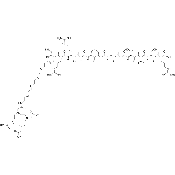 DOTA-TMVP1446
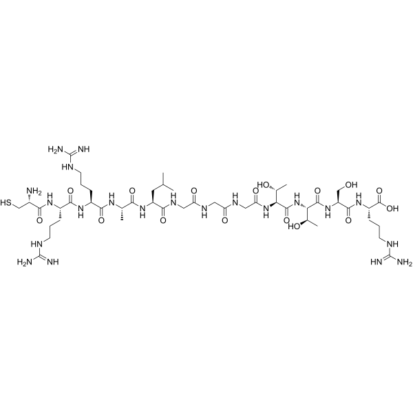 TMVP1446
 Girancitug
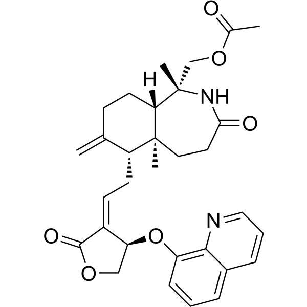 AGW-11
InvivoChem的所有产品仅用于作科学研究,不面向患者销售
Copyright 2020 InvivoChem LLC | All Rights Reserved 粤ICP备20063088号-1


 463611831
