| 规格 | 价格 | 库存 | 数量 |
|---|---|---|---|
| 1mg |
|
||
| 5mg |
|
||
| Other Sizes |
|
| 靶点 |
Dopamine D1 Receptor
|
|---|---|
| 体外研究 (In Vitro) |
A68930盐酸盐(1 μM;5-60 分钟;16HBE14o- 或 NCI-H292 细胞)可显着增加 cAMP 反应元件结合 (CREB) 蛋白的磷酸化[1]。用 1 μM A68930盐酸盐处理 NCI-H292 细胞 48 小时,导致 MUC5AC 蛋白表达以及 MUC5AC mRNA 表达增加[1]。 A68930 盐酸盐(1 μM;20 分钟;NCI-H292 细胞)可显着提高细胞内 cAMP 水平[1]。
多巴胺D1受体激动剂诱导16HBE14o-细胞和NCI-H292细胞的cAMP活性[1] 多巴胺D1受体通过刺激腺苷酸环化酶诱导cAMP的产生,腺苷酸环化酶被Gs蛋白激活。因此,我们研究了多巴胺D1受体的激活是否会增加人气道上皮细胞(16HBE14o-细胞和NCI-H292细胞)的细胞内cAMP水平。多巴胺(1 μM)及多巴胺d1样受体激动剂(SKF38393或A68930;1 μM)显著增加16HBE14o-细胞内cAMP水平(多巴胺;P < 0.001, skf38393;P < 0.001, A68930;P < 0.01, n = 6)和NCI-H292细胞(多巴胺;P < 0.01, skf38393;P < 0.01, A68930;P < 0.01, n = 6)(图3a)。多巴胺D1受体拮抗剂SCH23390 (1 μM)预处理显著逆转A68930 (1 μM)诱导的环AMP生成(16HBE14o-细胞:P < 0.001, n = 6;NCI-H292细胞:P < 0.05, n = 6)或SCH39166 (1 μM) (16HBE14o-细胞:P < 0.001, n = 6;NCI-H292细胞:P < 0.05, n = 6)(图3b)。 多巴胺D1受体激动剂诱导16HBE14o-细胞和NCI-H292细胞的CREB磷酸化[1] 多巴胺D1受体激活后cAMP升高,诱导蛋白激酶A (PKA)活化,从而诱导CREB磷酸化。MEK-ERK信号也参与CREB磷酸化。此外,多巴胺D1受体通过β-阻滞蛋白激活MEK-ERK信号。因此,我们检测了多巴胺D1受体激动剂A68930是否在16HBE14o-细胞和NCI-H292细胞中通过PKA和/或MEK磷酸化CREB。A68930 (1 μM, 20 min)显著增加16HBE14o-细胞(P < 0.05, n = 3)和NCI-H292细胞(P < 0.05, n = 7)的CREB磷酸化(图4a和b)。16HBE14o-细胞的磷酸化在20 - 30min达到最大水平,60 min内缓慢下降至基础水平,而NCI-H292细胞的磷酸化在60 min时保持升高。为了证实A68930通过PKA或MEK磷酸化CREB,我们用PKA抑制剂H89 (10 μM;30 min)或MEK抑制剂U0126 (5 μM;120分钟)。A68930 (1 μm;H89 (P < 0.001, n = 4)或U0126 (P < 0.05, n = 4)显著抑制NCI-H292细胞中CREB磷酸化(图4c)。这些结果证实,多巴胺D1受体激动剂诱导的CREB磷酸化通过PKA和MEK/ERK信号传导进行。 多巴胺D1受体激动剂诱导NCI-H292细胞MUC5AC mRNA表达[1] CREB先前被证明可以介导包括NCI-H292细胞在内的气道上皮细胞MUC5AC的转录调节。我们检测了多巴胺或多巴胺D1受体激动剂A68930是否诱导NCI-H292细胞MUC5AC mRNA的表达。多巴胺(1 μM)、A68930 (1 μM)和香烟烟雾提取物(10%)显著增加NCI-H292细胞MUC5AC mRNA的表达。Gs蛋白偶联的β2肾上腺素受体激动剂异丙肾上腺素(1 μM)也显著诱导MUC5AC mRNA表达(图5a)。先前对气道上皮细胞的研究使用的最终CSE浓度范围为1%至30%。MTT细胞活力分析证实,用10%或更高浓度(20%)的CSE处理NCI-H292细胞48小时,并没有降低NCI-H292细胞活力,这表明10%或20%的CSE处理没有细胞毒性(图5b)。 多巴胺D1受体激动剂诱导NCI-H292细胞MUC5AC蛋白表达[1] 我们使用免疫荧光法进一步研究了多巴胺D1受体激动剂A68930或多巴胺对MUC5AC蛋白表达的影响。与MUC5AC mRNA数据一致,多巴胺(1 μM)、A68930(1 μM)和CSE(10%)均增加了NCI-H292细胞中MUC5AC蛋白的表达(图6)。这些结果提示多巴胺D1受体的激活刺激MUC5AC的表达。 |
| 细胞实验 |
蛋白质印迹分析[1]
细胞类型: 16HBE14o- 或 NCI-H292 细胞 测试浓度: 1 μM 孵育时间: 5~60分钟 实验结果:CREB磷酸化显着增加。 RT-PCR[1] 细胞类型: NCI-H292 细胞 测试浓度: 1 μM 孵育时间: > 48 小时 实验结果:诱导 MUC5AC mRNA 表达。 免疫荧光[1] 细胞类型: NCI-H292 细胞 测试浓度: 1 μM 孵育时间:48小时 实验结果:MUC5AC的mRNA数据、MUC5AC蛋白表达量增加。 为了分析CREB磷酸化,我们将16HBE14o-细胞或NCI-H292细胞血清饥饿24 h,然后用多巴胺D1受体激动剂A68930 (1 μM)处理指定时间(5-60 min)。在单独的实验中,NCI-H292细胞最初用10 μM H89 (PKA抑制剂)预处理;30分钟)或5 μM U0126 (MEK抑制剂;A68930 (1 μM;20分钟)。处理后,细胞用冰冷的PBS洗涤两次,并在加入1 mM苯甲磺酰氟和1:20 00稀释的蛋白酶抑制剂鸡尾酒III的冰冷RIPA缓冲液中裂解。收集每个裂解的细胞样本,在4℃下,15000g离心15min,取上清液进行蛋白质分析。每个样品的蛋白浓度采用Pierce BCA试剂测定,以BSA为对照,样品保存于- 80°C。[1] <人力资源> 根据制造商的说明,使用HitHunter™cAMP Assay for Small Molecules试剂盒检测16HBE14o-和NCI-H292细胞系中cAMP的产量。简单地说,细胞在白壁96孔板中生长,用温PBS(37°C)洗涤两次。用多巴胺(1 μM)或多巴胺d1样受体激动剂(A68930或SKF38393) (1 μM)在37℃下孵育20 min。本研究中使用的多巴胺d1样受体激动剂的浓度和持续时间是在前人研究的基础上确定的。在单独的实验中,用多巴胺d1样受体拮抗剂[SCH23390 (1 μM)或SCH39166 (1 μM)]或载体(PBS)预处理细胞30 min,然后用A68930 (1 μM)在37℃下孵育20 min。每孔加入cAMP抗体试剂,再加入cAMP工作液(酶供体/裂解缓冲液/Emerald II/Galacton混合液),室温孵育60 min。细胞与酶受体试剂在室温下孵育3小时,用多模微孔板读取仪检测发光信号。[1] 采用RNeasy Mini Kit,分别用多巴胺(1 μM)、A68930 (1 μM)、异丙肾上腺素(1 μM)或CSE(10%)处理NCI-H292细胞48 h后提取总RNA。按照制造商的说明,使用ReverTra Ace qPCR RT Kit将总RNA转录成cDNA。在CFX96实时荧光定量PCR检测系统上使用雷鸟SYBR qPCR试剂盒进行实时荧光定量PCR。MUC5AC和GAPDH引物序列如表1所示。通过熔化曲线分析证实了扩增的特异性。CFX管理软件确定的所有样本的Ct值归一化为管家基因GAPDH,并通过比较Ct (ΔΔCt)方法计算与未处理对照的相对折叠诱导。[1] 按照上述方法对NCI-H292细胞MUC5AC蛋白进行免疫荧光染色,并做一些修改。简单地说,将NCI-H292细胞接种在8室显微镜载玻片上,血清饥饿24小时。饥饿后,细胞暴露于A68930 (1 μM)、多巴胺(1 μM)或CSE(10%)中48小时。细胞在室温下用4%多聚甲醛固定15分钟,并用PBS洗涤3次。通透化(0.2% Triton X-100在PBS中浸泡5分钟)和阻断(1%牛血清白蛋白在0.1% Triton X-100中浸泡15分钟)后,细胞与Alexa Fluor 488偶联MUC5AC抗体在4C下孵育过夜。用PBS洗涤细胞两次后,用含DAPI的延长金抗褪色试剂盖上载玻片,用倒置荧光显微镜观察。使用MetaMorph软件捕获数字化图像。在采集图像时,我们保持图像采集时间(300ms)、图像强度增益、图像增强和样本间图像黑电平不变。[1] |
| 参考文献 | |
| 其他信息 |
背景:多巴胺受体包含两个亚组,即Gs蛋白偶联的“D1样”受体(D1、D5)和Gi蛋白偶联的“D2样”受体(D2、D3、D4)。在气道中,多巴胺D1和D2受体均表达于气道平滑肌上,并调节气道平滑肌的收缩力。然而,多巴胺D1受体在气道上皮细胞上的功能性表达尚未被证实。Gs偶联受体的激活可刺激腺苷酸环化酶,进而产生环磷酸腺苷(cAMP),已知cAMP可通过气道上皮细胞中的cAMP反应元件结合蛋白(CREB)诱导黏液过度分泌。我们探究多巴胺D1受体是否表达于气道上皮细胞,以及它是否促进CREB磷酸化和MUC5AC的表达。方法:我们采用免疫组织化学和免疫印迹法评估了天然人呼吸道上皮细胞和三种培养的人呼吸道上皮细胞(包括原代培养的呼吸道上皮细胞、支气管上皮细胞系 (16HBE14o-) 和肺黏液表皮样癌细胞系 (NCI-H292))上多巴胺D1受体的蛋白表达。为了表征多巴胺D1受体对cAMP的刺激作用,我们在测定cAMP之前,用多巴胺或多巴胺D1受体激动剂(SKF38393或A68930)处理16HBE14o-细胞和NCI-H292细胞。通过免疫印迹法检测了A68930在16HBE14o-细胞和NCI-H292细胞中对CREB的磷酸化作用。本研究分别采用实时定量PCR和免疫荧光染色法评估了多巴胺或A68930对NCI-H292细胞中MUC5AC mRNA和蛋白表达的影响。
结果:在天然人呼吸道上皮细胞和三种来源的培养人呼吸道上皮细胞中均检测到了多巴胺D1受体蛋白。多巴胺或多巴胺D1样受体激动剂可刺激16HBE14o-细胞和NCI-H292细胞中cAMP的产生,而选择性多巴胺D1样受体拮抗剂(SCH23390或SCH39166)可逆转这一作用。A68930显著增加了16HBE14o-细胞和NCI-H292细胞中CREB的磷酸化水平,而PKA抑制剂(H89)和MEK抑制剂(U0126)可减弱这一作用。在NCI-H292细胞中,多巴胺或A68930均可增加MUC5AC mRNA和蛋白的表达。结论:这些结果表明,激活人呼吸道上皮细胞上的多巴胺D1受体可诱导黏液过度分泌,从而加重呼吸道阻塞症状。[1] |
| 分子式 |
C16H18CLNO3
|
|---|---|
| 分子量 |
307.77
|
| 精确质量 |
307.098
|
| 元素分析 |
C, 62.44; H, 5.90; Cl, 11.52; N, 4.55; O, 15.59
|
| CAS号 |
130465-39-3
|
| 相关CAS号 |
A68930;130465-45-1
|
| PubChem CID |
9904672
|
| 外观&性状 |
White to yellow solid powder
|
| 熔点 |
250ºC
|
| LogP |
3.913
|
| tPSA |
75.71
|
| 氢键供体(HBD)数目 |
4
|
| 氢键受体(HBA)数目 |
4
|
| 可旋转键数目(RBC) |
2
|
| 重原子数目 |
21
|
| 分子复杂度/Complexity |
319
|
| 定义原子立体中心数目 |
2
|
| SMILES |
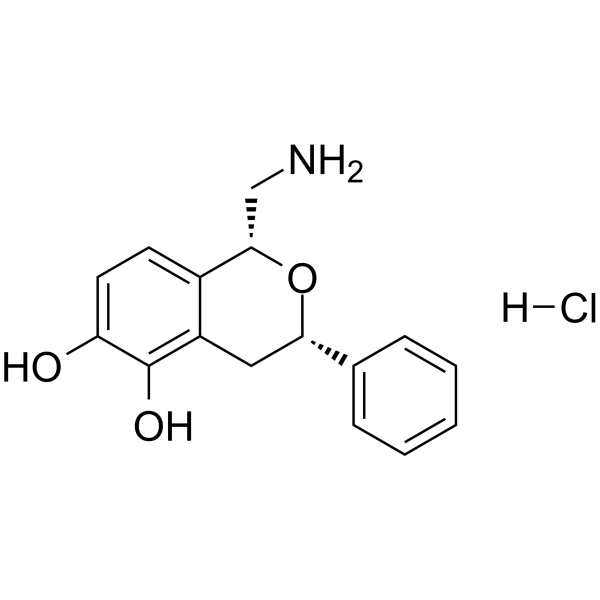
C1=CC=C(C=C1)[C@H]2CC3=C(C(=CC=C3[C@@H](CN)O2)O)O.Cl
|
| InChi Key |
PQPGUUQPTSMLKU-YYLIZZNMSA-N
|
| InChi Code |
InChI=1S/C16H17NO3.ClH/c17-9-15-11-6-7-13(18)16(19)12(11)8-14(20-15)10-4-2-1-3-5-10;/h1-7,14-15,18-19H,8-9,17H2;1H/t14-,15-;/m0./s1
|
| 化学名 |
(1R,3S)-1-(aminomethyl)-3-phenyl-3,4-dihydro-1H-isochromene-5,6-diol;hydrochloride
|
| 别名 |
130465-39-3; A-68930 hydrochloride; A 68930 hydrochloride; 1H-2-Benzopyran-5,6-diol, 1-(aminomethyl)-3,4-dihydro-3-phenyl-, hydrochloride (1:1), (1R,3S)-rel-; ...; A68930 (hydrochloride);
|
| HS Tariff Code |
2934.99.9001
|
| 存储方式 |
Powder -20°C 3 years 4°C 2 years In solvent -80°C 6 months -20°C 1 month 注意: 请将本产品存放在密封且受保护的环境中,避免吸湿/受潮。 |
| 运输条件 |
Room temperature (This product is stable at ambient temperature for a few days during ordinary shipping and time spent in Customs)
|
| 溶解度 (体外实验) |
May dissolve in DMSO (in most cases), if not, try other solvents such as H2O, Ethanol, or DMF with a minute amount of products to avoid loss of samples
|
|---|---|
| 溶解度 (体内实验) |
注意: 如下所列的是一些常用的体内动物实验溶解配方,主要用于溶解难溶或不溶于水的产品(水溶度<1 mg/mL)。 建议您先取少量样品进行尝试,如该配方可行,再根据实验需求增加样品量。
注射用配方
注射用配方1: DMSO : Tween 80: Saline = 10 : 5 : 85 (如: 100 μL DMSO → 50 μL Tween 80 → 850 μL Saline)(IP/IV/IM/SC等) *生理盐水/Saline的制备:将0.9g氯化钠/NaCl溶解在100 mL ddH ₂ O中,得到澄清溶液。 注射用配方 2: DMSO : PEG300 :Tween 80 : Saline = 10 : 40 : 5 : 45 (如: 100 μL DMSO → 400 μL PEG300 → 50 μL Tween 80 → 450 μL Saline) 注射用配方 3: DMSO : Corn oil = 10 : 90 (如: 100 μL DMSO → 900 μL Corn oil) 示例: 以注射用配方 3 (DMSO : Corn oil = 10 : 90) 为例说明, 如果要配制 1 mL 2.5 mg/mL的工作液, 您可以取 100 μL 25 mg/mL 澄清的 DMSO 储备液,加到 900 μL Corn oil/玉米油中, 混合均匀。 View More
注射用配方 4: DMSO : 20% SBE-β-CD in Saline = 10 : 90 [如:100 μL DMSO → 900 μL (20% SBE-β-CD in Saline)] 口服配方
口服配方 1: 悬浮于0.5% CMC Na (羧甲基纤维素钠) 口服配方 2: 悬浮于0.5% Carboxymethyl cellulose (羧甲基纤维素) 示例: 以口服配方 1 (悬浮于 0.5% CMC Na)为例说明, 如果要配制 100 mL 2.5 mg/mL 的工作液, 您可以先取0.5g CMC Na并将其溶解于100mL ddH2O中,得到0.5%CMC-Na澄清溶液;然后将250 mg待测化合物加到100 mL前述 0.5%CMC Na溶液中,得到悬浮液。 View More
口服配方 3: 溶解于 PEG400 (聚乙二醇400) 请根据您的实验动物和给药方式选择适当的溶解配方/方案: 1、请先配制澄清的储备液(如:用DMSO配置50 或 100 mg/mL母液(储备液)); 2、取适量母液,按从左到右的顺序依次添加助溶剂,澄清后再加入下一助溶剂。以 下列配方为例说明 (注意此配方只用于说明,并不一定代表此产品 的实际溶解配方): 10% DMSO → 40% PEG300 → 5% Tween-80 → 45% ddH2O (或 saline); 假设最终工作液的体积为 1 mL, 浓度为5 mg/mL: 取 100 μL 50 mg/mL 的澄清 DMSO 储备液加到 400 μL PEG300 中,混合均匀/澄清;向上述体系中加入50 μL Tween-80,混合均匀/澄清;然后继续加入450 μL ddH2O (或 saline)定容至 1 mL; 3、溶剂前显示的百分比是指该溶剂在最终溶液/工作液中的体积所占比例; 4、 如产品在配制过程中出现沉淀/析出,可通过加热(≤50℃)或超声的方式助溶; 5、为保证最佳实验结果,工作液请现配现用! 6、如不确定怎么将母液配置成体内动物实验的工作液,请查看说明书或联系我们; 7、 以上所有助溶剂都可在 Invivochem.cn网站购买。 |
| 制备储备液 | 1 mg | 5 mg | 10 mg | |
| 1 mM | 3.2492 mL | 16.2459 mL | 32.4918 mL | |
| 5 mM | 0.6498 mL | 3.2492 mL | 6.4984 mL | |
| 10 mM | 0.3249 mL | 1.6246 mL | 3.2492 mL |
1、根据实验需要选择合适的溶剂配制储备液 (母液):对于大多数产品,InvivoChem推荐用DMSO配置母液 (比如:5、10、20mM或者10、20、50 mg/mL浓度),个别水溶性高的产品可直接溶于水。产品在DMSO 、水或其他溶剂中的具体溶解度详见上”溶解度 (体外)”部分;
2、如果您找不到您想要的溶解度信息,或者很难将产品溶解在溶液中,请联系我们;
3、建议使用下列计算器进行相关计算(摩尔浓度计算器、稀释计算器、分子量计算器、重组计算器等);
4、母液配好之后,将其分装到常规用量,并储存在-20°C或-80°C,尽量减少反复冻融循环。
计算结果:
工作液浓度: mg/mL;
DMSO母液配制方法: mg 药物溶于 μL DMSO溶液(母液浓度 mg/mL)。如该浓度超过该批次药物DMSO溶解度,请首先与我们联系。
体内配方配制方法:取 μL DMSO母液,加入 μL PEG300,混匀澄清后加入μL Tween 80,混匀澄清后加入 μL ddH2O,混匀澄清。
(1) 请确保溶液澄清之后,再加入下一种溶剂 (助溶剂) 。可利用涡旋、超声或水浴加热等方法助溶;
(2) 一定要按顺序加入溶剂 (助溶剂) 。
InvivoChem的所有产品仅用于作科学研究,不面向患者销售
Copyright 2020 InvivoChem LLC | All Rights Reserved 粤ICP备20063088号-1


 463611831
