| 规格 | 价格 | |
|---|---|---|
| 500mg | ||
| 1g | ||
| Other Sizes |

| 靶点 |
Anandamide membrane transporter (AMT)
|
|---|---|
| 体外研究 (In Vitro) |
VR1香草酸(辣椒素)受体的一些合成激动剂也抑制内源性大麻素anandamide(花生四烯酸乙醇酰胺,AEA)向细胞内的促进转运。在这里,我们测试了几种含有各种衍生苯基或不同烷基链的AEA衍生物,作为完整细胞中AEA膜转运蛋白(AMT)的抑制剂,或转染人VR1的HEK细胞中VR1香草酸受体的功能激动剂。我们发现,四种已知的AMT抑制剂AM404、arvanil、olvanil和linvanil在比抑制AMT所需浓度低400-10000倍的浓度下激活VR1受体。然而,我们还发现了三种新的AEA衍生物,分别命名为VDM11、VDM12和VDM13,它们对AMT的抑制作用与AM404一样强,但在hVR1上几乎没有激动剂活性。这些化合物是AEA酶水解的弱抑制剂,CB(1)/CB(2)受体配体较差。我们首次表明,尽管AMT抑制剂和VR1激动剂的化学部分存在重叠,但可以开发出不激活VR1的选择性AEA摄取抑制剂(例如VDM11)[1]。
|
| 体内研究 (In Vivo) |
关于摄取抑制剂VDM11(N-(4-羟基-2-甲基苯基)花生四烯酸酰胺)在多大程度上能够通过脂肪酸酰胺水解酶(FAAH)抑制内源性大麻素anandamide(AEA)的代谢,存在一些争议。鉴于最近的一项研究表明,密切相关的化合物AM404(N-(4-羟基苯基)花生四酰亚胺)是FAAH的底物,我们重新研究了VDM11与FAAH的相互作用。[1]
在无脂肪酸的牛血清白蛋白(BSA,0.125%w v−1)存在的情况下,AM404和VDM11均以相似的效力抑制了大鼠脑FAAH对AEA的代谢(IC50值分别为2.1和2.6μM)。这些化合物作为细胞溶质单酰基甘油脂肪酶(MAGL)代谢2-油酰甘油(2-OG)的抑制剂的效力低约10倍。[1] VDM11对FAAH的效力取决于无脂肪酸牛血清白蛋白(BSA)的测定浓度。因此,在没有不含脂肪酸的BSA的情况下,抑制FAAH的IC50值降低了约两倍(从2.9μM降至1.6μM)。该化合物(从14μM降至6μM)和花生四烯酸丝氨酸醇(从24μM降至13μM)抑制膜结合MAGL的IC50值也出现了类似的降低。[1] 建立了一种高效液相色谱法来测定4-氨基间甲酚,这是FAAH催化VDM11水解的假设产物。4-氨基间甲酚的洗脱保留时间约为2.4分钟,但对化合物的降解呈时间依赖性,洗脱峰分别为5.6和8 min。在用VDM11孵育膜后,也发现了具有相同保留时间的峰值,但在加入VDM11之前,用FAAH抑制剂URB597(3′-氨基甲酰基联苯-3-基环己基氨基甲酸酯)和CAY10401(1-恶唑并[4,5-b]吡啶-2-基-9-十八碳炔-1-酮)预孵育膜时,没有发现峰值。VDM11的代谢率估计约为anandamide的15-20%。[1] 结论是,在这里使用的测定条件下,VDM11是FAAH的抑制剂,这种抑制可能至少部分是该化合物作为替代底物的结果[2]。 |
| 酶活实验 |
FAAH活性测定[1]
如前所述和其中引用的参考文献培养N18TG2细胞)。通过使用由N18TG2细胞制备的膜,在37°C下用50 mM Tris-HCl(pH 9)中浓度递增的化合物孵育30分钟,研究了化合物对[14C]AEA(6μM)酶水解的影响。在用2体积的CHCl3/CH3OH 2:1(体积比)提取孵育混合物后,通过闪烁计数水相来测量由[14C]AEA水解产生的[14C]乙醇胺。 AEA转运蛋白测定[1] 通过修改之前描述的方法,研究了化合物对RBL-2H3细胞摄取AEA的影响,除了使用更高浓度(4μM)的[14C]AEA外,与所述方案类似。在存在或不存在不同浓度抑制剂的情况下,细胞在37°C下与[14C]AEA一起孵育5分钟。用CHCl3/CH3OH 2:1(体积比)提取后,通过闪烁计数测定培养基中残留的[14C]AEA,用作细胞吸收AEA的指标。我们也将相同的方案应用于C6大鼠胶质瘤细胞,这些细胞也含有AEA的膜转运蛋白。数据表示为用GraphPad计算的对AEA摄取产生50%抑制的浓度(IC50)。 CB1和CB2受体结合试验[1] 通过使用[3H]SR141716A(0.4 nM,55 Ci/mmol)作为高亲和力配体,并采用之前描述的过滤技术,在冷冻雄性CD大鼠脑的膜制剂(0.4 mg/管)上,在100μM PMSF的存在下,对CB1受体进行置换分析。用1μM SR141716A计算特异性结合率为84.0%。CD大鼠的脾脏用于制备膜(0.4 mg/管),以使用[3H]WIN55212-2(0.8 nM,50.8 Ci/mmol)进行CB2结合测定,如前所述,并再次在100μM PMSF的存在下进行。用1μM HU-348计算特异性结合率为75.0%。在所有情况下,通过将Cheng-Prusoff方程应用于通过增加测试化合物的浓度来置换结合的放射性配体的IC50值来计算K i值。 |
| 细胞实验 |
Ca2+内流测定[1]
通过使用Fluo-3(一种选择性细胞内Ca2+荧光探针)测定这些物质对Ca2+流入细胞的影响。在实验前四天,将细胞转移到涂有聚-L-赖氨酸的六孔培养皿中,并在上述培养基中生长。实验当天,细胞(每孔50-60 000个)在25°C下用含0.04%Pluoronic的DMSO中的4μM Fluo-3甲酯装载2小时。装载后,用Tyrode pH=7.4洗涤细胞,并在连续混合下将胰蛋白酶悬浮在荧光检测器的试管中。在添加不同浓度的测试化合物之前和之后,通过在25°C(λEX=488nm,λEM=540nm)下测量细胞荧光进行实验。在试验化合物前30或10分钟分别加入辣椒素(1-5μM)或EGTA(4 mM)。数据表示为使用GraphPad软件计算的产生半最大效应的浓度(EC50)。通过将其与4μM离子霉素观察到的类似效果进行比较来确定其疗效。 |
| 动物实验 |
VDM11 的代谢稳定性 [2]
将成年 Sprague-Dawley 大鼠小脑的膜组分(100 μg 蛋白)在含有乙醇、50–100 μM 4-氨基间甲酚或 400 μM VDM11 的 Tris-HCl 缓冲液(10 mM,pH 7.2)中于 37°C 孵育,孵育时间见正文和图示。反应通过加入 400 μl 氯仿:甲醇(1:1 v/v)终止。通过离心(10 分钟,2500 rpm)分离各相。当使用 FAAH 抑制剂时,在加入 4-氨基间甲酚或 VDM11 之前,先将抑制剂与膜组分在 37°C 预孵育 15 分钟。将甲醇/缓冲相样品(20 μl)注入高效液相色谱(HPLC)系统。该系统由泵和紫外吸收检测器(Waters 486,法国)组成,检测波长设置为250 nm(4-氨基间甲酚在250 nm处具有最高的紫外吸收)。色谱分离采用Chromolith Performance RP-18e 4.6 × 100 mm色谱柱。流动相为水/乙腈(95 : 5 v/v),流速为2.0 ml min⁻¹。进样量为20 μl。在此条件下,未改变的4-氨基间甲酚的保留时间约为2.4 min,检测限为20 ng,相当于浓度约为8 μM。 |
| 药代性质 (ADME/PK) |
VDM11 的代谢稳定性 [2] FAAH 催化的 VDM11 水解预计会生成花生四烯酸和 4-氨基间甲酚(图 1)。因此,我们建立了 HPLC 检测方法来检测 4-氨基间甲酚。在所用的检测条件下,4-氨基间甲酚的保留时间约为 2.4 分钟(检测限为 20 ng)。向膜制备物(100 μg 蛋白)中加入 100 μM 4-氨基间甲酚并立即进行检测,得到了预期的峰,以及在约 5.6 分钟和约 11 分钟保留时间处的次要峰(图 3a)。对于添加乙醇载体而非 4-氨基间甲酚的样品,也观察到了约 11 分钟的保留时间(数据未显示)。为了定量分析早期峰,降低了增益。结果发现,随着样品孵育时间的延长,两个峰的比值发生了变化。因此,膜孵育0、10、30、60和120分钟后,约5.6分钟峰的AUC值占约2.4分钟峰AUC值的百分比分别为8%、13%、19%、30%和42%(数据未显示)。如果将实验中使用的4-氨基间甲酚溶液在室温下放置过夜,然后注入高效液相色谱仪(HPLC)进行分析,也能观察到约5.6分钟处的峰(数据未显示),因此推测该峰代表了该化合物的非酶促氧化产物。在120分钟时间点,观察到保留时间约为8分钟处出现另一个峰(数据未显示)。在两项独立的实验中,将膜组分(100 μg 蛋白)与 4-氨基间甲酚孵育 24 小时。在这两种情况下,主要峰均出现在约 8 分钟处(见图 3b)。当 4-氨基间甲酚与蒸馏水在 37°C 下孵育 24 小时时,未观察到该峰;但当与 pH 7.2 的 Tris 缓冲液孵育时,则观察到了该峰,这再次表明该峰为非酶促氧化产物。无论是用水、缓冲液还是仅与乙醇孵育的膜,均未观察到此类峰(数据未显示)。在加入 4-氨基间甲酚之前,先用 3 μM URB597 预孵育膜 15 分钟,并未影响主要峰(图 3c)。
向膜中加入 VDM11 (400 μM) 并孵育后,除孵育 0 或 10 分钟时在约 11 分钟处出现峰外,未观察到其他明显峰(数据未显示)。然而,在孵育 30 和 60 分钟时,观察到保留时间约为 2.4 分钟处的峰,到 120 分钟时,在约 5.4 分钟处出现一个小的附加峰(图 3d)。将三份样品分别与 400 μM VDM11 孵育 120 分钟和 180 分钟,对约 2.4 分钟处的峰值进行定量,并与相同样品与 50 μM 4-氨基间甲酚孵育 0 分钟后提取并测定所得的约 2.4 分钟和约 5.4 分钟处的峰值进行比较。根据这些数据,VDM11 的分解速率确定为 160±12 pmol min⁻¹ mg 蛋白⁻¹(数据未显示)。作为比较,表 1 中对照样品的 2 μM [³H]AEA 水解速率为 560±46 pmol min⁻¹ mg 蛋白⁻¹。 虽然 1-3 小时的孵育足以证明 VDM11 的代谢,但峰值太小,无法进行 VDM11 抑制剂敏感性的研究。因此,采用了较长的孵育时间(24 小时)。在此条件下,在与向样品中添加 4-氨基间甲酚时相同的保留时间处观察到了清晰的峰(图 3e)。当 VDM 与水或缓冲液在 37°C 下孵育 24 小时时,未观察到这些峰(数据未显示);更重要的是,当膜在加入 VDM11 之前先用 3 μM URB597 预孵育 15 分钟,然后再孵育 24 小时时,也未观察到这些峰(图 3f)。初步实验表明,1 μM URB597 可完全阻断 VDM11 的代谢,而较低浓度(10 和 100 nM)的 URB597 则可观察到残余的代谢活性(数据未显示)。 尽管 URB597 对 FAAH 的选择性远高于大麻素受体和 MAGL(Kathuria 等,2003),但已有报道称其可抑制其他丝氨酸水解酶(Lichtman 等,2004),这提示可能存在与 FAAH 无关的 URB597 敏感活性参与 VDM11 的代谢。因此,我们测试了表 1 所示的一系列 FAAH 抑制剂抑制 VDM11 代谢的能力。鉴于VDM11的测定浓度相对于其对FAAH的效力(以及预测的Km值)而言较高,应将这些化合物对VDM11的抑制敏感性与表1中所示的高浓度[3H]AEA的数据进行比较。结果与这些数据一致。因此,CAY10401对VDM11代谢产生浓度依赖性抑制作用,在1 μM(图4)、10 μM、50 μM和100 μM(数据未显示)的浓度下观察到完全阻断。OTMK(100 μM)也降低了VDM11代谢产生的峰值,而3 μM或30 μM的花生四烯酸血清素,以及1 μM或3 μM的OTMK均未观察到对VDM11代谢的明显抑制作用(数据未显示)。在所用最高浓度下,所有化合物均未影响4-氨基间甲酚产生的峰值(数据未显示)。 |
| 参考文献 |
|
| 其他信息 |
尽管多不饱和脂肪酸链是有效激活hVR1的必要前提,但仅靠它本身并不足以实现有效激活。事实上,我们发现大麻素受体的另一种内源性配体2-花生四烯酸甘油酯和另一种AEA类似物花生四烯酸甘氨酸对hVR1受体几乎没有活性(表1)。有趣的是,这两种化合物作为[14C]AEA摄取抑制剂的活性也很弱或无活性,这凸显了AMT和hVR1配体识别特性的相似性。然而,通过在AM404的邻位引入甲基或羟基,我们合成了两种化合物VDM11和VDM12,它们虽然仍然具有很强的AMT抑制剂活性(表1),但对hVR1的激动活性要么完全没有,要么效力大大降低(图1b)。 AM404 的邻甲基衍生物 VDM11,浓度高达 40 μM 时,对 hVR1 转染细胞的 Ca2+ 内流几乎没有明显影响,但其对 AMT 的抑制 IC50 值约为 10–11 μM。AM404 的邻羟基衍生物 VDM12,仅在 40 μM 浓度下才能在 hVR1 转染细胞中诱导有效的激动剂反应,而当其浓度达到 [14C]AEA 转运至细胞内半数抑制浓度时(表 1),对 hVR1 的激动剂作用却非常弱。VDM13(花生四烯酰-5-甲氧基色胺)即使在高剂量下也完全不激活 VR1,但其对 AMT 的效力与 VDM11 和 VDM12 相当。在相同的实验条件下,AM404 作为 AMT 抑制剂与 VDM11、VDM12 和 VDM13 的效力相当(表 1)。此外,这三种新化合物均不拮抗辣椒素对 hVR1 受体的作用(浓度为 1 μM,预孵育 30 分钟,数据未显示)。因此,与应用最广泛的 AMT 抑制剂 AM404 相比,VDM11、VDM12 和 VDM13 对 AEA 促进的转运的选择性远高于香草醛受体。我们还评估了这些新化合物对 FAAH 和 CB1/CB2 大麻素受体的亲和力。VDM11、VDM12 和 VDM13 对 N18TG2 细胞膜上 [14C]AEA 的水解抑制作用较弱(IC50>50 或分别为 25±1.6 μM 和 27±0.9 μM)。在相同条件下,AM404 的效力略高,IC50 为 22±0.7 μM。最后,在用大鼠脑膜或脾膜进行的内源性大麻素受体结合试验中,VDM11、VDM12 和 VDM13 在浓度高达 5–10 μM 时,配体置换率均低于 50%(所有情况下 Ki > 5–10 μM)。因此,这些化合物除了对 CB2 受体无活性外,其对 CB1 受体的配体活性也弱于 AM404、arvanil 或 linvanil,后三者对 CB1 受体的 Ki 值分别为 1.76、0.5–2.6 和 3.4 μM [13, 14, 35]。总之,由于这些新型化合物(尤其是VDM11)在所有实验中均表现出比先前报道的AMT抑制剂更高的选择性,因此可作为研究AMT在AEA生理终止作用中的作用的有效药理学工具。与这些化合物的高选择性相一致,我们发现,与AM404不同,VDM11和VDM12不会舒张大鼠肠系膜动脉(该过程由VR1和内源性大麻素受体介导),也不会抑制人乳腺癌细胞增殖(CB1介导的作用),除非剂量非常高(V. Di Marzo、D. Melck、Z. Járai、T. Bisogno和G. Kunos,未发表的观察结果)。总之,我们提供了hVR1和AMT配体识别特性部分重叠的证据。基于这些研究,我们已确定了可能使 AEA 类似物区分 hVR1 和 AMT 的化学修饰,并开发了后者蛋白的新型选择性抑制剂,这些抑制剂有望成为研究体内 AEA 失活的基本工具。[1]
本研究有三个概念上简单的目标,即确定:(a) 不同实验室观察到的 FAAH 对 VDM11 的敏感性差异是否与检测中是否存在不含脂肪酸的 BSA 有关;(b) VDM11 是否像 AM404 一样是 FAAH 的底物;以及 (c) VDM11 是否抑制 MAGL。从本文提供的数据可以看出,不含脂肪酸的BSA的存在会降低FAAH(以及MAGL)对VDM11的敏感性,这可能在一定程度上是由于避免了这种高粘性分子与移液器吸头等结合,从而提高了检测中AEA的浓度。然而,2-OG似乎没有出现这个问题,但MAGL的效价却发生了同样的变化,因此,检测底物绝对浓度的变化可能并非不含脂肪酸的BSA产生影响的全部原因。鉴于BSA与花生四烯酸基团的结合能力很强(Bojesen & Bojesen, 1994; Bojesen & Hansen, 2003),可以合理推测BSA也能与VDM11的花生四烯酰基结合,从而降低其游离浓度,尽管还需要进一步的实验来证实这一点。总之,由于文献报道中对VDM11和AM404抑制FAAH活性灵敏度最高的检测方法均含有不含脂肪酸的BSA(见引言),因此,检测中是否存在该试剂显然并非造成检测间差异的原因。此外,VDM11作为FAAH抑制剂的效力随底物浓度增加而降低这一发现也可排除作为造成差异的主要原因(尽管竞争性抑制模式与VDM11作为竞争性底物的作用相符,见下文),因为所用底物浓度相对于同一实验室先前报道的Km值差异不大(见Maurelli等人,1995;Jonsson等人,2001)。因此,这种变异仍然是一个无法解释的现象。 值得一提的是,本研究提供了VDM11和AM404与MAGL相互作用的数据,并证实VDM11确实是FAAH的底物。关于后者,本研究表明,VDM11与小脑膜孵育后,HPLC峰的模式(a)依赖于FAAH的活性,因为URB597和CAY10401可以抑制这些峰的出现;(b)这些峰的保留时间与假定的VDM11分解产物4-氨基间甲酚的HPLC模式相同。众所周知,4-氨基间甲酚在与生物材料孵育时容易被氧化并产生多种代谢物(Eggenreich等人,2004),因此出现多个峰并不令人意外。有人认为,较长的孵育时间和较高的VDM11浓度限制了数据的相关性。然而,由于4-氨基间甲酚的检测限相对较高,这些条件是必要的,而且在60分钟内即可观察到VDM11的代谢。通过120分钟和180分钟的孵育,估算出400 μM VDM11的分解速率为160 pmol mg蛋白⁻¹ min⁻¹。该值可与制备物中观察到的2 μM [³H]AEA的水解速率(560 pmol mg蛋白⁻¹ min⁻¹)进行比较。鉴于我们实验中 AEA 的 Km 值约为 1 μM(参见 Jonsson 等,2001),并假设在如此高的 VDM11 浓度(相对于其对 FAAH 的亲和力)下测得的是 Vmax 值,则现有数据表明 VDM11 代谢的 Vmax 值约为 AEA 代谢的 15-20%。相比之下,在 COS-7 细胞中表达的大鼠 FAAH 代谢肉豆蔻酰胺、棕榈酰胺和油酰胺(100 μM)的速率分别为 100 μM AEA 代谢速率的 5.8%、9.9% 和 24%(Cravatt 等,1996)。这表明,FAAH 对 VDM11 的代谢速率虽然明显不如对 AEA 的代谢速率,但与其他该酶的替代底物相比,其速率范围相当。 Fegley 等人的研究…… (2004)年,研究人员利用高效液相色谱/质谱联用技术(HPLC/MS)测定了AM404的浓度,结果表明,在与野生型小鼠的细胞膜孵育30分钟后,添加的低浓度AM404(0.1–1 nmol)能被有效去除,但在FAAH−/−小鼠的细胞膜中则不能。他们的研究表明底物会因FAAH依赖性而丢失,而我们的研究则表明推测产物的出现对URB597和CAY10401敏感,两者相辅相成。当然,由于采用了不同的方法,我们无法比较VDM11和AM404作为FAAH底物的相对速率。然而,无论它们的绝对kcat值如何,这两种化合物的作用方式都符合它们作为FAAH底物的特征,这意味着它们必然会与FAAH相互作用,从而由于底物竞争而降低AEA的代谢。这是否能完全解释它们的抑制模式尚待阐明。 关于AM404和VDM11与MAGL的相互作用,Saario等人(2004)近期报道,1 mM AM404在25°C下测定时,并不抑制大鼠小脑膜组分对50 μM 2-AG(在0.5% BSA存在下测定)的水解。相反,我们在此发现,在与100 μM AM404孵育后,胞质MAGL对2 μM 2-OG的代谢完全抑制。花生四烯酸三氟甲基酮也表现出类似的敏感性差异。Saario等人(2004)的研究表明,该化合物能以66 μM的IC50值抑制膜结合的2-AG代谢,而我们的研究则发现其在胞质2-OG代谢中表现出更强的抑制活性(IC50值为2.9 μM)(Ghafouri等人,2004),Dinh等人(2002)的研究也得出了类似的结果(IC50值为2.9 μM)。这表明,FAAH的检测敏感性差异在MAGL中也同样存在,强调了化合物间的比较必须在同一实验室进行。 AM404和VDM11与MAGL的相互作用值得关注。它们能够相互作用并不意味着它们是底物,这与FAAH的情况不同——事实上,在我们实验中,AEA(不被MAGL代谢,Dinh等人,2002)能够抑制大鼠小脑可溶性组分对2-OG的代谢,其IC50值为60 μM(即远低于其对FAAH的亲和力),花生四烯酸也观察到了类似的IC50值(Ghafouri等人,2004)。AM404和VDM11的情况可能也类似。当然,这些化合物可能通过降低 2-AG 的代谢速率间接干扰 2-AG 的再吸收,但 2-AG 吸收和/或 2-AG 水平对这些化合物的敏感性(Bifulco 等,2004;Hájos 等,2004;Melis 等,2004)更有可能反映出这些化合物对吸收过程本身的作用。 最后一点是关于当前数据与 AEA 吸收机制这一棘手问题的相关性。本文的目的并非阐明这一问题,而仅仅是确定 VDM11 是否与内源性大麻素代谢酶相互作用。很明显,虽然 FAAH 在吸收过程中起着重要作用(Day 等,2001;Deutsch 等,2001),但它绝不是唯一涉及的机制,因为在 FAAH−/− 小鼠中可以证明 AEA 的吸收和 AEA 吸收抑制剂的体内作用(Fegley 等,2004;Ligresti 等,2004;Ortega-Gutiérrez 等,2004),而且像 UCM707 和 OMDM-2 这样的化合物,无论使用何种检测方法,与 FAAH 的相互作用都很弱(López-Rodríguez 等,2003;Ortar 等,2003;Fowler 等,2004),都能增强 AEA 在体内的作用(de Lago 等,2002;2004)。显然,关于这种难以捉摸的运输者的争论还将继续。[2] |
| 分子式 |
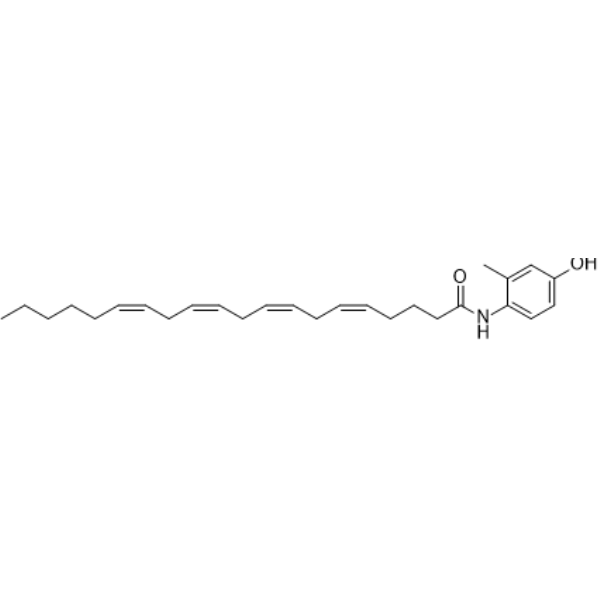
C27H39NO2
|
|---|---|
| 分子量 |
409.60406
|
| 精确质量 |
409.298079
|
| CAS号 |
313998-81-1
|
| PubChem CID |
9887748
|
| 外观&性状 |
Colorless to light yellow liquid
|
| 密度 |
1.0±0.1 g/cm3
|
| 沸点 |
586.6±50.0 °C at 760 mmHg
|
| 闪点 |
308.5±30.1 °C
|
| 蒸汽压 |
0.0±1.7 mmHg at 25°C
|
| 折射率 |
1.553
|
| LogP |
6.7
|
| tPSA |
49.3 Ų
|
| 氢键供体(HBD)数目 |
2
|
| 氢键受体(HBA)数目 |
2
|
| 可旋转键数目(RBC) |
15
|
| 重原子数目 |
30
|
| 分子复杂度/Complexity |
547
|
| 定义原子立体中心数目 |
0
|
| SMILES |
CCCCC/C=C\C/C=C\C/C=C\C/C=C\CCCC(=O)NC1=CC=C(C=C1C)O
|
| InChi Key |
WUZWFRWVRHLXHZ-ZKWNWVNESA-N
|
| InChi Code |
InChI=1S/C27H39NO2/c1-3-4-5-6-7-8-9-10-11-12-13-14-15-16-17-18-19-20-27(30)28-26-22-21-25(29)23-24(26)2/h7-8,10-11,13-14,16-17,21-23,29H,3-6,9,12,15,18-20H2,1-2H3,(H,28,30)/b8-7-,11-10-,14-13-,17-16-
|
| 化学名 |
(5Z,8Z,11Z,14Z)-N-(4-hydroxy-2-methylphenyl)icosa-5,8,11,14-tetraenamide
|
| 别名 |
313998-81-1; VDM 11; (5Z,8Z,11Z,14Z)-N-(4-HYDROXY-2-METHYLPHENYL)-5,8,11,14-EICOSATETRAENAMIDE; VDM-11; (5Z,8Z,11Z,14Z)-N-(4-Hydroxy-2-methylphenyl)icosa-5,8,11,14-tetraenamide; VDM11; VDM-11 (Solution in Ethanol); (5Z,8Z,11Z,14Z)-N-(4-Hydroxy-2-methylphenyl)-5,8,11,14-eicosatetraenamide;
|
| HS Tariff Code |
2934.99.9001
|
| 存储方式 |
Powder -20°C 3 years 4°C 2 years In solvent -80°C 6 months -20°C 1 month |
| 运输条件 |
Room temperature (This product is stable at ambient temperature for a few days during ordinary shipping and time spent in Customs)
|
| 溶解度 (体外实验) |
May dissolve in DMSO (in most cases), if not, try other solvents such as H2O, Ethanol, or DMF with a minute amount of products to avoid loss of samples
|
|---|---|
| 溶解度 (体内实验) |
注意: 如下所列的是一些常用的体内动物实验溶解配方,主要用于溶解难溶或不溶于水的产品(水溶度<1 mg/mL)。 建议您先取少量样品进行尝试,如该配方可行,再根据实验需求增加样品量。
注射用配方
注射用配方1: DMSO : Tween 80: Saline = 10 : 5 : 85 (如: 100 μL DMSO → 50 μL Tween 80 → 850 μL Saline)(IP/IV/IM/SC等) *生理盐水/Saline的制备:将0.9g氯化钠/NaCl溶解在100 mL ddH ₂ O中,得到澄清溶液。 注射用配方 2: DMSO : PEG300 :Tween 80 : Saline = 10 : 40 : 5 : 45 (如: 100 μL DMSO → 400 μL PEG300 → 50 μL Tween 80 → 450 μL Saline) 注射用配方 3: DMSO : Corn oil = 10 : 90 (如: 100 μL DMSO → 900 μL Corn oil) 示例: 以注射用配方 3 (DMSO : Corn oil = 10 : 90) 为例说明, 如果要配制 1 mL 2.5 mg/mL的工作液, 您可以取 100 μL 25 mg/mL 澄清的 DMSO 储备液,加到 900 μL Corn oil/玉米油中, 混合均匀。 View More
注射用配方 4: DMSO : 20% SBE-β-CD in Saline = 10 : 90 [如:100 μL DMSO → 900 μL (20% SBE-β-CD in Saline)] 口服配方
口服配方 1: 悬浮于0.5% CMC Na (羧甲基纤维素钠) 口服配方 2: 悬浮于0.5% Carboxymethyl cellulose (羧甲基纤维素) 示例: 以口服配方 1 (悬浮于 0.5% CMC Na)为例说明, 如果要配制 100 mL 2.5 mg/mL 的工作液, 您可以先取0.5g CMC Na并将其溶解于100mL ddH2O中,得到0.5%CMC-Na澄清溶液;然后将250 mg待测化合物加到100 mL前述 0.5%CMC Na溶液中,得到悬浮液。 View More
口服配方 3: 溶解于 PEG400 (聚乙二醇400) 请根据您的实验动物和给药方式选择适当的溶解配方/方案: 1、请先配制澄清的储备液(如:用DMSO配置50 或 100 mg/mL母液(储备液)); 2、取适量母液,按从左到右的顺序依次添加助溶剂,澄清后再加入下一助溶剂。以 下列配方为例说明 (注意此配方只用于说明,并不一定代表此产品 的实际溶解配方): 10% DMSO → 40% PEG300 → 5% Tween-80 → 45% ddH2O (或 saline); 假设最终工作液的体积为 1 mL, 浓度为5 mg/mL: 取 100 μL 50 mg/mL 的澄清 DMSO 储备液加到 400 μL PEG300 中,混合均匀/澄清;向上述体系中加入50 μL Tween-80,混合均匀/澄清;然后继续加入450 μL ddH2O (或 saline)定容至 1 mL; 3、溶剂前显示的百分比是指该溶剂在最终溶液/工作液中的体积所占比例; 4、 如产品在配制过程中出现沉淀/析出,可通过加热(≤50℃)或超声的方式助溶; 5、为保证最佳实验结果,工作液请现配现用! 6、如不确定怎么将母液配置成体内动物实验的工作液,请查看说明书或联系我们; 7、 以上所有助溶剂都可在 Invivochem.cn网站购买。 |
| 制备储备液 | 1 mg | 5 mg | 10 mg | |
| 1 mM | 2.4414 mL | 12.2070 mL | 24.4141 mL | |
| 5 mM | 0.4883 mL | 2.4414 mL | 4.8828 mL | |
| 10 mM | 0.2441 mL | 1.2207 mL | 2.4414 mL |
1、根据实验需要选择合适的溶剂配制储备液 (母液):对于大多数产品,InvivoChem推荐用DMSO配置母液 (比如:5、10、20mM或者10、20、50 mg/mL浓度),个别水溶性高的产品可直接溶于水。产品在DMSO 、水或其他溶剂中的具体溶解度详见上”溶解度 (体外)”部分;
2、如果您找不到您想要的溶解度信息,或者很难将产品溶解在溶液中,请联系我们;
3、建议使用下列计算器进行相关计算(摩尔浓度计算器、稀释计算器、分子量计算器、重组计算器等);
4、母液配好之后,将其分装到常规用量,并储存在-20°C或-80°C,尽量减少反复冻融循环。
计算结果:
工作液浓度: mg/mL;
DMSO母液配制方法: mg 药物溶于 μL DMSO溶液(母液浓度 mg/mL)。如该浓度超过该批次药物DMSO溶解度,请首先与我们联系。
体内配方配制方法:取 μL DMSO母液,加入 μL PEG300,混匀澄清后加入μL Tween 80,混匀澄清后加入 μL ddH2O,混匀澄清。
(1) 请确保溶液澄清之后,再加入下一种溶剂 (助溶剂) 。可利用涡旋、超声或水浴加热等方法助溶;
(2) 一定要按顺序加入溶剂 (助溶剂) 。
 AZ513
AZ513
 ASP 8477
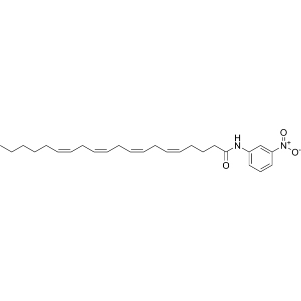 Arachidonoyl m-nitroaniline
 MK-3168 (12C)
InvivoChem的所有产品仅用于作科学研究,不面向患者销售
Copyright 2020 InvivoChem LLC | All Rights Reserved 粤ICP备20063088号-1


 463611831
