| 规格 | 价格 | |
|---|---|---|
| 500mg | ||
| 1g | ||
| Other Sizes |
| 靶点 |
Vesicular acetylcholine transport; Sigma 1/2 Receptor
|
|---|---|
| 体外研究 (In Vitro) |
阻断乙酰胆碱转运至突触小泡以及随后乙酰胆碱的量子释放被认为是(±)-Vesamicol HCl的药理作用[1]。
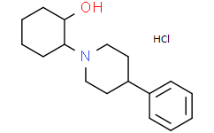
为了讨论的目的,将Vesamicol的结构分为三个主要片段: A、 B和C(图1)。尽管已经合成了许多维生素C醇类似物,以扩大我们对与囊泡乙酰胆碱转运蛋白结合的结构-活性关系的理解,但绝大多数含有未修饰的B片段,其余仅含有该片段的单点修饰。因此,人们对分子的这一区域与囊泡乙酰胆碱转运蛋白之间的相互作用知之甚少。在之前的研究中, 我们和其他人利用构象限制策略(在片段a和/或C中)为囊泡乙酰胆碱转运蛋白开发了选择性高亲和力配体。为了测试这种策略的局限性,我们选择将我们的研究扩展到片段B。[1] 在我们的第一次尝试中,我们用托帕尼基部分替换了哌啶基片段B。从形式上讲,托烷可以被视为2,6-乙哌啶。由于乙烯桥阻止了哌啶构象的相互转化,因此托烷酰基片段可以被视为构象受限的哌啶基残基。在一项平行的工作中,我们还合成了1-甲基螺[1H-吲哚啉-3,4'-哌啶](7)的几种衍生物,作为构象受限的Vesamicol类似物。后一项研究是我们之前对3−5等肺活量类似物的研究的延伸。测试了目标化合物与鱼雷突触小泡的囊泡乙酰胆碱转运蛋白的结合。还评估了与σ受体的结合,因为Vesamicol及其一些类似物对这些位点显示出中度至高度的亲和力。11还测试了这些化合物与单胺转运体的结合情况,因为在许多单胺再摄取抑制剂中都发现了3β-苯基托烷酰部分。最后,由于啮齿动物的行为效应,评估了这些化合物对多巴胺D2受体的亲和力(数据未显示)。由于参考配体[125I]NCQ298不能区分多巴胺D2和D3受体,因此用这种放射性配体获得的数据反映了与两种多巴胺受体亚型的结合。[1] 尽管许多托烷类似物对囊泡乙酰胆碱转运蛋白显示出适度的亲和力,但没有一种新化合物比Vesamicol更有效。事实上,与相应的哌啶基类似物相比,大多数新化合物的表现相当差。因此,虽然化合物10a的亲和力比Vesamicol低4倍,但10b−d的效力分别比苯并Vesamicol(2)、25和26低至少2个数量级(表1)。即使环己基部分被羟乙基取代,这些差异仍然存在(27 vs 10f-k),但当N-烷基取代基变得更加灵活时,这些差异消失了(28 vs 10e)。综上所述,上述内容表明,双碳桥(将哌啶基转化为托烷酰片段)对配体-受体相互作用极为不利。随着双碳桥增加片段B的空间体积,对该桥不利影响的一个合理解释可能是片段B适合结合位点内的一个狭窄口袋。[1] 尽管3−5等螺酰胺醇对囊泡乙酰胆碱转运蛋白的亲和力与Vesamicol相当或更高,但7的所有类似物的效力都明显低于Vesamicol。此外,新化合物的效力通常比相应的碳类似物低1-2个数量级(比较18a与4,19a与19b,20a与20b,21a与21b)。虽然这两个系列化合物之间的差异被环己基的缺失所掩盖(比较22a与22b和23a与23b),但很明显,7不是Vesamicol中a-B片段的合适替代品。 |
| 体内研究 (In Vivo) |
(±)-盐酸Vesamicol(3 mg/kg;腹腔注射;一次;雄性 Wistar 大鼠)治疗可增加除纹状体外区域外所有区域的脑内胞质乙酰胆碱 (ACh) 水平,并降低脑内水泡 ACh 水平 [2] 。
Vesamicol已知在体外抑制乙酰胆碱(ACh)向突触小泡的转运,但在体内对其在大脑中的作用知之甚少。为了评估韦沙醇在体内的作用,我们检查了胆碱能参数,如ACh的亚细胞分布、酶的活性、胆碱的摄取以及大鼠纹状体、海马和大脑皮层中的毒蕈碱受体结合,这些参数是在腹腔注射韦沙醇(3 mg/kg)或韦沙醇联合DDVP(5 mg/kg)30和60分钟后测定的,这些参数在韦沙醇前10分钟给药。注射维生素C后,脑所有区域的细胞质ACh水平都升高,而除纹状体外,所有区域的囊泡ACh水平均降低。注射维生素C后,DDVP诱导的纹状体细胞外ACh和细胞质ACh水平的增加通常会增强,而先前注射DDVP时,Vesamicol并未降低囊泡ACh的水平。Vesamicol没有诱导三个区域的酶活性、胆碱摄取或[6H]苯并咪唑奎核环酯与毒蕈碱ACh受体的结合发生任何显著变化。DDVP与维沙唑的联合给药不能逆转DDVP引起的胆碱能参数的变化。目前的研究结果表明,韦沙醇可以在体内抑制ACh向脑组织突触小泡的转运,尽管它不能逆转在韦沙醇之前注射的敌敌畏的作用。[2] 亚细胞乙酰胆碱水平[2] 如图1所示,注射后30分钟和60分钟,Vesamicol没有引起大脑三个区域细胞外ACh水平的显著变化。注射Vesamicol后30和/或60分钟,纹状体、海马和皮质中的细胞质ACh水平升高。相比之下,注射Vesamicol后30分钟和60分钟,海马和皮质中的囊泡ACh水平降低,但纹状体中的水平没有降低。在纹状体中,注射维沙莫醇30和60分钟后,囊泡ACh水平没有明显变化(图1)。 在40和70分钟时,DDVP的给药增加了纹状体中细胞外ACh、纹状体和海马中细胞质ACh以及纹状体中囊泡ACh的水平(图1)。注射敌敌畏后,大脑任何区域的囊泡ACh水平都没有变化,尽管注射敌敌敌畏40分钟后纹状体和海马的囊泡AC水平没有显著增加。 如图1所示,与对照组和DDVP治疗的大鼠相比,联合施用Vesamicol和DDVP导致纹状体细胞外ACh和细胞质ACh水平显著升高。在注射DDVP后40或70分钟,对照组大鼠和注射DDVP和韦沙莫醇的大鼠在大脑的三个区域的囊泡ACh水平没有显著差异。 3.2.乙酰胆碱酯酶和胆碱乙酰转移酶的活性、胆碱的高亲和力摄取(HACU)和毒蕈碱受体的结合[2] 图2显示了注射Vesamicol、DDVP或DDVP加Vesamicol的大鼠大脑三个区域的AChE活性。注射后30或60分钟,单独使用Vesamicol不会导致任何大脑区域的活动发生任何变化。注射敌敌畏后,在40和70分钟时,大脑三个区域的AChE活性显著降低。服用维沙莫醇对之前注射过敌敌畏的大鼠脑三个区域AChE活动的降低没有任何影响。 如图4所示,注射Vesamicol后30或60分钟,对照组和大鼠大脑三个区域的HACU Km(图4A)和Vmax(图4B)值没有显著变化。然而,注射后40分钟和70分钟,DDVP导致大脑三个区域HACU的两个参数都增加。同时接受DDVP和维沙莫醇的大鼠大脑各区域的HACU与单独注射DDVP的大鼠大致相同。 图5显示了注射生理盐水(对照组)、Vesamicol、DDVP或DDVP加Vesamicol的大鼠大脑三个区域中[3H]QNB与mAChRs的结合。对照组和单独注射韦沙醇30和60分钟后检查的大鼠在所有区域的Kd(图5A)和Bmax(图5B)值没有显著差异。注射敌敌畏通常会降低Kd和Bmax值。接受DDVP加Vesamicol的大鼠大脑各区域的这些参数值与单独注射DDVP的大鼠的值大致相同。 |
| 酶活实验 |
囊泡乙酰胆碱转运蛋白结合。[1]
通过Rogers等人的方法,在22°C下孵育24小时后,通过与[3H]Vesamicol与电器官突触小泡的结合竞争来确定新化合物的解离常数。6 σ受体结合。根据已发表的程序,在豚鼠脑膜中用σ1-选择性放射性配体[3H]-(+)-戊佐辛标记σ1结合位点。在(+)-戊佐辛(100 nM)存在下,用[3H]DTG在大鼠肝膜中测定了33,34σ-2位点,这是这些位点的丰富来源。 膜制备。[1] 从冷冻的豚鼠脑中减去小脑制备粗P2膜部分。在均质化之前,让大脑在冰上慢慢解冻。还从雄性Sprague−Dawley大鼠(175−225 g)的肝脏制备了粗P2膜组分。动物被斩首处死,肝脏被取出并切碎,然后进行均质化。 在4°C下,使用Porter Elvehjem组织研磨机在10 mL/g组织重量的10 mM Tris-HCl/0.32 M蔗糖(pH 7.4)中进行组织均质化。将粗匀浆在1000g下离心10分钟,将上清液保存在冰上。通过涡旋将沉淀物重新悬浮在2 mL/g组织重量冰冷的10 mM Tris-HCl/0.32 M蔗糖(pH 7.4)中。在1000g下离心10分钟后,丢弃沉淀物,合并上清液,在31000g下离心15分钟。通过涡旋将沉淀物重新悬浮在3 mL/g 10 mM Tris-HCl(pH 7.4)中,并使悬浮液在25°C下孵育15分钟。在31000g上离心15分钟后,将沉淀物轻轻均质至1.53 mL/g,再悬浮在10 mM Tris-HaCl(pH 7.3)中,等分试样储存在-80°C下直至使用。悬浮液的蛋白质浓度通过Bradford的方法测定,通常在6-11mg蛋白质/mL的范围内。 σ1结合试验。[1] 将豚鼠膜(100μg蛋白质)与3 nM[3H]-(+)-戊佐辛(31.6 Ci/mmol)在pH 8.0的50 mM Tris-HCl中在25°C下孵育120或240分钟。将测试化合物溶解在乙醇中,然后在缓冲液中稀释,使总孵育体积为0.5 mL。通过加入冰冷的10 mM Tris-HHCl(pH 8.0)终止测定,然后使用Brandel收割机通过Whatman GF/B玻璃过滤器(预浸在0.5%聚乙烯亚胺中)快速过滤。用5mL冰冷的缓冲液洗涤过滤器两次。在10μM(+)-戊佐辛存在下测定非特异性结合。使用Beckman LS 6000IC光谱仪在Ecolite(+)中进行液体闪烁计数,计数效率为50%。典型的计数为总结合70 dpm/μg蛋白质,非特异性结合6 dpm/µg,特异性结合64 dpm/微克。 σ2结合试验。[1] 在100 nM(+)-戊佐辛的存在下,用3 nM[3H]DTG(38.3 Ci/mmol)孵育大鼠肝膜(35μg蛋白质)或豚鼠脑膜(360μg),以掩盖σ1位点。在25°C下,在pH 8.0的50 mM Tris-HCl中孵育120分钟,总孵育体积为0.5 mL。通过加入冰冷的10 mM Tris-HHCl(pH 8.0)终止测定,然后使用Brandel收割机通过Whatman GF/B玻璃过滤器(预浸在0.5%聚乙烯亚胺中)快速过滤。然后用0.5mL冰冷的缓冲液洗涤过滤器两次。在5μM DTG存在下测定非特异性结合。使用Beckman LS 6000IC光谱仪在Ecolite(+)中进行液体闪烁计数,计数效率为50%。大鼠肝脏的典型计数为总结合297 dpm/μg蛋白质,非特异性结合11 dpm/µg蛋白质,特异性结合286 dpm/微克蛋白质。豚鼠脑的典型计数为总结合16dpm/μg蛋白质,非特异性结合2dpm/μg,特异性结合14dpm/µg。 数据分析。σ位点的IC50值通过使用JMP的非线性回归分析分三次测定,每种乙酰胆碱化合物的浓度为5-10。Ki值使用Cheng-Prusoff方程35计算,表示平均值±SEM。除非另有说明,否则所有测定均一式三份。对于放射性配体,采用了以下之前报道的33,34 Kd值: [3H]DTG,17.9 nM(大鼠肝脏);[3H]-(+)-戊佐辛,4.8 nM(豚鼠脑)。豚鼠脑中[3H]DTG的Kd值通过Scatchard分析确定为21.6 nM。 多巴胺D2/D3受体结合试验。[1] Perry Molinoff博士提供了表达大鼠多巴胺D2受体的草地贪夜蛾(Sf9)昆虫细胞的冷冻膜制剂。如前所述制备大鼠纹状体匀浆。如前所述,使用0.2 nM[125I]NCQ298(用于Sf9/D2受体)或0.05 nM[127I]NCQ2 98(用于纹状体匀浆中的D2/D3受体)作为放射性配体,在这些细胞膜中进行了36次竞争实验,并使用8−10浓度(10-10−10-5 M)的竞争药物(在含有0.1%BSA的Tris-HCl缓冲液中连续稀释)。37非特异性结合用1μM spiperone定义。在37°C下孵育30分钟。通过用1%聚乙烯亚胺预浸的玻璃纤维过滤器过滤,将结合的放射性配体与游离的放射性配体分离,从而终止反应。然后用3mL冰冷的20mM Tris缓冲液洗涤过滤器三次,并在γ计数器中以70%的效率计数。使用迭代非线性最小二乘曲线拟合程序LIGAND对竞争实验进行了分析。 多巴胺和血清素转运体结合试验。[1] 如前所述,[125I]IPT分别在大鼠纹状体和皮质匀浆中与多巴胺和血清素转运体结合。39竞争实验在含有50 mM Tris-HCl、pH 7.4和120 mM NaCl的缓冲液中进行,缓冲液中含有0.2 M[125I]IPT(用于纹状体匀浆)或0.5 M[125I]IPT,以及8−10浓度(10-10−10-5 M)的竞争药物。非特异性结合是在40μM(−)可卡因存在的情况下定义的。使用迭代非线性最小二乘曲线拟合程序LIGAND对竞争实验进行了分析。 |
| 动物实验 |
Animal/Disease Models: Male Wistar rats (120-300 g)[2]
Doses: 3 mg/kg Route of Administration: intraperitoneal (ip)injection; once Experimental Results: The levels of cytosolic acetylcholine (ACh) increased in all regions of the brain, while those of vesicular ACh diminished in all regions except for the striatum. In the present experiments, cholinergic parameters were determined for regions of the brain of rats that had been treated with Vesamicol alone or combination with DDVP. Rats received a single dose (3 mg/5 ml saline/kg, IP) of Vesamicol [(±)-Vesamicol hydrochloride) and they were sacrificed 30 or 60 min later. Control rats were injected with 5 ml/kg of saline and killed 30 min later. Some animals received DDVP (5 mg/kg, SC; Kobayashi et al.) 10 min prior to Vesamicol or saline and were sacrificed 40 or 70 min later. A dose of Vesamicol of 3 mg/kg was chosen because a higher dose (5 mg/kg) killed two of five rats tested and the LD50 of this drug (administered IP) has been reported to be 4.2 mg/kg in mice [2]. |
| 药代性质 (ADME/PK) |
Metabolism / Metabolites
Copper is mainly absorbed through the gastrointestinal tract, but it can also be inhalated and absorbed dermally. It passes through the basolateral membrane, possibly via regulatory copper transporters, and is transported to the liver and kidney bound to serum albumin. The liver is the critical organ for copper homoeostasis. In the liver and other tissues, copper is stored bound to metallothionein, amino acids, and in association with copper-dependent enzymes, then partitioned for excretion through the bile or incorporation into intra- and extracellular proteins. The transport of copper to the peripheral tissues is accomplished through the plasma attached to serum albumin, ceruloplasmin or low-molecular-weight complexes. Copper may induce the production of metallothionein and ceruloplasmin. The membrane-bound copper transporting adenosine triphosphatase (Cu-ATPase) transports copper ions into and out of cells. Physiologically normal levels of copper in the body are held constant by alterations in the rate and amount of copper absorption, compartmental distribution, and excretion. (L277, L279) |
| 毒性/毒理 (Toxicokinetics/TK) |
Toxicity Summary
Excess copper is sequestered within hepatocyte lysosomes, where it is complexed with metallothionein. Copper hepatotoxicity is believed to occur when the lysosomes become saturated and copper accumulates in the nucleus, causing nuclear damage. This damage is possibly a result of oxidative damage, including lipid peroxidation. Copper inhibits the sulfhydryl group enzymes such as glucose-6-phosphate 1-dehydrogenase, glutathione reductase, and paraoxonases, which protect the cell from free oxygen radicals. It also influences gene expression and is a co-factor for oxidative enzymes such as cytochrome C oxidase and lysyl oxidase. In addition, the oxidative stress induced by copper is thought to activate acid sphingomyelinase, which lead to the production of ceramide, an apoptotic signal, as well as cause hemolytic anemia. Copper-induced emesis results from stimulation of the vagus nerve. (L277, T49, A174, L280) 212231 mouse LD50 oral 48931 ug/kg PERIPHERAL NERVE AND SENSATION: FLACCID PARALYSIS WITHOUT ANESTHESIA (USUALLY NEUROMUSCULAR BLOCKAGE) European Journal of Pharmacology., 8(93), 1969 [PMID:4243404] 212231 mouse LD50 intravenous 5874 ug/kg PERIPHERAL NERVE AND SENSATION: FLACCID PARALYSIS WITHOUT ANESTHESIA (USUALLY NEUROMUSCULAR BLOCKAGE) European Journal of Pharmacology., 8(93), 1969 [PMID:4243404] |
| 参考文献 |
|
| 其他信息 |
As part of our ongoing structure-activity studies of the vesicular acetylcholine transporter ligand 2-(4-phenylpiperidino)cyclohexanol (Vesamicol, 1), 22 N-hydroxy(phenyl)alkyl derivatives of 3 beta-phenyltropane, 6, and 1-methylspiro[1H-indoline-3,4'-piperidine], 7, were synthesized and tested for binding in vitro. Although a few compounds displayed moderately high affinity for the vesicular acetylcholine transporter, no compound was more potent than the prototypical vesicular acetylcholine transporter ligand Vesamicol. However, a few derivatives of 6 displayed higher affinity for the dopamine transporter than cocaine. We conclude that modification of the piperidyl fragment of 1 will not lead to more potent vesicular acetylcholine transporter ligands. [1]
In summary, 3β-phenyltropanyl derivatives of Vesamicol exhibit lower affinity for the vesicular acetylcholine transporter than the parent compound. Consequently, the introduction of a two-carbon bridge across the C2 and C6 positions of the piperidyl moiety of Vesamicol is not a suitable strategy for enhancing affinity for the vesicular acetylcholine transporter or increasing selectivity for the vesicular acetylcholine transporter relative to σ receptors. The introduction of an aminomethyl bridge into the C fragment of Vesamicol (to yield analogues of 7) is also unsuitable for similar reasons. Since the results obtained with the tropanes suggest that fragment B binds to a narrow pocket within the binding site, we suggest that further modification of this fragment should be limited to single-point substitution. [1] The observation that the binding of QNB to mAChRs was unaffected 30 and 60 min after injection of Vesamicol indicates that this drug is unable to modify the mAChRs that have high affinity for ACh. A kinetic model has suggested that ACh has high affinity for the vesicular ACh transporter, through which ACh is transported and to which vesamicol binds at an allosteric site, referred to as the Vesamicol receptor. Kd and Bmax express the affinity for the ligand ACh and the density of mAChRs. Administration of DDVP caused an increase in Kd and a decrease in Bmax in the present experiments, suggesting that accumulation of ACh must have occurred around mAChRs. Vesamicol failed to reverse the effects of DDVP on the ability of QNB to bind to mAChRs. In conclusion, the present study of cholinergic parameters in three regions of the rat brain demonstrated that Vesamicol, an inhibitor of the transport of ACh into synaptic vesicles in vitro and in situ caused reduced levels of vesicular ACh and increased levels of cytoplasmic ACh in vivo after systemic administration. Thus, vesamicol apears to inhibit the uptake of ACh in the cytoplasm into vesicles in vivo. Other parameters, such as HACU, the activity of ChAT and levels of mAChRs, appear not to be involved directly in the effects of vesamicol under the present experimental conditions.[2] |
| 分子式 |
C17H26CLNO
|
|---|---|
| 分子量 |
295.85
|
| 精确质量 |
295.17
|
| 元素分析 |
C, 69.02; H, 8.86; Cl, 11.98; N, 4.73; O, 5.41
|
| CAS号 |
23965-53-9
|
| 相关CAS号 |
22232-64-0 (Parent); 120447-62-3; 112709-59-8; 23965-53-9
|
| PubChem CID |
212231
|
| 外观&性状 |
Typically exists as solid at room temperature
|
| tPSA |
23.5
|
| 氢键供体(HBD)数目 |
2
|
| 氢键受体(HBA)数目 |
2
|
| 可旋转键数目(RBC) |
2
|
| 重原子数目 |
20
|
| 分子复杂度/Complexity |
266
|
| 定义原子立体中心数目 |
0
|
| SMILES |
C1CCC(C(C1)N2CCC(CC2)C3=CC=CC=C3)O.Cl
|
| InChi Key |
XJNUHVMJVWOYCW-UHFFFAOYSA-N
|
| InChi Code |
InChI=1S/C17H25NO.ClH/c19-17-9-5-4-8-16(17)18-12-10-15(11-13-18)14-6-2-1-3-7-14;/h1-3,6-7,15-17,19H,4-5,8-13H2;1H
|
| 化学名 |
2-(4-phenylpiperidin-1-yl)cyclohexan-1-ol;hydrochloride
|
| 别名 |
AH-5183 hydrochloride; AH 5183 hydrochloride; VESAMICOL HYDROCHLORIDE; 120447-62-3; AH 5183 HCl; 23965-53-9; Cyclohexanol, 2-(4-phenylpiperidino)-, hydrochloride; 2-(4-Phenylpiperidino)cyclohexanol hydrochloride; (+-)-Vesamicol HCl; vesamicol hydrochloride, (+,-)-; Vesamicol hydrochloride
|
| HS Tariff Code |
2934.99.9001
|
| 存储方式 |
Powder -20°C 3 years 4°C 2 years In solvent -80°C 6 months -20°C 1 month |
| 运输条件 |
Room temperature (This product is stable at ambient temperature for a few days during ordinary shipping and time spent in Customs)
|
| 溶解度 (体外实验) |
May dissolve in DMSO (in most cases), if not, try other solvents such as H2O, Ethanol, or DMF with a minute amount of products to avoid loss of samples
|
|---|---|
| 溶解度 (体内实验) |
注意: 如下所列的是一些常用的体内动物实验溶解配方,主要用于溶解难溶或不溶于水的产品(水溶度<1 mg/mL)。 建议您先取少量样品进行尝试,如该配方可行,再根据实验需求增加样品量。
注射用配方
注射用配方1: DMSO : Tween 80: Saline = 10 : 5 : 85 (如: 100 μL DMSO → 50 μL Tween 80 → 850 μL Saline)(IP/IV/IM/SC等) *生理盐水/Saline的制备:将0.9g氯化钠/NaCl溶解在100 mL ddH ₂ O中,得到澄清溶液。 注射用配方 2: DMSO : PEG300 :Tween 80 : Saline = 10 : 40 : 5 : 45 (如: 100 μL DMSO → 400 μL PEG300 → 50 μL Tween 80 → 450 μL Saline) 注射用配方 3: DMSO : Corn oil = 10 : 90 (如: 100 μL DMSO → 900 μL Corn oil) 示例: 以注射用配方 3 (DMSO : Corn oil = 10 : 90) 为例说明, 如果要配制 1 mL 2.5 mg/mL的工作液, 您可以取 100 μL 25 mg/mL 澄清的 DMSO 储备液,加到 900 μL Corn oil/玉米油中, 混合均匀。 View More
注射用配方 4: DMSO : 20% SBE-β-CD in Saline = 10 : 90 [如:100 μL DMSO → 900 μL (20% SBE-β-CD in Saline)] 口服配方
口服配方 1: 悬浮于0.5% CMC Na (羧甲基纤维素钠) 口服配方 2: 悬浮于0.5% Carboxymethyl cellulose (羧甲基纤维素) 示例: 以口服配方 1 (悬浮于 0.5% CMC Na)为例说明, 如果要配制 100 mL 2.5 mg/mL 的工作液, 您可以先取0.5g CMC Na并将其溶解于100mL ddH2O中,得到0.5%CMC-Na澄清溶液;然后将250 mg待测化合物加到100 mL前述 0.5%CMC Na溶液中,得到悬浮液。 View More
口服配方 3: 溶解于 PEG400 (聚乙二醇400) 请根据您的实验动物和给药方式选择适当的溶解配方/方案: 1、请先配制澄清的储备液(如:用DMSO配置50 或 100 mg/mL母液(储备液)); 2、取适量母液,按从左到右的顺序依次添加助溶剂,澄清后再加入下一助溶剂。以 下列配方为例说明 (注意此配方只用于说明,并不一定代表此产品 的实际溶解配方): 10% DMSO → 40% PEG300 → 5% Tween-80 → 45% ddH2O (或 saline); 假设最终工作液的体积为 1 mL, 浓度为5 mg/mL: 取 100 μL 50 mg/mL 的澄清 DMSO 储备液加到 400 μL PEG300 中,混合均匀/澄清;向上述体系中加入50 μL Tween-80,混合均匀/澄清;然后继续加入450 μL ddH2O (或 saline)定容至 1 mL; 3、溶剂前显示的百分比是指该溶剂在最终溶液/工作液中的体积所占比例; 4、 如产品在配制过程中出现沉淀/析出,可通过加热(≤50℃)或超声的方式助溶; 5、为保证最佳实验结果,工作液请现配现用! 6、如不确定怎么将母液配置成体内动物实验的工作液,请查看说明书或联系我们; 7、 以上所有助溶剂都可在 Invivochem.cn网站购买。 |
| 制备储备液 | 1 mg | 5 mg | 10 mg | |
| 1 mM | 3.3801 mL | 16.9005 mL | 33.8009 mL | |
| 5 mM | 0.6760 mL | 3.3801 mL | 6.7602 mL | |
| 10 mM | 0.3380 mL | 1.6900 mL | 3.3801 mL |
1、根据实验需要选择合适的溶剂配制储备液 (母液):对于大多数产品,InvivoChem推荐用DMSO配置母液 (比如:5、10、20mM或者10、20、50 mg/mL浓度),个别水溶性高的产品可直接溶于水。产品在DMSO 、水或其他溶剂中的具体溶解度详见上”溶解度 (体外)”部分;
2、如果您找不到您想要的溶解度信息,或者很难将产品溶解在溶液中,请联系我们;
3、建议使用下列计算器进行相关计算(摩尔浓度计算器、稀释计算器、分子量计算器、重组计算器等);
4、母液配好之后,将其分装到常规用量,并储存在-20°C或-80°C,尽量减少反复冻融循环。
计算结果:
工作液浓度: mg/mL;
DMSO母液配制方法: mg 药物溶于 μL DMSO溶液(母液浓度 mg/mL)。如该浓度超过该批次药物DMSO溶解度,请首先与我们联系。
体内配方配制方法:取 μL DMSO母液,加入 μL PEG300,混匀澄清后加入μL Tween 80,混匀澄清后加入 μL ddH2O,混匀澄清。
(1) 请确保溶液澄清之后,再加入下一种溶剂 (助溶剂) 。可利用涡旋、超声或水浴加热等方法助溶;
(2) 一定要按顺序加入溶剂 (助溶剂) 。