| 规格 | 价格 | 库存 | 数量 |
|---|---|---|---|
| 1mg |
|
||
| 2mg |
|
||
| 5mg |
|
||
| 10mg |
|
||
| 25mg |
|
||
| 100mg |
|
||
| Other Sizes |
|
| 靶点 |
PD-1/PD-L1 (EC50 = 253 nM)
BMS-1001 (CAS#: 2113650-03-4) targets human PD-L1 (programmed death-ligand 1). [2] |
|---|---|
| 体外研究 (In Vitro) |
BMS-1001 可降低可溶性 PD-L1 对 T 细胞受体介导的 T 淋巴细胞活化的抑制作用。BMS-1001 能有效降低 PD-L1 相关细胞表面蛋白的抑制作用。[1]
BMS-1001 (CAS#: 2113650-03-4) 与 PD-L1 结合,并破坏 PD-1/PD-L1 相互作用,这已通过基于核磁共振的 AIDA(拮抗剂诱导解离分析)证实。[2] 细胞毒性:针对 PD-1 效应细胞(修饰的 Jurkat T 细胞)的 EC50 为 33.4 μM(处理 48 小时,代谢活性测定)。 [2]在 PD-1/PD-L1 检查点检测(PD-1 效应细胞和 PD-L1+ 人工抗原呈递细胞的共培养)中,BMS-1001 (CAS#: 2113650-03-4) 以剂量依赖的方式恢复 TCR 介导的荧光素酶报告活性,EC50 值在三位数纳摩尔范围内(未指定确切值)。 [2] 在可溶性PD-L1 (sPD-L1) 效应分析中,BMS-1001 (CAS#: 2113650-03-4)(浓度分别为0.3、1.2和3 μM;与sPD-L1的摩尔比分别为1:2、2:1和5:1)呈剂量依赖性地逆转sPD-L1诱导的抗CD3刺激T细胞活化的抑制作用,并在3 μM浓度下(摩尔浓度为sPD-L1的5倍)完全恢复活化。[2] 流式细胞术显示,BMS-1001 (CAS#: 2113650-03-4)在1 μM浓度下显著降低荧光标记的sPD-L1与表达PD-1的细胞的结合,与抗PD-L1抗体的作用相似。 [2] 交联和尺寸排阻色谱实验表明,BMS-1001 (CAS#: 2113650-03-4) 可诱导溶液中可溶性 PD-L1 的二聚化。[2] 晶体结构 (PDB 5NIU) 显示,BMS-1001 (CAS#: 2113650-03-4) 结合于 PD-L1 同源二聚体的界面,诱导构象变化(Tyr56 侧链的旋转),从而形成疏水通道。(2R)-2-氨基-3-羟基丙酸部分与 Asp122 侧链形成氢键,并通过水介导与 Lys124 和 Tyr123 形成氢键;3-氰基苄基取代基与 Tyr123 和 Arg125 形成疏水接触。[2] |
| 体内研究 (In Vivo) |
NP19(BMS-1001类似物)在H22肝癌小鼠模型中的体内抗肿瘤活性[3]
鉴于NP19在黑色素瘤B16-F10肿瘤模型中表现出优异的体内抗肿瘤疗效,且PD-1/PD-L1抑制剂具有广谱抗肿瘤活性,我们进一步利用BALB/c小鼠H22肝癌肿瘤模型评估了化合物NP19的体内抗肿瘤疗效。每只小鼠右侧腹部皮下注射80万个H22细胞。待肿瘤体积达到约100 mm³后,将小鼠随机分组,并分别腹腔注射NP19或载体溶液,持续14天。如图 8 所示,NP19 在 25 mg/kg 剂量下表现出显著的体内抗肿瘤疗效,肿瘤生长抑制率 (TGI) 为 76.5%(图 8A、8B、8C)。此外,NP19 未引起明显的体重下降(图 8D),表明该化合物具有良好的耐受性。 NP19(BMS-1001 的类似物)在 B16-F10 小鼠黑色素瘤模型中的体内抗肿瘤活性[3] 为了确定新合成化合物的体外抗 PD-1/PD-L1 活性是否能转化为体内疗效,我们测试了化合物 NP19 在小鼠黑色素瘤 B16-F10 肿瘤模型中的抗肿瘤活性。由于NP19合成简便且细胞毒性较低(表9),与效力更强的化合物NP2或效力相当的化合物NP12相比,NP19被选用于体内疗效研究。我们用载体对照组和NP19(25 mg/kg、50 mg/kg、100 mg/kg)治疗携带黑色素瘤的BALB/c小鼠,NP19通过灌胃法每日一次给药,持续15天。如图6所示,治疗15天后,NP19治疗显著抑制了黑色素瘤的生长。 NP19的体内药代动力学特性[BMS-1001类似物][3] 由于化合物NP19在体外显示出高活性,接下来通过静脉注射和口服给药,在雄性Sprague-Dawley大鼠中评估了其药代动力学(PK)特征。表8总结了口服和静脉给药的关键药代动力学参数。单次静脉注射1 mg/kg化合物NP19后,NP19的半衰期(t1/2)、清除率(CL)和表观分布容积(Vss)分别为1.5 ± 0.5 h、0.9 ± 0.2 L/h/kg和2.1 ± 0.5 L/kg。口服10 mg/kg NP19后,观察到口服吸收良好(Tmax = 0.6 ± 0.2 h)、半衰期较长(t1/2 = 10.9 ± 7.7 h)和口服生物利用度较高(F = 5%)。此外,在大鼠中未观察到明显的不良反应。与静脉注射半衰期(1.5 h)相比,NP19经灌胃给药后的半衰期(10.9 h)显著延长。这可能是由于NP19的高亲脂性(logP = 7.9)或较差的水溶性所致。因此,NP19表现出翻转药代动力学特征。这种翻转药代动力学特征有时会出现在水溶性差的化合物中,例如瑞巴派特,其t1/2(po)/t1/2(iv)比值为13.5,这是由于其水溶性差(7.6 μg/mL)。另一个例子是李建明等人报道的亲脂性化合物IAT(一种抗微管蛋白药物,水溶性为19 μg/mL),其t1/2(po)/t1/2(iv)比值约为5,与NP19[t1/2(po)/t1/2(iv) = 7.1]相似。由于化合物NP19的口服生物利用度较低,我们推测需要高剂量才能达到足够的药物浓度以发挥抗肿瘤疗效。因此,我们进一步研究了化合物NP19的体内活性。 |
| 酶活实验 |
百时美施贵宝(BMS)近期公开了首个针对PD-1/PD-L1通路的非肽类小分子抑制剂,这些抑制剂在均相时间分辨荧光(HTRF)结合试验中显示出活性;然而,并未提供进一步的数据来支持其活性。
\n\n体外PD-1/PD-L1结合试验[3] \n本研究采用PD-1/PD-L1均相时间分辨荧光(HTRF)结合试验,考察了化合物抑制PD-1/PD-L1相互作用的能力。PD-1/PD-L1结合试验试剂盒购自Cisbio公司。实验按照操作手册进行,该手册可从 https://www.cisbio.com/usa/drug-discovery/human-pd1pd-l1-biochemical-interaction-assay 下载。\n \n\n\nBMS 近期公开了首个针对 PD-1/PD-L1 通路的非肽类小分子抑制剂,这些抑制剂在均相时间分辨荧光 (HTRF) 结合实验中显示出活性;然而,并未提供进一步的数据来支持其活性。 \n\n核磁共振 (NMR) 测量[2] \n通过在以 15NH4Cl 为唯一氮源的 M9 最小培养基中表达蛋白质,获得了均匀的 15N 标记。进行 NMR 测量时,通过凝胶过滤将缓冲液更换为 pH 7.4 的 PBS (PD-L1) 或 pH 6.4 的含 100 mM NaCl 的 25 mM 磷酸钠缓冲液 (PD-1)。向样品中加入10% (v/v) 的D₂O以提供锁定信号。所有谱图均使用Bruker Avance III 600 MHz核磁共振波谱仪在300 K下记录。通过监测化合物滴定过程中¹H-¹⁵N二维HMQC谱图中核磁共振化学位移的变化,评估化合物与PD-L1的相互作用。使用拮抗剂诱导解离实验评估受试化合物解离PD-L1/PD-1的能力。简而言之,用未标记的PD-L1略微过量滴定¹⁵N标记的PD-1 (0.2 mM)。将化合物分装到所得混合物中。实验过程中,使用HMQC监测1H-15N信号。\n \n\nPD1/PD-L1检查点检测[2] \n在检测前24小时,将aAPC以每孔10,000个细胞的密度接种于白色96孔板中,并置于培养基中。检测当天,将抗体在含1% FBS的RPMI 1640培养基中进行3.5倍系列稀释。将BMS化合物[BMS-1001/strong>]在DMSO中进行系列稀释,并配制于含1% FBS的RPMI 1640培养基中。这样,所有样品中DMSO的浓度均保持恒定。从孔中移除95 μl培养基,并在细胞上覆盖40 μl化合物稀释液。将 20,000 个内皮细胞 (EC) 加入到每个孔中,孔内含有 40 μl 含 1% FBS 的 RPMI 1640 培养基。在 37°C 孵育 6 小时后,将培养板在室温下平衡 10 分钟,然后向每个孔中加入 80 μl Bio-Glo 试剂。孵育 10 分钟后,使用 FlexStation 3 定量发光值。通过将 Hill 方程拟合到实验数据来确定半数最大有效浓度 (EC50) 和最大发光值 (RLUmax)。 \nPD-1/sPD-L1 效应测定[2] \n为了评估 BMS 对可溶性 PD-L1 抑制 T 细胞的影响,在重组人 sPD-L1 存在的情况下,用抗 CD3 抗体刺激内皮细胞。为此,将96孔白色平底板在4°C下用5 μg/ml的抗CD3抗体或同型对照抗体PBS溶液包被过夜。去除抗体溶液后,用PBS洗涤孔板3次并干燥。将sPD-L1(氨基酸18-134)用添加了青霉素/链霉素溶液(终浓度均为100 U/ml)的PBS稀释,并加入BMS化合物[BMS-1001]或等体积的DMSO。然后,将15 μl该溶液加入到已包被抗体的孔板的每个孔中。将内皮细胞离心并稀释至50,000个/ml,然后将60 μl细胞悬液加入到每个孔中。sPD-L1的终浓度为10 μg/ml(0.6 μM)。 BMS化合物[BMS-1001]的最终浓度分别为:0.12、0.3、1.2和3 μM,对应的BMS与sPD-L1的摩尔比分别为:1:5、1:2、2:1和5:1。细胞培养24小时后,使用Bio-Glo荧光素酶检测试剂盒,按照制造商的说明进行荧光素酶活性测定。 采用核磁共振波谱法评估了BMS-1001(CAS号:2113650-03-4)与PD-L1的相互作用。用该化合物滴定均匀标记的15N PD-L1(残基18-134),并在300 K下使用600 MHz核磁共振波谱仪记录1H-15N二维HMQC谱。谱线展宽表明化合物诱导形成了更高分子量的复合物。[2] 使用拮抗剂诱导解离试验 (AIDA) 评估了化合物解离预先形成的 PD-1/PD-L1 复合物的能力。用未标记的 PD-L1 滴定 15N 标记的 PD-1 (0.2 mM),然后加入化合物。PD-1 的 1H-15N HMQC 信号的恢复表明复合物被破坏。[2] 结晶:将纯化的 PD-L1(氨基酸残基 18-134)以 5 mg/ml 的浓度溶于 10 mM Tris pH 8.0、20 mM NaCl 缓冲液中,并与 BMS-1001 (CAS#: 2113650-03-4) 以 1:3 的摩尔比(蛋白质:化合物)混合。在室温下,从含有 0.2 M 氯化镁和 30% (w/v) PEG 4000 的 0.1 M Tris pH 8.5 溶液中获得了衍射质量的晶体。X射线衍射数据在 PETRA III P11 光束线(德国电子同步加速器中心,汉堡)上收集。使用 Phaser 程序通过分子置换法解析结构,并在 Phenix 程序中进行精修。最终分辨率为 2.01 Å。[2] 交联:将 hPD-L1(10 μM,溶于 pH 7.4 的 PBS 缓冲液)与 BMS-1001(CAS 编号:2113650-03-4)按 1:1 的摩尔比混合,然后加入 BS3 交联剂(0.25-1 mM),在室温下反应 30 分钟。反应用 25 mM Tris pH 7.5 终止。SDS-PAGE 分析显示单体和二聚体条带。 [2] 尺寸排阻色谱:使用 Superdex S75 10/30 HR 色谱柱,以 10 mM Tris pH 8.0、20 mM NaCl 缓冲液平衡。将 hPD-L1 (4 mg/ml) 与抑制剂以 3:1(抑制剂:蛋白)的摩尔比孵育 15 分钟,然后进行分析。与 apo-PD-L1 相比,保留时间缩短,表明形成了二聚体。[2] 分子对接:使用 Hex 8.0.0 进行。从共晶结构中提取的 PD-L1 原聚体作为受体;提取的 BMS 化合物作为刚性配体。[2] |
| 细胞实验 |
BMS-1001 可降低可溶性 PD-L1 对 T 细胞受体介导的 T 淋巴细胞活化的抑制作用。BMS-1001 可有效降低细胞表面相关 PD-L1 的抑制作用。[1] 为了评估 BMS 对可溶性 PD-L1 抑制 T 细胞的影响,我们用抗 CD3 抗体在重组人 sPD-L1 存在下刺激内皮细胞 (EC)。具体操作为:将 5 μg/ml 的抗 CD3 抗体或同型对照抗体溶液(溶于 PBS)包被于 96 孔白色平底板,4°C 孵育过夜。待板干燥并用 PBS 洗涤三次后,去除抗体溶液。在BMS-1001或等体积DMSO存在的情况下,将PBS缓冲液补充青霉素/链霉素溶液(终浓度均为100 U/ml),并用sPD-L1(氨基酸18-134)稀释。然后将该溶液以15 μl的体积加入到抗体包被板的每个孔中。在向每个孔中加入60 μl细胞悬液之前,将内皮细胞离心并稀释至50,000个/ml。最终sPD-L1的浓度为10 μg/ml(0.6 μM)。BMS-1001的最终浓度分别为0.12、0.3、1.2和3 μM,对应于BMS与sPD-L1的摩尔比分别为1:5、1:2、2:1和5:1。利用 Bio-Glo 荧光素酶检测系统,细胞培养 24 小时后进行荧光素酶活性检测。
\n\n细胞系[2] \n为了验证 BMS 化合物 [BMS-1001] 抑制 PD-1/PD-L1 相互作用的效力,我们使用了基于细胞的 PD-1/PD-L1 免疫检查点阻断模型。该检测使用了两种模型细胞系:人工抗原呈递细胞(PD-L1+ aAPC/CHO-K1 细胞,简称 aAPC),其过表达 TCR 配体和 PD-L1;以及 T 细胞替代物,即过表达 PD-1 并携带在 NFAT 启动子控制下的荧光素酶报告基因的修饰 Jurkat T 细胞系(PD-1 效应细胞,简称 EC)。细胞购自Promega公司,并在添加了10%胎牛血清、100 U/ml青霉素和100 U/ml链霉素的RPMI 1640培养基中培养。此外,细胞在持续存在潮霉素B(50 μg/ml)和G418(250 μg/ml)的培养基中培养,以确保导入基因构建体的稳定表达。实验中省略了后两种抗生素。通过流式细胞术验证了内皮细胞(ECs)上PD-1和抗原呈递细胞(aAPCs)上PD-L1的过表达(数据未显示),并通过监测抗CD3抗体刺激后的荧光素酶活性验证了荧光素酶表达基因的存在。抗生素筛选、流式细胞术和报告基因表达作为细胞系鉴定方法。定期检测细胞,使用基于PCR的方法检测支原体污染,结果均为阴性。\n\n \n细胞毒性试验[2] \n将5000个内皮细胞接种于透明的96孔板中,并在浓度递增的BMS化合物[BMS-1001]或DMSO(作为对照,所有样品中DMSO的浓度均保持不变)存在下培养48小时。处理后,根据制造商的说明,使用Biolog Redox Dye Mix MB进行代谢活性测试。\n \n\n流式细胞术测量[2] \n通过流式细胞术评估sPD-L1(氨基酸18-134)与内皮细胞的结合情况。将His标签的PD-L1蛋白或其突变体与NTA-Atto 647 N荧光染料在22°C下以8:1的摩尔比(蛋白:染料)孵育2小时。将PD-L1-Atto与待测化合物或抗体配制于150 μl PBS中。样品在4°C避光孵育30分钟。同时,将内皮细胞(ECs)离心,用PBS洗涤,并重悬于新鲜PBS中,浓度为1 × 10⁶个细胞/ml。向每个样品中加入50 μl ECs,并在冰上孵育60分钟。各组分的最终浓度为:PD-L1(1.5 μM)25 μg/ml,抗PD-L1抗体和对照抗体125 μg/ml,以及BMS化合物[BMS-1001] 1 μM。使用 BD FACS Verse 流式细胞仪和 BD FACSuite v1.0.6 软件分析样本。 细胞毒性试验:将 5,000 个 PD-1 效应细胞(修饰的 Jurkat T 细胞)接种于 96 孔板中,并用递增浓度的 BMS-1001(CAS#: 2113650-03-4) 或 DMSO 对照培养 48 小时。根据制造商的说明,使用 Biolog Redox Dye Mix MB 测定代谢活性。通过拟合确定 EC50 值。[2] PD-1/PD-L1 检查点试验:将人工抗原呈递细胞(aAPC,过表达 TCR 配体和 PD-L1 的 CHO-K1 细胞)以 10,000 个/孔的密度接种于白色 96 孔板中,培养 24 小时。将BMS-1001 (CAS#: 2113650-03-4)进行系列稀释,稀释液为含1% FBS(DMSO浓度恒定)的RPMI 1640培养基。移除95 μl培养基后,加入40 μl化合物稀释液,再加入40 μl含20,000个PD-1效应细胞(EC)。37°C孵育6小时,室温平衡10分钟,然后加入80 μl Bio-Glo试剂。10分钟后使用FlexStation 3定量发光值。EC50和RLUmax通过Hill方程拟合确定。[2] 对于PD-1/sPD-L1效应细胞检测:将96孔白色板用5 μg/ml抗CD3抗体PBS溶液包被,4°C过夜。洗涤后,将 sPD-L1(氨基酸 18-134)用含青霉素/链霉素的 PBS 缓冲液稀释,并加入 BMS-1001(CAS#: 2113650-03-4,最终浓度分别为 0.12、0.3、1.2 和 3 μM(与 sPD-L1 的摩尔比分别为 1:5、1:2、2:1 和 5:1)。向每个孔中加入 15 μl 该溶液。向每个孔中加入 60 μl ECs(50,000/ml)。最终 sPD-L1 浓度为 10 μg/ml(0.6 μM)。细胞培养 24 小时后,使用 Bio-Glo 荧光素酶检测试剂盒测定荧光素酶活性。 [2] 流式细胞术结合实验:His标签的sPD-L1用NTA-Atto 647 N荧光染料标记(摩尔比8:1,22℃孵育2小时)。将PD-L1-Atto(1.5 μM)溶于150 μl PBS中,加入1 μM的BMS-1001(CAS号:2113650-03-4)或抗体,于4℃避光孵育30分钟。加入内皮细胞(ECs,1×10^6个细胞/ml,50 μl),冰上孵育60分钟。使用BD FACS Verse流式细胞仪分析样品,并测量MFI(几何平均荧光强度)。[2] |
| 动物实验 |
雄性Sprague-Dawley大鼠药代动力学研究[3]
本研究采用雄性Sprague-Dawley大鼠(200-220 g)研究化合物NP19(BMS-1001类似物)的药代动力学。实验前12小时禁食,但可自由饮水。分别于口服(10 mg/kg)或静脉注射(1 mg/kg)化合物NP19后0.0833、0.25、0.5、1、1.5、2、4、6、8、12和24小时,从尾静脉采集血样(0.3 mL)至肝素化1.5 mL聚乙烯管中。该化合物溶于 5% DMSO 和 95% PEG-300 的混合溶液中用于静脉注射,或悬浮于 0.5% 羧甲基纤维素钠 (CMC-Na) 溶液中用于口服。样品立即以 3000 g 离心 10 分钟。所得血浆 (100 μL) 储存于 -20 °C 直至分析。使用 DAS(药物与统计)软件,通过非房室模型分析,根据个体动物数据确定药代动力学 (PK) 参数。PK 研究的仪器和分析条件:使用配备电喷雾电离 (ESI) 接口的 ACQUITY I-Class UPLC 和 XEVO TQD 三重四极杆质谱仪(Waters 公司,美国马萨诸塞州米尔福德)的 UPLC-MS/MS 系统分析血液样本。UPLC 系统由二元溶剂管理器 (BSM) 和带流通针的样品管理器 (SM-FTN) 组成。使用 Masslynx 4.1 软件(Waters 公司)进行数据采集和仪器控制。采用多反应监测 (MRM) 模式,NP19 的 m/z 555.35 → 181.03 和卡马西平的 m/z 237 → 194.1 进行定量分析。 小鼠 B16F10 黑色素瘤模型体内疗效研究[3] 使用 6-8 周龄的 BALB/c 小鼠研究 NP19(BMS-1001 的类似物)对皮下移植黑色素瘤细胞模型的抑制作用。将处于对数生长期的小鼠 B16F10 黑色素瘤细胞悬浮于 PBS 中,浓度为 2 × 10⁶ 个/mL。每只小鼠皮下接种 200 μL 含有 4 × 10⁵ 个细胞的细胞悬液。当肿瘤体积达到约100 mm³时,将小鼠随机分为四组(n = 10),分别给予NP19(25、50、100 mg/kg)和溶剂对照。药物通过灌胃法每日一次给药,持续15天。溶剂对照组灌胃0.5%羧甲基纤维素钠(CMC-Na)。在整个实验期间监测动物的活动和体重,以评估急性毒性。治疗开始16天后处死小鼠,收集肿瘤组织和主要器官(肝脏、脾脏、胸腺和肾脏)样本。收集的肿瘤组织和器官(肝脏、肾脏)用4%多聚甲醛固定,常规石蜡包埋,苏木精-伊红(H&E)染色,并在显微镜下观察。肿瘤生长抑制值 (TGI) 的计算公式为:TGI(%) = [1 – Wt/Wv] × 100%,其中 Wt 和 Wv 分别为治疗组和载体对照组的平均肿瘤重量。 小鼠 H22 肝癌模型体内疗效研究[3] 使用 6-8 周龄雄性 BALB/c 小鼠。根据肿瘤移植研究方案,将 8 × 10⁵ 个 H22 细胞接种到每只小鼠的右侧腹部。NP19(BMS-1001 的类似物)溶解于 5% DMSO、40% PEG-200 和 55% 生理盐水中,配制成所需浓度。对照组小鼠腹腔注射 200 μL 载体溶液。每隔2天使用可溯源的电子数显卡尺测量肿瘤体积,并使用公式a × b² × 0.5计算,其中a和b分别代表肿瘤的长径和短径。治疗结束后处死小鼠,切除肿瘤并称重。 |
| 毒性/毒理 (Toxicokinetics/TK) |
BMS-1001 (CAS#: 2113650-03-4)对PD-1效应细胞的细胞毒性较低,EC50为33.4 μM。本文未报告其他毒性数据(例如,体内毒性、肝毒性、蛋白结合)。[2]
|
| 参考文献 |
|
| 其他信息 |
近年来,靶向PD-1/PD-L1免疫检查点的抗体在抗癌治疗中取得了显著成功。然而,迄今为止,尚未有具有细胞活性的小分子药物被报道。本文提供的证据表明,小分子药物可以缓解PD-1/PD-L1免疫检查点介导的Jurkat T淋巴细胞耗竭。由百时美施贵宝公司开发的两种优化的小分子PD-1/PD-L1相互作用抑制剂BMS-1001和BMS-1166,在分离的蛋白质上进行测试时,能够与人PD-L1结合并阻断其与PD-1的相互作用。这些化合物对所测试的细胞系毒性较低,并且能够阻断可溶性PD-L1与细胞表面表达的PD-1之间的相互作用。因此,BMS-1001和BMS-1166能够减轻可溶性PD-L1对T细胞受体介导的T淋巴细胞活化的抑制作用。此外,这些化合物能有效减弱细胞表面相关PD-L1的抑制作用。我们还解析了BMS-1001和BMS-1166与PD-L1复合物的X射线晶体结构,揭示了这些化合物相比其前体效力增强的可能原因。预计未来的研究将开发基于口服免疫检查点抑制剂的抗癌疗法。[1]
利用单克隆抗体阻断PD-1/PD-L1免疫检查点通路在癌症治疗领域取得了显著进展。然而,基于抗体的免疫疗法也存在一些缺点,例如成本高、半衰期短和免疫原性。由于该通路的结构信息尚不完整,能够克服这些缺陷的小分子PD-1/PD-L1抑制剂的研发进展缓慢。百时美施贵宝公司近期宣布了首批化学PD-1/PD-L1抑制剂的上市。本文介绍了这两种抑制剂的核磁共振(NMR)和X射线晶体结构表征结果。PD-L1/抑制剂复合物的X射线晶体结构显示,抑制剂分子位于PD-L1同源二聚体的中心,填充在两个PD-L1分子之间的深疏水通道状口袋中。(2-甲基-3-联苯)甲醇衍生物的结构阻碍了通道的一侧,而基于[3-(2,3-二氢-1,4-苯并二恶英-6-基)-2-甲基苯基]甲醇的化合物则诱导了更大的相互作用界面,从而在PD-L1二聚体中形成开放的“面背式”通道。 [2] BMS-1001 (CAS#: 2113650-03-4) 是百时美施贵宝公司专利(WO2015034820、WO2015160641)中的一种优化化合物。其与 PD-L1 复合物的晶体结构(PDB 5NIU)显示,Tyr56 的旋转诱导形成结合通道。它通过与二聚体界面结合诱导 PD-L1 二聚化。保留 PD-L1 结合能力的最小片段被鉴定为双芳环体系(BMS-1166 分解产物中的化合物 4)。[2] |
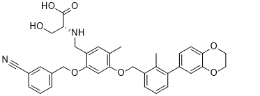
| 分子式 |
C35H34N2O7
|
|
|---|---|---|
| 分子量 |
594.65
|
|
| 精确质量 |
594.24
|
|
| 元素分析 |
C, 70.69; H, 5.76; N, 4.71; O, 18.83
|
|
| CAS号 |
2113650-03-4
|
|
| 相关CAS号 |
|
|
| PubChem CID |
131839624
|
|
| 外观&性状 |
White to off-white solid powder
|
|
| tPSA |
130Ų
|
|
| 氢键供体(HBD)数目 |
3
|
|
| 氢键受体(HBA)数目 |
9
|
|
| 可旋转键数目(RBC) |
12
|
|
| 重原子数目 |
44
|
|
| 分子复杂度/Complexity |
957
|
|
| 定义原子立体中心数目 |
1
|
|
| SMILES |
CC1=CC(=C(C=C1OCC2=C(C(=CC=C2)C3=CC4=C(C=C3)OCCO4)C)OCC5=CC(=CC=C5)C#N)CN[C@H](CO)C(=O)O
|
|
| InChi Key |
UWNXGZKSIKQKAH-SSEXGKCCSA-N
|
|
| InChi Code |
InChI=1S/C35H34N2O7/c1-22-13-28(18-37-30(19-38)35(39)40)33(43-20-25-6-3-5-24(14-25)17-36)16-32(22)44-21-27-7-4-8-29(23(27)2)26-9-10-31-34(15-26)42-12-11-41-31/h3-10,13-16,30,37-38H,11-12,18-21H2,1-2H3,(H,39,40)/t30-/m1/s1
|
|
| 化学名 |
(2-((3-Cyanobenzyl)oxy)-4-((3-(2,3-dihydrobenzo[b][1,4]dioxin-6-yl)-2-methylbenzyl)oxy)-5-methylbenzyl)-D-serine
|
|
| 别名 |
|
|
| HS Tariff Code |
2934.99.9001
|
|
| 存储方式 |
Powder -20°C 3 years 4°C 2 years In solvent -80°C 6 months -20°C 1 month |
|
| 运输条件 |
Room temperature (This product is stable at ambient temperature for a few days during ordinary shipping and time spent in Customs)
|
| 溶解度 (体外实验) |
|
|||
|---|---|---|---|---|
| 溶解度 (体内实验) |
注意: 如下所列的是一些常用的体内动物实验溶解配方,主要用于溶解难溶或不溶于水的产品(水溶度<1 mg/mL)。 建议您先取少量样品进行尝试,如该配方可行,再根据实验需求增加样品量。
注射用配方
注射用配方1: DMSO : Tween 80: Saline = 10 : 5 : 85 (如: 100 μL DMSO → 50 μL Tween 80 → 850 μL Saline)(IP/IV/IM/SC等) *生理盐水/Saline的制备:将0.9g氯化钠/NaCl溶解在100 mL ddH ₂ O中,得到澄清溶液。 注射用配方 2: DMSO : PEG300 :Tween 80 : Saline = 10 : 40 : 5 : 45 (如: 100 μL DMSO → 400 μL PEG300 → 50 μL Tween 80 → 450 μL Saline) 注射用配方 3: DMSO : Corn oil = 10 : 90 (如: 100 μL DMSO → 900 μL Corn oil) 示例: 以注射用配方 3 (DMSO : Corn oil = 10 : 90) 为例说明, 如果要配制 1 mL 2.5 mg/mL的工作液, 您可以取 100 μL 25 mg/mL 澄清的 DMSO 储备液,加到 900 μL Corn oil/玉米油中, 混合均匀。 View More
注射用配方 4: DMSO : 20% SBE-β-CD in Saline = 10 : 90 [如:100 μL DMSO → 900 μL (20% SBE-β-CD in Saline)] 口服配方
口服配方 1: 悬浮于0.5% CMC Na (羧甲基纤维素钠) 口服配方 2: 悬浮于0.5% Carboxymethyl cellulose (羧甲基纤维素) 示例: 以口服配方 1 (悬浮于 0.5% CMC Na)为例说明, 如果要配制 100 mL 2.5 mg/mL 的工作液, 您可以先取0.5g CMC Na并将其溶解于100mL ddH2O中,得到0.5%CMC-Na澄清溶液;然后将250 mg待测化合物加到100 mL前述 0.5%CMC Na溶液中,得到悬浮液。 View More
口服配方 3: 溶解于 PEG400 (聚乙二醇400) 请根据您的实验动物和给药方式选择适当的溶解配方/方案: 1、请先配制澄清的储备液(如:用DMSO配置50 或 100 mg/mL母液(储备液)); 2、取适量母液,按从左到右的顺序依次添加助溶剂,澄清后再加入下一助溶剂。以 下列配方为例说明 (注意此配方只用于说明,并不一定代表此产品 的实际溶解配方): 10% DMSO → 40% PEG300 → 5% Tween-80 → 45% ddH2O (或 saline); 假设最终工作液的体积为 1 mL, 浓度为5 mg/mL: 取 100 μL 50 mg/mL 的澄清 DMSO 储备液加到 400 μL PEG300 中,混合均匀/澄清;向上述体系中加入50 μL Tween-80,混合均匀/澄清;然后继续加入450 μL ddH2O (或 saline)定容至 1 mL; 3、溶剂前显示的百分比是指该溶剂在最终溶液/工作液中的体积所占比例; 4、 如产品在配制过程中出现沉淀/析出,可通过加热(≤50℃)或超声的方式助溶; 5、为保证最佳实验结果,工作液请现配现用! 6、如不确定怎么将母液配置成体内动物实验的工作液,请查看说明书或联系我们; 7、 以上所有助溶剂都可在 Invivochem.cn网站购买。 |
| 制备储备液 | 1 mg | 5 mg | 10 mg | |
| 1 mM | 1.6817 mL | 8.4083 mL | 16.8166 mL | |
| 5 mM | 0.3363 mL | 1.6817 mL | 3.3633 mL | |
| 10 mM | 0.1682 mL | 0.8408 mL | 1.6817 mL |
1、根据实验需要选择合适的溶剂配制储备液 (母液):对于大多数产品,InvivoChem推荐用DMSO配置母液 (比如:5、10、20mM或者10、20、50 mg/mL浓度),个别水溶性高的产品可直接溶于水。产品在DMSO 、水或其他溶剂中的具体溶解度详见上”溶解度 (体外)”部分;
2、如果您找不到您想要的溶解度信息,或者很难将产品溶解在溶液中,请联系我们;
3、建议使用下列计算器进行相关计算(摩尔浓度计算器、稀释计算器、分子量计算器、重组计算器等);
4、母液配好之后,将其分装到常规用量,并储存在-20°C或-80°C,尽量减少反复冻融循环。
计算结果:
工作液浓度: mg/mL;
DMSO母液配制方法: mg 药物溶于 μL DMSO溶液(母液浓度 mg/mL)。如该浓度超过该批次药物DMSO溶解度,请首先与我们联系。
体内配方配制方法:取 μL DMSO母液,加入 μL PEG300,混匀澄清后加入μL Tween 80,混匀澄清后加入 μL ddH2O,混匀澄清。
(1) 请确保溶液澄清之后,再加入下一种溶剂 (助溶剂) 。可利用涡旋、超声或水浴加热等方法助溶;
(2) 一定要按顺序加入溶剂 (助溶剂) 。
Structures and the PD-1/PD-L1 blocking potential of BMS compounds.Oncotarget.2017Aug 7;8(42):72167-72181. |
|---|
Cytotoxicity and activity of BMS compounds in PD-1/PD-L1 checkpoint assay.Oncotarget.2017Aug 7;8(42):72167-72181. |
BMS compounds restore the sPD-L1-supressed activation of Jurkat T-cells.Oncotarget.2017 |
BMS-1166 induces binding cleft opening.Oncotarget.2017Aug 7;8(42):72167-72181. |
|---|
Decomposition of BMS-1166.Oncotarget.2017Aug 7;8(42):72167-72181. |
he prediction of BMS-1001 and −1166 binding sites on PD-L1 surface.Oncotarget.2017Aug 7;8(42):72167-72181. |
 GR-1405
 PD-L1-IN-10
 H-20
H-20
 PL120131 acetate
InvivoChem的所有产品仅用于作科学研究,不面向患者销售
Copyright 2020 InvivoChem LLC | All Rights Reserved 粤ICP备20063088号-1


 COA
COA


 463611831
