Daporinad sulfate

别名: Daporinad sulfate; 658084-64-1; FK866; FK-866; APO-866; APO866; Daporinad [INN]; ...; 201034-75-5;
目录号: V19176 纯度: ≥98%
Daporinad硫酸盐(APO866;FK866)是一种新型NAD合成酶烟酰胺磷酸核糖基转移酶(NAMPT)抑制剂,具有潜在的抗癌和抗血管生成活性。
Daporinad sulfate CAS号: 201034-75-5
产品类别: New1
产品仅用于科学研究,不针对患者销售
规格 价格
500mg
1g
Other Sizes

Other Forms of Daporinad sulfate:

  • 达珀利奈
  • 达珀利奈盐酸盐
点击了解更多
InvivoChem产品被CNS等顶刊论文引用
产品描述
Daporinad硫酸盐(APO866;FK866)是一种新型NAD合成酶烟酰胺磷酸核糖转移酶(NAMPT)抑制剂,具有潜在的抗癌和抗血管生成活性。它通过结合并抑制烟酰胺磷酸核糖基转移酶 (NMPRTase) 发挥作用,从而抑制烟酰胺(维生素 B3)生物合成烟酰胺腺嘌呤二核苷酸 (NAD+)。这可能会耗尽代谢活跃的肿瘤细胞中的能量储备并诱导肿瘤细胞凋亡。此外,该药物还可抑制肿瘤细胞产生血管内皮生长因子(VEGF),从而抑制肿瘤血管生成。
生物活性&实验参考方法
靶点
NAMPT/Nicotinamide phosphoribosyltransferase (IC50 = 0.09 nM)
体外研究 (In Vitro)
通过选择性杀死 MM 细胞,(E)-Daporinad (FK866)抑制 Nampt 会导致细胞内 NAD+ 大幅减少。 MM 细胞中的基线 NAD+ 水平高于正常 PBMC,从而产生 (E)-Daporinad (FK866) 敏感性。 (E)-Daporinad (FK866) 诱导的细胞死亡与 Nampt 活性的抑制有关,而不是与蛋白质表达有关。 (E)-Daporinad (FK866) 消除了骨髓微环境的生存优势[1]。 (E)-Daporinad (FK866) 降低 Jurkat 和激活的 PBL 中 TG 响应性 Ca2+ 储存的 Ca2+ 含量,并抑制各种有丝分裂原引起的 [Ca2+]i 升高。 (E)-Daporinad (FK866) 会降低 Jurkat 细胞中 TG 响应性 Ca2+ 储备的 Ca2+ 含量,但在 Bcl2-Jurkat 细胞中不会降低 [2]。使用 p53 乙酰化途径,通过 (E)-Daporinad (FK866) 抑制 NAMPT 或通过烟酰胺抑制 SIRT 可减少增殖并导致 293T 细胞死亡[3]。
Nampt特异性化学抑制剂Daporinad (FK866)在MM细胞系和患者MM细胞中引发细胞毒性,但在正常供体和MM患者PBMCs中不会引发细胞毒性。重要的是,FK866以剂量依赖的方式引发了对传统和新型抗MM疗法耐药的MM细胞的细胞毒性,并克服了细胞因子(IL-6、IGF-1)和骨髓基质细胞的保护作用。RNAi敲除Nampt证实了其在维持MM细胞活力和细胞内NAD(+)储存中的关键作用。有趣的是,FK866的细胞毒性会触发自噬,但不会引发细胞凋亡。自噬介导的FK866 MM细胞毒性的转录依赖性(TFEB)和非依赖性(PI3K/mTORC1)激活。[1]
通过Daporinad(FK866)介导的烟酰胺磷酸核糖基转移酶抑制降低[NAD(+)](i),降低了Jurkat细胞和活化T淋巴细胞中丝裂原诱导的[Ca(2+)](i)升高。因此,在FK866存在的情况下,这些细胞中thapsigargin敏感的Ca(2+)库的Ca(++)含量大大降低。[2]
以293T细胞系(用大T抗原转化的HEK293细胞)为模型,我们提供了证据,表明p53是参与Daporinad(FK866)介导的293T细胞杀伤的关键下游靶点之一。在这些细胞中敲除p53后,凋亡率降低,S期细胞数量增加。FK866抑制NAMPT或烟酰胺抑制SIRT会降低涉及p53乙酰化途径的293T细胞的增殖并引发其死亡。此外,敲除p53减弱了FK866对细胞增殖、凋亡和细胞周期阻滞的影响。这里提供的数据揭示了两个重要事实:(1)293T细胞中的p53在NAMPT通路抑制剂FK866的存在下是活跃的;(2)FK866在293T细胞中诱导的凋亡与p53在Lys382处的乙酰化增加有关,这是p53功能活性所必需的[3]。
体内研究 (In Vivo)
在 CB17-SCID 小鼠中,(E)-Daporinad (FK866)(30 mg/kg,腹腔注射)可减轻肿瘤负荷,并显着降低肿瘤组织中 ERK 磷酸化和 LC3 的蛋白水解裂解[1]。
Daporinad/FK866的体内抗MM疗效[1]
为了研究Daporinad/FK866对Nampt的抑制是否可以抑制体内MM细胞的生长,我们使用了皮下移植MM.1S细胞的CB17-SCID小鼠。14只荷瘤小鼠被随机分配接受30mg/kg的Daporinad/FK866腹腔注射(每天两次,连续4天,每个周期休息3天,重复3周)或载体对照。图7A显示了接受载体对照组(n=7)与治疗组(n=6)MM.1S增长的比较。与对照组小鼠相比,早在治疗的第7天就观察到肿瘤负荷显著降低,并在第21天得到证实。(分别为P=0.0061和P=0.0067)。重要的是,治疗耐受性良好,没有明显的体重减轻或神经系统变化,并导致总体生存期显著延长(图7B;P=0.0014)。对照组的中位总生存期为19.5天,而FK866治疗组为45天。收集肿瘤,对裂解物进行蛋白质印迹分析,以评估ERK和LC3B的磷酸化(图7C)。与我们的体外结果一致,与对照组相比,FK866治疗小鼠的肿瘤组织ERK磷酸化和LC3蛋白水解切割显著降低。这些结果为FK866治疗多发性骨髓瘤的研究提供了体内概念验证。
抗肿瘤活性[4]
没有发现客观的反应。在接受治疗的24名患者中,4名患者病情稳定至少3个月(前列腺4个月,黑色素瘤5个月,间皮瘤3个月,口咽5个月)。通过影像学检查,癌症前列腺患者病情稳定,但由于PSA升高,4个月后退出研究。口咽癌患者有一些临床益处,疼痛减轻。患者的CT扫描在首次疾病评估时是稳定的;然而,在5个月时出现了进展。
血管内皮生长因子测量[4]
在第二队列和更高队列的患者中测量了VEGF血清水平。在给药前、输注48小时和96小时再次记录VEGF水平。在MTD治疗的6名患者中,有5名患者的VEGF水平下降。由于收集的样本数量较少,VEGF的减少没有达到统计学意义(图1)。
小鼠静脉(IV)药代动力学(PK)研究的应用[5]
开发的LC-qTOF-MS方法成功应用于以5、10和30mg/kg的剂量静脉注射后,获得小鼠血浆中Daporinad(FK866)的PK参数。除早期时间点样品的一些PK样品外,大多数PK样品的浓度都在合格的校准曲线内。因此,早期时间点PK样品用空白小鼠血浆适当稀释,以确保每个样品浓度在校准曲线范围内。Daporinad (FK866)的浓缩时间曲线如图3所示。使用WinNonlin(版本8.0.0)通过非房室分析(NCA)计算PK参数,结果汇总在表4中。
酶活实验
首先,通过混合NADPH溶液a和B、UDPGA、GSH和Daporinad(FK866)的工作溶液(2mg/mL)制备辅因子化合物混合物。将上述辅因子化合物混合物在37°C下预孵育5分钟。然后将380µL预孵育混合物转移到1.7 mL聚丙烯管中,并施加20µL的20 mg/mL小鼠或人肝微粒体。微粒体和辅因子化合物混合物在37°C下孵育0和120分钟,通过加入450µL ACN进行蛋白质沉淀来停止反应。在8000×g rpm下离心10分钟后,将550µL上清液转移到新鲜试管中,并在真空下使用旋转蒸发器蒸发至干。完全干燥的试管用110µL 30%ACN的DW溶液和0.1%FA复溶。复溶样品以12000×g rpm离心5分钟,其上清液转移到LC小瓶中进行体外Met-ID分析。
小鼠PK样品中体内Met-ID的样品制备[5]
对于体内Met-ID分析,分别根据汉密尔顿合并法合并30mg/kg静脉注射PK组的血浆样本。将210µL合并的血浆样品转移到聚丙烯管中,加入800µL ACN进行蛋白质沉淀。然后,将上述样品在10000×g rpm下离心10分钟,并在旋转蒸发器中真空蒸发900µL上清液至干。完全干燥的试管用110µL 30%ACN的DW溶液和0.1%FA复溶。复溶后的样品涡旋并以12000×g rpm离心5分钟,上清液转移到LC小瓶中用于体内Met-ID分析。
荧光法测定细胞内钙水平[2]
PBL或Jurkat细胞(2×10⁶/ml),无论是否用植物血凝素(PHA)(5μg/ml)刺激和/或在有或没有33nmDaporinad(FK866)或0.1mm NAM、NA或NMN的情况下处理24小时,在RPMI培养基中于37°C下用10μm FLUO-3AM或Fura-2AM加载45分钟,用含Ca2+的Hanks平衡盐溶液(HBSS)洗涤,并以2×10⁶个细胞/ml的浓度重新悬浮在相同的溶液中。或者,在某些实验中,细胞被洗涤并重新悬浮在无Ca2+的HBSS中。在添加thapsigargin(TG)之前。在96孔板(105个细胞/孔)中用负载Fluo-3的细胞进行[Ca2+]i测量。调节基础荧光(激发,485 nm;发射,520 nm),使每个孔的基础强度相当(在±10%范围内)。每3秒用荧光板读数器测量一次荧光。将发射光的强度绘制为时间的函数。使用公式Δ/baseal×100计算每条迹线的钙变化,其中Δ是添加刺激后的最大荧光与基础荧光(基础)之间的差值,归一化为基础荧光(基本)。 将Fura-2负载的细胞接种在涂有聚赖氨酸的玻璃底细胞培养皿上,并在37°C下孵育20分钟。[Ca2+]i测量和校准使用微荧光系统进行。
细胞内NAD+和cADPR水平的测定[2]
如上所述,将PBL或Jurkat细胞培养24小时,并在存在或不存在Daporinad(FK866)(33 nm)、NAM(0.1、1或10 mm)、NA(0.1 mm)或NMN(0.1mm)的情况下,用PHA(5μg/ml)刺激或不刺激。每次孵育结束时,取出1ml细胞,在16000×g下离心15秒。细胞颗粒在4°C下用0.3ml 0.6m高氯酸溶解。细胞提取物在16000×g下离心3分钟;收集上清液,将等分试样在pH 8.0的100mm磷酸钠缓冲液中稀释200倍,以测定NAD+含量。通过将水样与4倍体积的含有1,1,2-三氯三氟乙烷和三正辛胺的溶液混合,从剩余的上清液中去除高氯酸,并测定cADPR含量。NAD+和cADPR值根据Bradford测定法测定的蛋白质浓度进行归一化。
细胞内ATP含量的测定[2]
如上所述,在存在或不存在<强>Daporinad(FK866)(33nm)、NAM(0.1mm)、NA(0.1mm。孵育结束时,取出1ml细胞,在16000×g下离心15秒。细胞颗粒在4°C下在0.3ml 0.6m高氯酸中裂解,中和的提取物通过HPLC进行分析(11)。ATP值根据Bradford测定法测定的蛋白质浓度进行归一化。
细胞实验
Daporinad (FK866)对BM旁分泌MM细胞生长的影响 在有或没有药物的情况下,MM1S细胞(2×10⁴个细胞/孔)在BMSC包被的96孔板中培养72和96小时。通过(3H)-胸苷摄取来测量DNA合成,在培养的最后8小时内添加(3H)–胸苷(0.5μCi/孔)。
细胞死亡的特征[1]
细胞与Daporinad (FK866)(0.1nM-33nM)一起培养96小时。对于胱天蛋白酶抑制试验,在加入FK866之前,用泛胱天蛋白酶抑制剂(zVAD-fmk)、胱天蛋白酶3-(zDEVD-fmk”)和胱天蛋白酶9-(zLEHD-fmk)抑制剂预处理细胞至少2小时。为了抑制自噬,在FK866治疗前,将细胞与抑制剂渥曼青霉素(0.25μM)、LY294002(5μM),3-甲基腺嘌呤(3MA 100μM)和氯喹(20μM)一起孵育至少30分钟。根据制造商的方案,使用膜联蛋白-V–FITC染色和流式细胞术分析对细胞凋亡进行定量。
免疫荧光[1]
U266和RPMI8226/S细胞用pEGFP-LC3B瞬时转染,并用载体或Daporinad(FK866)10nM处理24和48小时。使用配备Coolsnap CF彩色相机的尼康E800落射荧光显微镜记录GFP荧光。为了定量,使用10个字段,每个字段由40-100个GFP阳性细胞组成,计算GFP-LC3B斑点细胞相对于GFP阳性细胞总数的数量。
动物实验
Murine xenograft model of human MM [1]
CB17-SCID mice (28-35 days old) were used. All animal studies were conducted according to protocols approved by the Animal Ethics Committee of the Dana-Farber Cancer Institute. Mice were irradiated (200 cGy), and then inoculated subcutaneously in the right flank with 3 × 10⁶ MM1S cells in 100 μL RPMI 1640. After detection of tumor (∼ 2 weeks after the injection), 7 mice were treated intraperitoneally with either vehicle or Daporinad (FK866) (30 mg/kg body weight) twice a day for 4 days, repeated weekly over 3 weeks. Caliper measurements of the longest perpendicular tumor diameters were performed twice a week to estimate the tumor volume using the following formula: length × width2 × 0.5. Tumor growth inhibition (TGI) was calculated using the formula (Δcontrol average volume − Δ treated average volume) × 100 (Δcontrol average volume). Animals were killed when tumors reached 2 cm3 or the mice appeared moribund. Survival was evaluated from the first day of treatment until death. The images were captured with a Canon IXY digital 700 camera. Excised tumors from mice were collected and assessed by Western blotting.
Male ICR mice (30 ± 3 g) were housed in groups of 6~8 per cage and given standard rodent chow. The animals were fasted overnight with free access to water for at least 12 h before administration. Mice were distributed into three different dosing groups (n = 3 for each dosing group; 5, 10 and 30 mg/kg). The blood sampling time points were 2, 10, 30, 60, 120, 240 and 420 min after administration. The sampling blood samples were centrifuged at 12,000× g rpm for 5 min and the plasma was transferred to another tube and then stored at −20 °C until further analysis. [5]

Pharmacokinetic analysis [4]
Plasma samples were obtained prior to dosing and serially up to 152 h after the initiation of the infusion to determine the concentration and clearance of Daporinad (FK866) both during the continuous infusion and after the completion of the infusion. The concentrations of FK866 and its metabolite, FK866-N-oxide (FR247684), were measured in plasma samples using HPLC (high performance liquid chromatography) with tandem mass spectroscopy (LS/MS/MS) by BioProof AG for DL 1 and 2, and by Japan Clinical Laboratories, Inc. for DL three to 6. The lower limits of quantification (LLOQ) of FK866 and FR247684 in plasma were 0.04 ng/ml. The mean and the standard deviation (SD) of concentrations of FK866 and FR247684 in plasma were calculated at each dose levels. The concentrations below LLOQ were treated as zero. If the number of measurable concentration data was less than two at each treatment group, SD was not calculated. Plasma concentration data were excluded from all pharmacokinetic calculations if they were clearly recognized as outliers due to a fairly large deviation from the individual concentration-time curve.
The following pharmacokinetic parameters of Daporinad (FK866) and FR247684, except for plasma concentration at steady-state (CSS), were calculated for each subject based on a model-independent approach by the computer program WinNonlin® Standard version 4.1. All statistical analyses were performed by SAS® software release 8.02 .
Dissolved in 0.9% saline; 20 mg/kg; i.p. injection
C.B.-17 SCID mice xenograft models of human AML, lymphoblastic lymphoma, and leukemia.
药代性质 (ADME/PK)
Pharmacokinetics [4]
Pharmacokinetics of Daporinad (FK866) and its metabolite FK866-N-Oxide had a large inter-individual variability. Intra-individual variability between cycles for each patient was rather limited (Table 4 and Fig. 2). The Css of FK866 and FK866-N-Oxide was reached 48 h after the start of the infusion; the Css of the N-Oxide metabolite was 10-fold lower than that of FK866. Both were eliminated rapidly after stopping the infusion. The Css of both compounds increased with dose escalation, see Tables 4 and 5.
The mean Css (+SD) and AUC0–t of Daporinad (FK866) at MTD were 5.51 + 2.57 ng/ml (14 nM) and 505.9 + 249.8 ng.hr/mL, respectively. The apparent terminal t 1/2 of FK866 was estimated to be 7.9–76.5 h. The Css of FK866 can be regarded as an appropriate predictor of drug exposure, because it closely correlated with its AUC0–t and the Css and AUC0–t of its metabolite. The pharmacokinetics of FK866 was considered to be roughly dose-dependent, based upon the 95% confidence intervals of the multiplier in the power equation for Css of FK866 against infusion rate and absolute amount used.
The major dose limiting toxicity, thrombocytopenia, was compared to drug levels in Fig. 3. This figure shows a proportional decline in platelets as the concentration of Daporinad (FK866) increases. Further comparisons between hematologic parameters, specifically lymphocyte count and hemoglobin, and FK866 levels confirmed a dose relationship of FK866 to toxicity (data not shown).
Daporinad (FK866) is one of the highly specific inhibitors of nicotinamide phosphoribosyl transferase (NAMPT) and known to have its unique mechanism of action that induces the tumor cell apoptosis. In this study, a simple and sensitive liquid chromatography–quadrupole-time-of-flight–mass spectrometric (LC-qTOF-MS) assay has been developed for the evaluation of drug metabolism and pharmacokinetics (DMPK) properties of Daporinad in mice. A simple protein precipitation method using acetonitrile (ACN) was used for the sample preparation and the pre-treated samples were separated by a C18 column. The calibration curve was evaluated in the range of 1.02~2220 ng/mL and the quadratic regression (weighted 1/concentration2) was used for the best fit of the curve with a correlation coefficient ≥ 0.99. The qualification run met the acceptance criteria of ±25% accuracy and precision values for QC samples. The dilution integrity was verified for 5, 10 and 30-fold dilution and the accuracy and precision of the dilution QC samples were also satisfactory within ±25% of the nominal values. The stability results indicated that Daporinad was stable for the following conditions: short-term (4 h), long-term (2 weeks), freeze/thaw (three cycles). This qualified method was successfully applied to intravenous (IV) pharmacokinetic (PK) studies of Daporinad in mice at doses of 5, 10 and 30 mg/kg. As a result, it showed a linear PK tendency in the dose range from 5 to 10 mg/kg, but a non-linear PK tendency in the dose of 30 mg/kg. In addition, in vitro and in vivo metabolite identification (Met ID) studies were conducted to understand the PK properties of Daporinad and the results showed that a total of 25 metabolites were identified as ten different types of metabolism in our experimental conditions. In conclusion, the LC-qTOF-MS assay was successfully developed for the quantification of Daporinad in mouse plasma as well as for its in vitro and in vivo metabolite identification.[5]
In this study, an LC-qTOF-MS assay was developed and qualified for the quantification of Daporinad (FK866) in mouse plasma. The calibration curves were acceptable over the concentration range from 1.02 to 2200 ng/mL for Daporinad using the quadratic regression with a correlation coefficient ≥ 0.99. Daporinad was stable in mouse plasma under the several preliminary stability test conditions, such as short-term (4 h), freeze–thaw (three cycles), and long-term (2 weeks), and achieved the dilution integrity. This method was successfully applied to quantify the in vivo IV PK mouse plasma samples.
The PK results suggest that Daporinad has low to moderate clearance values depending on the 5 to 30 mg/kg administered dose range. It was interesting that in the dose range from 10 to 30 mg/kg, the Cmax was dose-proportional, but the AUC appeared to increase supra proportions. There are several possibilities regarding this result and based on the clearance mechanism, we hypothesize that either metabolic enzymes or transporters related to the elimination pathway of Daporinad might play a role. This triggers in vitro and in vivo MetID studies, and as a result, twenty-five metabolites were newly identified under the current experimental conditions. The results suggest that ten different metabolic pathways were identified for Daporinad, and most of them were phase Ⅰ metabolic reactions, such as amide hydrolysis, oxidation and desaturation. Although many interesting metabolites were newly identified in this experiment, no significant difference from in vivo metabolites perspectives were observed from different dose levels conducted in this study (data not shown) and, therefore, it appeared that other mechanism such as saturation of transporters etc might likely play a role for this phenomenon of clearance change we observed from the in vivo mouse PK. Further experiments such as semi-mass balance study to understand the elimination pathway or in vitro transporter assays to identify the responsible transporters would be necessary.[5] In conclusion, we developed a sensitive, simple and reproducible LC-qTOF-MS assay to evaluate Daporinad in mouse PK samples, and also evaluated the in vitro and in vivo metabolite profiling of Daporinad with several novel metabolites. This research would warrant further experiments to better understand the in vivo clearance mechanism of Daporinad.
毒性/毒理 (Toxicokinetics/TK)
Toxicity [4]
A toxicity was defined as an adverse event that was at least possibly related to the study drug. Daporinad (FK866) was generally well tolerated with thrombocytopenia less than 25,000/μl being the unique dose limiting toxicity (DLT). This occurred in two patients in the cohort at 0.144 mg/m2/h and in one patient at 0.126 mg/m2/h There was also a consistent drop in the lymphocyte counts. This lymphopenia, however, was never greater than grade 3 and there were no cases of opportunistic infections. Neutropenia, other than one patient with grade 1, was not observed. Non-hematology toxicities were relatively infrequent and mild. There was one grade 3 fatigue and two patients with grade 3 nausea/vomiting. The nausea and vomiting was well controlled with five HT3 antagonists. The only other grade 3 toxicity was hyperglycemia. All adverse events grade 2 or higher are listed by cohort in Table 3. One patient had a cerebral vascular accident while on study; this was assessed as not related to therapy. There were no complaints of visual acuity loss nor did repeated ophthalmologic evaluation, including ERG, show signs of retinopathy. There were no treatment-related deaths on study.
LTs and the MTD [4]
Two of the five patients treated at 0.144 mg/m2/h had thrombocytopenia-related DLTs. Both of these patients were transfused with platelets and recovered appropriately. According to the de-escalation design, a cohort was then treated at 0.108 mg/m2/h. There were no DLTs at this cohort and therefore the subsequent escalation tested an intermediate dose at 0.126 mg/m2/h. One out of six patients at this dose level experienced a DLT (thrombocytopenia). This dose is therefore the MTD.
参考文献

[1]. Targeting NAD+ salvage pathway induces autophagy in multiple myeloma cells via mTORC1 and extracellular signal-regulated kinase (ERK1/2) inhibition. Blood. 2012 Oct 25;120(17):3519-29.

[2]. NAD+ levels control Ca2+ store replenishment and mitogen-induced increase of cytosolic Ca2+ by Cyclic ADP-ribose-dependent TRPM2 channel gating in human T lymphocytes. J Biol Chem. 2012 Jun 15;287(25):21067-81.

[3]. Inhibition of NAMPT pathway by FK866 activates the function of p53 in HEK293T cells. Biochem Biophys Res Commun. 2012 Aug 3;424(3):371-7.

[4]. The pharmacokinetics, toxicities, and biologic effects of FK866, a nicotinamide adenine dinucleotide biosynthesis inhibitor. Invest New Drugs. 2008 Feb;26(1):45-51.

[5]. Quantitative Analysis of Daporinad (FK866) and Its In Vitro and In Vivo Metabolite Identification Using Liquid Chromatography-Quadrupole-Time-of-Flight Mass Spectrometry. Molecules. 2022 Mar 21;27(6):2011.

其他信息
FK-866 is a member of benzamides and a N-acylpiperidine.
Daporinad has been used in trials studying the treatment of Melanoma, Cutaneous T-cell Lymphoma, and B-cell Chronic Lymphocytic Leukemia.
Daporinad is a small molecule with potential antineoplastic and antiangiogenic activities. Daporinad binds to and inhibits nicotinamide phosphoribosyltransferase (NMPRTase), inhibiting the biosynthesis of nicotinamide adenine dinucleotide (NAD+) from niacinamide (vitamin B3), which may deplete energy reserves in metabolically active tumor cells and induce tumor cell apoptosis. In addition, this agent may inhibit tumor cell production of vascular endothelial growth factor (VEGF), resulting in the inhibition of tumor angiogenesis. The coenzyme NAD+ plays an essential role in cellular redox reactions, including the redox reaction linking the citric acid cycle and oxidative phosphorylation.
Malignant cells have a higher nicotinamide adenine dinucleotide (NAD(+)) turnover rate than normal cells, making this biosynthetic pathway an attractive target for cancer treatment. Here we investigated the biologic role of a rate-limiting enzyme involved in NAD(+) synthesis, Nampt, in multiple myeloma (MM). Nampt-specific chemical inhibitor FK866 triggered cytotoxicity in MM cell lines and patient MM cells, but not normal donor as well as MM patients PBMCs. Importantly, FK866 in a dose-dependent fashion triggered cytotoxicity in MM cells resistant to conventional and novel anti-MM therapies and overcomes the protective effects of cytokines (IL-6, IGF-1) and bone marrow stromal cells. Nampt knockdown by RNAi confirmed its pivotal role in maintenance of both MM cell viability and intracellular NAD(+) stores. Interestingly, cytotoxicity of FK866 triggered autophagy, but not apoptosis. A transcriptional-dependent (TFEB) and independent (PI3K/mTORC1) activation of autophagy mediated FK866 MM cytotoxicity. Finally, FK866 demonstrated significant anti-MM activity in a xenograft-murine MM model, associated with down-regulation of ERK1/2 phosphorylation and proteolytic cleavage of LC3 in tumor cells. Our data therefore define a key role of Nampt in MM biology, providing the basis for a novel targeted therapeutic approach. [1]
Intracellular NAD(+) levels ([NAD(+)](i)) are important in regulating human T lymphocyte survival, cytokine secretion, and the capacity to respond to antigenic stimuli. NAD(+)-derived Ca(2+)-mobilizing second messengers, produced by CD38, play a pivotal role in T cell activation. Here we demonstrate that [NAD(+)](i) modifications in T lymphocytes affect intracellular Ca(2+) homeostasis both in terms of mitogen-induced [Ca(2+)](i) increase and of endoplasmic reticulum Ca(2+) store replenishment. Lowering [NAD(+)](i) by FK866-mediated nicotinamide phosphoribosyltransferase inhibition decreased the mitogen-induced [Ca(2+)](i) rise in Jurkat cells and in activated T lymphocytes. Accordingly, the Ca(2+) content of thapsigargin-sensitive Ca(2+) stores was greatly reduced in these cells in the presence of FK866. When NAD(+) levels were increased by supplementing peripheral blood lymphocytes with the NAD(+) precursors nicotinamide, nicotinic acid, or nicotinamide mononucleotide, the Ca(2+) content of thapsigargin-sensitive Ca(2+) stores as well as cell responsiveness to mitogens in terms of [Ca(2+)](i) elevation were up-regulated. The use of specific siRNA showed that the changes of Ca(2+) homeostasis induced by NAD(+) precursors are mediated by CD38 and the consequent ADPR-mediated TRPM2 gating. Finally, the presence of NAD(+) precursors up-regulated important T cell functions, such as proliferation and IL-2 release in response to mitogens.[2]
Inhibitors of HDACs (HDACi) are a new class of therapeutic agents because they induce cytotoxicity in wide range of cancer cells. Reconstituting the gene expression programme by inhibition of HDACs is a potential underlying mechanism in the efficacy of HDACi. HDACi like SAHA and VPA, which primarily inhibit class I and II deacetylases, exhibit strong anti-tumor activity and have entered phase II clinical trials. The increasing evidence that SIRTs are critical regulators of major tumor-suppressor proteins, like p53 and FOXO3a, has recently lead to the development of drugs which can specifically target SIRTs. The fact that cancer cells require high turnover of NAD+ to maintain their growth, and SIRTs require NAD+ to maintain their activity, further highlights the importance of FK866 and its ability to specifically target cancer cells. The presented data contribute towards the understanding of mechanism(s) by which FK866 exerts its anti-cancer effects.
In a recent communication we have shown that FK866 upregulates the acetylation of FOXO3a protein in 293T cells, leading to apoptosis. However, this is the first evidence for the presence of functionally active p53 in 293T cells, a cell line known for the abnormal p53 function due to its interaction with large T-antigen. In conclusion, enhancing acetylation of p53 by inhibiting the NAMPT/SIRT pathway improves functional activity of p53 in cells transformed with large T-antigen, which has broad implications for malignancies characterized by p53 inactivation.[3]
Background: FK866 is a potent inhibitor or NAD synthesis. This first-in-human study was performed to determine the maximum-tolerated dose, toxicity profile, and pharmacokinetics on a 96-h continuous infusion schedule. Materials and methods: Twenty four patients with advanced solid tumor malignancies refractory to standard therapies were treated with escalating doses of FK866 as a continuous, 96-h infusion given every 28 days. Serial plasma samples were collected to characterize the pharmacokinetics of FK866. Further blood samples were collected for the measurement of plasma VEGF levels. Results: There were 12 women and 12 men with a median age of 61 (range 34-78) and a median KPS of 80%, received a 4-day of infusion of FK866 at dose levels of 0.018 mg/m2/h (n=3), 0.036 mg/m2/h (n=3), 0.072 mg/m2/h (n=3), 0.108 mg/m2/h (n=4), 0.126 mg/m2/h (n=6), and 0.144 mg/m2/h (n=5). Thrombocytopenia was the dose limiting toxicity, observed in two patients at the highest dose level and one patient at the recommended phase II dose of 0.126 mg/m2/h No other hematologic toxicities were noted other than mild lymphopenia and anemia. There was mild fatigue and grade 3 nausea; the latter was controlled with antiemetics and was not a DLT. Css (the mean of the 72 and 96 h plasma concentrations) increased in relation to the dose escalation. The study drug did not significantly affect plasma concentrations of VEGF. There were no objective responses, although four patients had stable disease (on treatment for 3 months or greater). Conclusions: The recommended phase II dose is 0.126 mg/m2/h given as a continuous 96-h infusion every 28 days. The dose limiting toxicity of FK866 is thrombocytopenia. Pharmacokinetic data suggest an increase in the plasma Css in relation to the escalation of FK866.[4]
*注: 文献方法仅供参考, InvivoChem并未独立验证这些方法的准确性
化学信息 & 存储运输条件
分子量
391.50596
精确质量
391.226
CAS号
201034-75-5
相关CAS号
658084-64-1;1785666-54-7 (HCl);201034-75-5;1198425-96-5 (deleted);
PubChem CID
6914657
外观&性状
Typically exists as solid at room temperature
LogP
4.262
tPSA
62.3
氢键供体(HBD)数目
1
氢键受体(HBA)数目
3
可旋转键数目(RBC)
8
重原子数目
29
分子复杂度/Complexity
534
定义原子立体中心数目
0
SMILES
C1CN(CCC1CCCCNC(=O)C=CC2=CN=CC=C2)C(=O)C3=CC=CC=C3
InChi Key
KPBNHDGDUADAGP-VAWYXSNFSA-N
InChi Code
InChI=1S/C24H29N3O2/c28-23(12-11-21-8-6-15-25-19-21)26-16-5-4-7-20-13-17-27(18-14-20)24(29)22-9-2-1-3-10-22/h1-3,6,8-12,15,19-20H,4-5,7,13-14,16-18H2,(H,26,28)/b12-11+
化学名
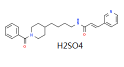
(E)-N-[4-(1-benzoylpiperidin-4-yl)butyl]-3-pyridin-3-ylprop-2-enamide
别名
Daporinad sulfate; 658084-64-1; FK866; FK-866; APO-866; APO866; Daporinad [INN]; ...; 201034-75-5;
HS Tariff Code
2934.99.9001
存储方式

Powder      -20°C    3 years

                     4°C     2 years

In solvent   -80°C    6 months

                  -20°C    1 month

运输条件
Room temperature (This product is stable at ambient temperature for a few days during ordinary shipping and time spent in Customs)
溶解度数据
溶解度 (体外实验)
May dissolve in DMSO (in most cases), if not, try other solvents such as H2O, Ethanol, or DMF with a minute amount of products to avoid loss of samples
溶解度 (体内实验)
注意: 如下所列的是一些常用的体内动物实验溶解配方,主要用于溶解难溶或不溶于水的产品(水溶度<1 mg/mL)。 建议您先取少量样品进行尝试,如该配方可行,再根据实验需求增加样品量。

注射用配方
(IP/IV/IM/SC等)
注射用配方1: DMSO : Tween 80: Saline = 10 : 5 : 85 (如: 100 μL DMSO 50 μL Tween 80 850 μL Saline)
*生理盐水/Saline的制备:将0.9g氯化钠/NaCl溶解在100 mL ddH ₂ O中,得到澄清溶液。
注射用配方 2: DMSO : PEG300Tween 80 : Saline = 10 : 40 : 5 : 45 (如: 100 μL DMSO 400 μL PEG300 50 μL Tween 80 450 μL Saline)
注射用配方 3: DMSO : Corn oil = 10 : 90 (如: 100 μL DMSO 900 μL Corn oil)
示例: 注射用配方 3 (DMSO : Corn oil = 10 : 90) 为例说明, 如果要配制 1 mL 2.5 mg/mL的工作液, 您可以取 100 μL 25 mg/mL 澄清的 DMSO 储备液,加到 900 μL Corn oil/玉米油中, 混合均匀。
View More

注射用配方 4: DMSO : 20% SBE-β-CD in Saline = 10 : 90 [如:100 μL DMSO 900 μL (20% SBE-β-CD in Saline)]
*20% SBE-β-CD in Saline的制备(4°C,储存1周):将2g SBE-β-CD (磺丁基-β-环糊精) 溶解于10mL生理盐水中,得到澄清溶液。
注射用配方 5: 2-Hydroxypropyl-β-cyclodextrin : Saline = 50 : 50 (如: 500 μL 2-Hydroxypropyl-β-cyclodextrin (羟丙基环胡精) 500 μL Saline)
注射用配方 6: DMSO : PEG300 : Castor oil : Saline = 5 : 10 : 20 : 65 (如: 50 μL DMSO 100 μL PEG300 200 μL Castor oil 650 μL Saline)
注射用配方 7: Ethanol : Cremophor : Saline = 10: 10 : 80 (如: 100 μL Ethanol 100 μL Cremophor 800 μL Saline)
注射用配方 8: 溶解于Cremophor/Ethanol (50 : 50), 然后用生理盐水稀释。
注射用配方 9: EtOH : Corn oil = 10 : 90 (如: 100 μL EtOH 900 μL Corn oil)
注射用配方 10: EtOH : PEG300Tween 80 : Saline = 10 : 40 : 5 : 45 (如: 100 μL EtOH 400 μL PEG300 50 μL Tween 80 450 μL Saline)


口服配方
口服配方 1: 悬浮于0.5% CMC Na (羧甲基纤维素钠)
口服配方 2: 悬浮于0.5% Carboxymethyl cellulose (羧甲基纤维素)
示例: 口服配方 1 (悬浮于 0.5% CMC Na)为例说明, 如果要配制 100 mL 2.5 mg/mL 的工作液, 您可以先取0.5g CMC Na并将其溶解于100mL ddH2O中,得到0.5%CMC-Na澄清溶液;然后将250 mg待测化合物加到100 mL前述 0.5%CMC Na溶液中,得到悬浮液。
View More

口服配方 3: 溶解于 PEG400 (聚乙二醇400)
口服配方 4: 悬浮于0.2% Carboxymethyl cellulose (羧甲基纤维素)
口服配方 5: 溶解于0.25% Tween 80 and 0.5% Carboxymethyl cellulose (羧甲基纤维素)
口服配方 6: 做成粉末与食物混合


注意: 以上为较为常见方法,仅供参考, InvivoChem并未独立验证这些配方的准确性。具体溶剂的选择首先应参照文献已报道溶解方法、配方或剂型,对于某些尚未有文献报道溶解方法的化合物,需通过前期实验来确定(建议先取少量样品进行尝试),包括产品的溶解情况、梯度设置、动物的耐受性等。

请根据您的实验动物和给药方式选择适当的溶解配方/方案:
1、请先配制澄清的储备液(如:用DMSO配置50 或 100 mg/mL母液(储备液));
2、取适量母液,按从左到右的顺序依次添加助溶剂,澄清后再加入下一助溶剂。以 下列配方为例说明 (注意此配方只用于说明,并不一定代表此产品 的实际溶解配方):
10% DMSO → 40% PEG300 → 5% Tween-80 → 45% ddH2O (或 saline);
假设最终工作液的体积为 1 mL, 浓度为5 mg/mL: 取 100 μL 50 mg/mL 的澄清 DMSO 储备液加到 400 μL PEG300 中,混合均匀/澄清;向上述体系中加入50 μL Tween-80,混合均匀/澄清;然后继续加入450 μL ddH2O (或 saline)定容至 1 mL;

3、溶剂前显示的百分比是指该溶剂在最终溶液/工作液中的体积所占比例;
4、 如产品在配制过程中出现沉淀/析出,可通过加热(≤50℃)或超声的方式助溶;
5、为保证最佳实验结果,工作液请现配现用!
6、如不确定怎么将母液配置成体内动物实验的工作液,请查看说明书或联系我们;
7、 以上所有助溶剂都可在 Invivochem.cn网站购买。
制备储备液 1 mg 5 mg 10 mg
1 mM 2.5542 mL 12.7711 mL 25.5421 mL
5 mM 0.5108 mL 2.5542 mL 5.1084 mL
10 mM 0.2554 mL 1.2771 mL 2.5542 mL

1、根据实验需要选择合适的溶剂配制储备液 (母液):对于大多数产品,InvivoChem推荐用DMSO配置母液 (比如:5、10、20mM或者10、20、50 mg/mL浓度),个别水溶性高的产品可直接溶于水。产品在DMSO 、水或其他溶剂中的具体溶解度详见上”溶解度 (体外)”部分;

2、如果您找不到您想要的溶解度信息,或者很难将产品溶解在溶液中,请联系我们;

3、建议使用下列计算器进行相关计算(摩尔浓度计算器、稀释计算器、分子量计算器、重组计算器等);

4、母液配好之后,将其分装到常规用量,并储存在-20°C或-80°C,尽量减少反复冻融循环。

计算器

摩尔浓度计算器可计算特定溶液所需的质量、体积/浓度,具体如下:

  • 计算制备已知体积和浓度的溶液所需的化合物的质量
  • 计算将已知质量的化合物溶解到所需浓度所需的溶液体积
  • 计算特定体积中已知质量的化合物产生的溶液的浓度
使用摩尔浓度计算器计算摩尔浓度的示例如下所示:
假如化合物的分子量为350.26 g/mol,在5mL DMSO中制备10mM储备液所需的化合物的质量是多少?
  • 在分子量(MW)框中输入350.26
  • 在“浓度”框中输入10,然后选择正确的单位(mM)
  • 在“体积”框中输入5,然后选择正确的单位(mL)
  • 单击“计算”按钮
  • 答案17.513 mg出现在“质量”框中。以类似的方式,您可以计算体积和浓度。

稀释计算器可计算如何稀释已知浓度的储备液。例如,可以输入C1、C2和V2来计算V1,具体如下:

制备25毫升25μM溶液需要多少体积的10 mM储备溶液?
使用方程式C1V1=C2V2,其中C1=10mM,C2=25μM,V2=25 ml,V1未知:
  • 在C1框中输入10,然后选择正确的单位(mM)
  • 在C2框中输入25,然后选择正确的单位(μM)
  • 在V2框中输入25,然后选择正确的单位(mL)
  • 单击“计算”按钮
  • 答案62.5μL(0.1 ml)出现在V1框中
g/mol

分子量计算器可计算化合物的分子量 (摩尔质量)和元素组成,具体如下:

注:化学分子式大小写敏感:C12H18N3O4  c12h18n3o4
计算化合物摩尔质量(分子量)的说明:
  • 要计算化合物的分子量 (摩尔质量),请输入化学/分子式,然后单击“计算”按钮。
分子质量、分子量、摩尔质量和摩尔量的定义:
  • 分子质量(或分子量)是一种物质的一个分子的质量,用统一的原子质量单位(u)表示。(1u等于碳-12中一个原子质量的1/12)
  • 摩尔质量(摩尔重量)是一摩尔物质的质量,以g/mol表示。
/

配液计算器可计算将特定质量的产品配成特定浓度所需的溶剂体积 (配液体积)

  • 输入试剂的质量、所需的配液浓度以及正确的单位
  • 单击“计算”按钮
  • 答案显示在体积框中
动物体内实验配方计算器(澄清溶液)
第一步:请输入基本实验信息(考虑到实验过程中的损耗,建议多配一只动物的药量)
第二步:请输入动物体内配方组成(配方适用于不溶/难溶于水的化合物),不同的产品和批次配方组成不同,如对配方有疑问,可先联系我们提供正确的体内实验配方。此外,请注意这只是一个配方计算器,而不是特定产品的确切配方。
+
+
+

计算结果:

工作液浓度 mg/mL;

DMSO母液配制方法 mg 药物溶于 μL DMSO溶液(母液浓度 mg/mL)。如该浓度超过该批次药物DMSO溶解度,请首先与我们联系。

体内配方配制方法μL DMSO母液,加入 μL PEG300,混匀澄清后加入μL Tween 80,混匀澄清后加入 μL ddH2O,混匀澄清。

(1) 请确保溶液澄清之后,再加入下一种溶剂 (助溶剂) 。可利用涡旋、超声或水浴加热等方法助溶;
            (2) 一定要按顺序加入溶剂 (助溶剂) 。

临床试验信息
NCT Number Recruitment interventions Conditions Sponsor/Collaborators Start Date Phases
NCT00435084 Completed Drug: APO866 B-cell Chronic Lymphocytic Leukemia Valerio Therapeutics February 2007 Phase 1
Phase 2
NCT00432107 Completed Drug: APO866 Melanoma Valerio Therapeutics July 2006 Phase 2
NCT00431912 Completed Drug: APO866 Cutaneous T-cell Lymphoma Valerio Therapeutics February 2007 Phase 2
相关产品
联系我们