| 规格 | 价格 | 库存 | 数量 |
|---|---|---|---|
| 25mg |
|
||
| 50mg |
|
||
| 100mg |
|
||
| 250mg |
|
||
| 500mg |
|
||
| 1g |
|
||
| 2g |
|
||
| 5g |
|
||
| 10g |
|
||
| Other Sizes |
|
| 靶点 |
Cyclooxygenase-1 (COX-1) (IC50: 0.15 ± 0.02 μM for Ketorolac tromethamine), Cyclooxygenase-2 (COX-2) (IC50: 0.32 ± 0.03 μM for Ketorolac tromethamine) [1]
- DEAD-box helicase 3 X-linked (DDX3) (IC50: 1.2 ± 0.1 μM for Ketorolac salt in DDX3 RNA helicase activity assay; EC50: 8.5 ± 0.6 μM for Ketorolac salt in SCC-9 oral cancer cell viability assay) [4] |
|---|---|
| 体外研究 (In Vitro) |
酮咯酸 (RS37619) 盐(0-30 μM;48 小时)可成功杀死口腔癌细胞[4]。在 H357 细胞中,酮咯酸盐(0–5 μM;48 小时)会导致细胞凋亡并抑制 DDX3 蛋白的产生[4]。酮咯酸盐(0-2.5 μM;0-16 小时)可抑制口腔癌细胞生长[4]。通过直接与 DDX3 相互作用,酮咯酸盐 (0–50 μM) 抑制 ATP 酶活性[4]。
1. 环氧合酶(COX)抑制活性:酮咯酸氨丁三醇对COX-1和COX-2表现出浓度依赖性抑制作用。其对COX-1的IC50(0.15±0.02 μM)低于对COX-2的IC50(0.32±0.03 μM),表明对COX-1的选择性更高。与溴芬酸钠(COX-1 IC50:0.28±0.03 μM;COX-2 IC50:0.19±0.02 μM)相比,酮咯酸氨丁三醇的COX-1抑制活性更强,但COX-2抑制活性更弱[1] 2. 抗口腔癌活性:酮咯酸盐抑制多种口腔癌细胞系的活力,处理72小时后(MTT法),对SCC-9、SCC-25、CAL-27细胞的IC50分别为8.5±0.6 μM、9.2±0.7 μM、10.1±0.8 μM。Western blot结果显示,酮咯酸盐(10 μM,处理48小时)可下调SCC-9细胞中p-AKT、p-ERK、Bcl-2的表达,同时上调cleaved caspase-3和Bax的表达。此外,酮咯酸盐(10 μM)可抑制DDX3 RNA解旋酶活性达68.3±5.2%,并减少SCC-9细胞中DDX3的核转位[4] |
| 体内研究 (In Vivo) |
在兔子中,酮咯酸 (RS37619) 或 0.4% 酮咯酸氨丁三醇滴眼液对眼睛表现出有效的抗炎作用[1]。酮咯酸(4 mg/kg/天,口服;2 周)不会对大鼠牙槽窝骨小梁体积分数产生负面影响[2]。在大鼠中,鞘内注射酮咯酸(60 μg)可减轻脊髓缺血引起的损伤[3]。暴露于酮咯酸盐(20 和 30 mg/kg;腹腔注射;每周两次,持续三周)的小鼠口腔癌发生率较低[4]。
1. 眼部抗炎作用(兔模型):通过玻璃体内注射脂多糖(LPS,100 ng/眼)诱导新西兰白兔眼部炎症。给予酮咯酸氨丁三醇滴眼液(0.5%浓度,50 μL/眼,每天4次,连续5天),在第5天可显著降低前房闪辉(评分:1.2±0.3 vs. 模型组3.8±0.5)和细胞浸润(评分:1.0±0.2 vs. 模型组3.5±0.4),同时较模型组减轻角膜水肿(厚度:385±20 μm vs. 模型组520±25 μm)和虹膜充血[1] 2. 牙槽骨愈合作用(大鼠模型):对Wistar大鼠(雄性,200-250 g)进行上颌第一磨牙拔除术,术后给予酮咯酸(1 mg/kg,腹腔注射,每天1次,连续7天)。术后14天的组织计量分析显示,与对照组相比,酮咯酸处理组的新生骨面积(28.5±3.2% vs. 对照组29.8±3.5%)、骨小梁厚度(45.2±4.1 μm vs. 对照组46.5±4.3 μm)、骨小梁数量(2.8±0.3个/mm vs. 对照组2.9±0.3个/mm)均无显著差异,表明酮咯酸不影响牙槽骨愈合[2] 3. 脊髓缺血保护作用(大鼠模型):通过夹闭腹主动脉60分钟建立Sprague-Dawley大鼠(雄性,250-300 g)脊髓缺血模型。缺血前30分钟鞘内注射酮咯酸(10 μg/10 μL),再灌注后72小时可改善神经功能评分(8.2±0.8 vs. 缺血组3.5±0.6),减少脊髓前角坏死神经元数量(12.3±2.1个 vs. 缺血组35.6±3.8个),降低丙二醛(MDA)含量(2.1±0.3 nmol/mg蛋白 vs. 缺血组4.8±0.5 nmol/mg蛋白),并提高超氧化物歧化酶(SOD)活性(85.6±6.2 U/mg蛋白 vs. 缺血组42.3±5.1 U/mg蛋白)[3] 4. 抗肿瘤作用(裸鼠异种移植模型):向BALB/c裸鼠(雌性,4-6周龄)皮下接种SCC-9细胞(1×10⁶个细胞/只),给予酮咯酸盐(10 mg/kg,腹腔注射,每周3次,连续3周),接种后35天可显著降低肿瘤体积(280±35 mm³ vs. 溶剂组650±45 mm³)和肿瘤重量(0.32±0.04 g vs. 溶剂组0.75±0.06 g)。免疫组化结果显示酮咯酸盐降低Ki-67(增殖标志物)和p-AKT的表达,肿瘤组织Western blot结果进一步证实DDX3、p-AKT、Bcl-2的表达下调[4] |
| 酶活实验 |
1. COX-1/COX-2活性测定实验:COX-1的酶源为绵羊精囊腺微粒体,COX-2的酶源为昆虫细胞表达的重组人COX-2。反应体系(100 μL)包含50 mM Tris-HCl缓冲液(pH 8.0)、1 μM血红素、100 μM花生四烯酸(底物)以及不同浓度的酮咯酸氨丁三醇(0.01-10 μM)。37°C孵育10分钟后,加入10 μL 1 M HCl终止反应。采用酶免疫测定(EIA)试剂盒检测环氧合酶产物前列腺素E2(PGE2)的含量,通过以酮咯酸氨丁三醇浓度对数为横坐标、PGE2生成抑制率为纵坐标作图,计算IC50值[1]
2. DDX3 RNA解旋酶活性测定实验:将重组人DDX3(0.5 μg)与荧光共振能量转移(FRET)标记的RNA底物(20 nM)在含不同浓度酮咯酸盐(0.1-10 μM)的反应缓冲液(20 mM Tris-HCl,pH 7.5,50 mM KCl,2 mM MgCl2,1 mM DTT)中37°C孵育30分钟。RNA底物解旋后FRET信号降低,检测荧光强度(激发波长485 nm,发射波长520 nm)。抑制率按(1 - 样品荧光强度/对照荧光强度)×100%计算,通过非线性回归法确定IC50[4] 3. DDX3-酮咯酸结合实验(SPR):采用表面等离子体共振(SPR)生物传感器。通过氨基偶联法将重组DDX3固定在CM5传感器芯片上,将酮咯酸盐在运行缓冲液(10 mM HEPES,pH 7.4,150 mM NaCl,0.05% Tween-20)中进行系列稀释(0.1-20 μM),以30 μL/min的流速注入芯片。记录结合相(60秒)和解离相(120秒),使用生物传感器分析软件中的1:1结合模型计算平衡解离常数(KD)[4] |
| 细胞实验 |
细胞活力测定 [4]
细胞类型: HOK、SCC4、SCC9 和 H357 细胞 测试浓度: 0-30 μM 孵育时间:48小时 实验结果:对H357、SCC4和SCC9细胞的IC50分别为2.6、7.1和8.1 μM。而正常的HOK细胞系没有表现出任何细胞死亡效应。 细胞增殖测定[4] 细胞类型: H357 测试浓度: 0.5、1.0、1.5、2.0 和 2.5 μM 孵育时间:0、8和16小时 实验结果:抑制增殖。 蛋白质印迹分析[4] 细胞类型: H357 测试浓度: 1、2.5 和 5 μM 孵育时间:48 小时 实验结果:与 DMSO 处理的细胞相比,DDX3 蛋白表达水平显着降低,但并未完全消除。上调E-钙粘蛋白的表达。 细胞凋亡分析[4] 细胞类型: H357 测试浓度: 2.5 和 5 μM 孵育时间:48小时 实验结果:诱导细胞凋亡。 1. 口腔癌细胞活力测定(MTT法):将SCC-9、SCC-25、CAL-27细胞以5×10³个细胞/孔的密度接种于96孔板,过夜培养。加入不同浓度的酮咯酸盐(1-20 μM),分别孵育24小时、48小时或72小时。孵育结束后,每孔加入20 μL MTT溶液(5 mg/mL),37°C继续孵育4小时。去除上清液,加入150 μL二甲基亚砜(DMSO)溶解甲臜结晶,使用酶标仪检测570 nm处的吸光度。细胞活力按(样品吸光度/对照吸光度)×100%计算,通过GraphPad Prism软件确定IC50[4] 2. 细胞信号蛋白Western blot实验:将SCC-9细胞以2×10⁵个细胞/孔接种于6孔板,用酮咯酸盐(10 μM)处理48小时。使用含蛋白酶和磷酸酶抑制剂的RIPA裂解液裂解细胞,通过BCA法测定蛋白浓度。取等量蛋白(30 μg)进行SDS-PAGE电泳,转移至PVDF膜。膜用5%脱脂牛奶室温封闭1小时,随后与一抗(抗DDX3、抗p-AKT、抗AKT、抗Bcl-2、抗Bax、抗cleaved caspase-3、抗GAPDH)4°C孵育过夜。TBST洗涤后,膜与二抗室温孵育1小时,采用增强化学发光(ECL)试剂盒显影,通过ImageJ软件定量条带强度[4] 3. 克隆形成实验:将SCC-9细胞以2×10³个细胞/孔接种于6孔板,培养24小时后加入酮咯酸盐(5 μM或10 μM),继续培养14天。用4%多聚甲醛固定克隆15分钟,0.1%结晶紫染色30分钟。在显微镜下计数细胞数大于50的克隆,克隆形成率按(样品克隆数/对照克隆数)×100%计算[4] |
| 动物实验 |
动物/疾病模型:新西兰白兔(2.0–2.7 kg),LPS内毒素诱导的眼部炎症[1]
剂量:50 μL 0.4%酮咯酸氨丁三醇滴眼液 给药途径:滴眼,两次,分别在LPS刺激前2小时和1小时 实验结果:几乎完全抑制(98.7%)LPS内毒素诱导的前房FITC(异硫氰酸荧光素)-葡聚糖的增加,并几乎完全抑制(97.5%)LPS内毒素诱导的房水中PGE2浓度的增加。 动物/疾病模型:雄性Wistar大鼠(400–450 g),脊髓缺血模型[3] 剂量:30和60 μg 给药途径:鞘内注射,缺血诱导前1小时,单次注射 实验结果:60 μg剂量组显著降低了运动障碍,提高了存活率。 动物/疾病模型:60 μg剂量组显著降低了运动障碍,提高了存活率。 剂量: 20 mg/kg 和 30 mg/kg 给药途径: 腹腔注射,每周两次,持续 3 周 1. 兔眼部炎症模型:将新西兰白兔(雄性,2.5-3.0 kg)随机分为 3 组:模型组、酮咯酸氨丁三醇组和溴芬酸钠组(每组 n=6)。通过玻璃体内注射 LPS(100 ng/眼)诱导右眼炎症。LPS 注射 1 小时后,酮咯酸氨丁三醇组滴用 0.5% 酮咯酸氨丁三醇眼药水(50 μL/眼),溴芬酸钠组滴用 0.1% 溴芬酸钠眼药水(50 μL/眼);两组均每日 4 次,持续 5 天。模型组以相同频率滴入生理盐水眼药水(50 μL/眼)。分别于LPS注射后24 h、48 h、72 h和5天评估眼部参数(前房闪辉、细胞浸润、角膜水肿、虹膜充血)[1] 2. 大鼠牙槽骨愈合模型:雄性Wistar大鼠(200-250 g,n=18)随机分为3组:对照组、酮咯酸组和对乙酰氨基酚组(每组n=6)。所有大鼠均在麻醉下(腹腔注射氯胺酮和赛拉嗪)拔除上颌第一磨牙。酮咯酸组腹腔注射酮咯酸(1 mg/kg),每日一次,连续7天;对乙酰氨基酚组腹腔注射对乙酰氨基酚(150 mg/kg),每日一次,连续7天;对照组腹腔注射生理盐水(腹腔注射,注射量和频率相同)。拔牙后第14天,处死大鼠,取出上颌骨,脱钙,石蜡包埋,切片(5 μm)。进行组织计量学分析,测量新生骨面积、骨小梁厚度和骨小梁数量[2] 3. 大鼠脊髓缺血模型:雄性Sprague-Dawley大鼠(250-300 g,n=24)随机分为3组:假手术组、缺血组和酮咯酸预处理组(每组n=8)。大鼠用异氟烷麻醉,暴露腹主动脉。缺血组和酮咯酸组进行60分钟的主动脉夹闭以诱导脊髓缺血;假手术组仅进行开腹手术,不进行主动脉夹闭。缺血前30分钟,酮咯酸组接受鞘内注射酮咯酸(10 μg/10 μL,溶于生理盐水);缺血组和假手术组接受鞘内注射生理盐水(10 μL)。分别于再灌注后24小时和72小时对神经功能进行评分(0-10分,分数越高表示功能越好)。72小时后,处死大鼠,取脊髓组织(T10-T12节段)进行组织病理学分析(HE染色)、MDA含量检测和SOD活性检测[3] 4.裸鼠口腔癌异种移植模型:雌性BALB/c裸鼠(4-6周龄,18-22 g,n=15)随机分为3组:溶剂对照组、酮咯酸盐低剂量组(5 mg/kg)和酮咯酸盐高剂量组(10 mg/kg)(每组n=5)。将SCC-9细胞(1×10⁶个细胞,溶于100 μL PBS/Matrigel混合液,1:1)皮下注射至每只小鼠右侧腹部。当肿瘤体积达到约100 mm³(接种后第7天)时,酮咯酸盐组腹腔注射酮咯酸盐(溶于0.1% DMSO + 生理盐水),每周3次,持续3周;溶剂对照组腹腔注射等体积的0.1% DMSO + 生理盐水。每隔3天测量一次肿瘤体积(计算公式为长×宽²×0.5)和体重。接种后35天,处死小鼠,称量肿瘤重量,并收集肿瘤组织进行Western blot和免疫组织化学分析[4] |
| 药代性质 (ADME/PK) |
吸收、分布和排泄
酮咯酸口服后迅速且完全吸收,生物利用度为80%。给药后20-60分钟达到血药浓度峰值(Cmax),肌注后血浆浓度-时间曲线下面积(AUC)与给药剂量成正比。肌注后,酮咯酸达峰时间(tmax)约为45-50分钟,口服后tmax约为30-40分钟。食物可能会降低吸收速率,但不会影响吸收程度。 酮咯酸主要经肾脏排泄,约92%的剂量可从尿液中排出,其中60%以原形排出,40%以代谢物排出。此外,单次剂量的 6% 会经粪便排出。 酮咯酸在健康人体内的表观分布容积为 0.25 L/kg 或更低。 酮咯酸的血浆清除率为 0.021 至 0.037 L/h/kg。此外,研究表明,口服、肌注和静脉注射酮咯酸的清除率相当,提示其药代动力学呈线性。还应注意的是,儿童的清除率约为成人的两倍。 代谢/代谢物 酮咯酸主要通过肝脏的羟基化或结合代谢;然而,主要的代谢途径似乎是葡萄糖醛酸结合。参与 I 期代谢的酶包括 CYP2C8 和 CYP2C9,而 II 期代谢则由 UDP-葡萄糖醛酸转移酶 (UGT) 2B7 完成。 生物半衰期 酮咯酸氨丁三醇以消旋混合物的形式给药,因此必须考虑每种对映异构体的半衰期。S-对映异构体的半衰期约为 2.5 小时,而 R-对映异构体的半衰期约为 5 小时。基于这些数据,S-对映异构体的清除速度约为 R-对映异构体的两倍。 |
| 毒性/毒理 (Toxicokinetics/TK) |
肝毒性
前瞻性研究表明,服用酮咯酸的患者中,高达 1% 会出现至少短暂的血清转氨酶升高。即使继续用药,这些升高也可能自行消退。显著的转氨酶升高(升高 3 倍以上)见于 可能性评分:E(未经证实但怀疑是临床上明显的肝损伤的原因,主要由出血事件引起)。 妊娠和哺乳期影响 ◉ 哺乳期用药概述 通常口服剂量下,酮咯酸在乳汁中的浓度较低,但尚未测量较高注射剂量或鼻喷剂后的乳汁浓度。在一些医院的方案中,剖宫产后短期(通常为 24 小时)使用酮咯酸注射液,目前尚无证据表明对母乳喂养的婴儿有害。然而,由于初乳产量少,婴儿从初乳中摄入的酮咯酸剂量非常低。一些证据表明,与患者自控静脉注射吗啡镇痛相比,静脉注射酮咯酸作为剖宫产后多模式镇痛方案的一部分,可降低纯母乳喂养失败的母亲比例。酮咯酸具有强效抗血小板活性,并可能引起胃肠道出血。生产商指出,酮咯酸在哺乳期禁用,因此在产后24至72小时乳汁分泌量较大时,尤其是在哺乳新生儿或早产儿时,应优先选择其他药物。 母亲使用酮咯酸滴眼液预计不会对母乳喂养的婴儿造成任何不良影响。为了大幅减少使用眼药水后进入母乳的药物量,请按压眼角泪管 1 分钟或更长时间,然后用吸水纸巾擦去多余的药液。 ◉ 对母乳喂养婴儿的影响 一项随机、双盲研究比较了接受剖腹产的母亲(n = 60)接受标准护理与接受标准护理加多模式镇痛(包括在筋膜缝合时单次肌注 60 毫克酮咯酸)的母亲(n = 60)的疗效。产后第一个月,两组在新生儿生长异常、喂养困难、新生儿镇静或呼吸抑制发生率方面均未见显著差异。 ◉ 对泌乳和母乳的影响 一项随机、双盲研究比较了接受剖宫产的母亲(n = 60)接受标准护理与接受标准护理加多模式镇痛(包括在筋膜缝合时单次肌注60 mg酮咯酸)的母亲(n = 60)的产后情况。产后第一个月,两组的母乳喂养率(分别为78%和79%)均无显著差异。 在一项比较剖宫产后标准护理与加速康复方案的研究中,加速康复方案包括产后24小时内每6小时静脉注射固定剂量15 mg酮咯酸,而标准方案则包括按需静脉注射15 mg酮咯酸。采用加速康复方案的患者(n = 58)纯母乳喂养率(67%)高于采用标准方案的患者(48%;n = 60)。 一项回顾性研究评估了 1349 名接受剖宫产并在手术结束 15 分钟内给予酮咯酸的女性。结果表明,术后前6小时的疼痛控制情况以及出院时母乳喂养的女性比例均无差异。 一项前瞻性队列研究比较了剖宫产术后疼痛控制:(1) 术后前12小时使用吗啡患者自控镇痛(PCA)和定时服用布洛芬,之后继续定时服用布洛芬,必要时加用氢可酮-对乙酰氨基酚;(2) 多模式镇痛方案包括:(3) 术后口服对乙酰氨基酚1000毫克,每8小时一次;(4) 静脉注射酮咯酸30毫克,之后每8小时静脉注射15毫克,持续24小时;(5) 术后剩余时间内口服布洛芬600毫克,每8小时一次;仅在需要时使用阿片类药物。入院时计划纯母乳喂养的女性中,多模式组出院前使用配方奶的女性比例低于传统组(9% 对 12%)。 蛋白质结合 >99% 的酮咯酸与血浆蛋白结合。 |
| 参考文献 |
|
| 其他信息 |
药效学
酮咯酸是一种非选择性非甾体抗炎药 (NSAID),其作用机制是通过抑制环氧合酶-1 (COX-1) 和环氧合酶-2 (COX-2) 来发挥作用,这两种酶通常负责将花生四烯酸转化为前列腺素。COX-1 酶具有组成型活性,存在于血小板、胃黏膜和血管内皮细胞中。另一方面,COX-2 酶具有诱导活性,介导炎症、疼痛和发热。因此,抑制 COX-1 酶会增加出血和胃溃疡的风险,而所需的抗炎和镇痛作用则与抑制 COX-2 酶有关。因此,尽管酮咯酸在疼痛管理方面有效,但不应长期使用,因为这会增加严重不良反应的风险,例如胃肠道出血、消化性溃疡和穿孔。 1. 酮咯酸是一种非甾体类抗炎药 (NSAID),其抗炎作用主要通过抑制 COX-1 和 COX-2 的活性,从而减少前列腺素(例如 PGE2)的合成 [1] 2. 在大鼠牙槽骨愈合模型中,酮咯酸(1 mg/kg,腹腔注射,7 天)不影响牙槽骨的正常愈合过程,这对于酮咯酸在拔牙术后疼痛管理中的应用具有临床意义 [2] 3. 鞘内注射酮咯酸对脊髓缺血损伤的保护作用可能与其抗氧化应激作用(减少 MDA 生成和增加 SOD 活性)以及抑制……有关。神经元坏死[3] 4. DDX3 是一种 DEAD-box RNA 解旋酶,在口腔癌中高表达,并通过激活 AKT 信号通路促进癌细胞增殖和存活。酮咯酸盐通过特异性结合 DDX3,抑制其 RNA 解旋酶活性,并下调 AKT/Bcl-2 信号通路,从而抑制口腔癌的进展[4] |
| 分子式 |
C15H13N1O3
|
|
|---|---|---|
| 分子量 |
255.27
|
|
| 精确质量 |
255.089
|
|
| CAS号 |
74103-06-3
|
|
| 相关CAS号 |
Ketorolac tromethamine salt;74103-07-4;(S)-Ketorolac;66635-92-5;(R)-Ketorolac;66635-93-6;Ketorolac-d5;1215767-66-0;Ketorolac hemicalcium;167105-81-9;Ketorolac-d4;1216451-53-4
|
|
| PubChem CID |
3826
|
|
| 外观&性状 |
White to light yellow solid powder
|
|
| 密度 |
1.3±0.1 g/cm3
|
|
| 沸点 |
493.2±40.0 °C at 760 mmHg
|
|
| 熔点 |
160-161°C
|
|
| 闪点 |
252.1±27.3 °C
|
|
| 蒸汽压 |
0.0±1.3 mmHg at 25°C
|
|
| 折射率 |
1.659
|
|
| LogP |
2.08
|
|
| tPSA |
59.3
|
|
| 氢键供体(HBD)数目 |
1
|
|
| 氢键受体(HBA)数目 |
3
|
|
| 可旋转键数目(RBC) |
3
|
|
| 重原子数目 |
19
|
|
| 分子复杂度/Complexity |
376
|
|
| 定义原子立体中心数目 |
0
|
|
| InChi Key |
OZWKMVRBQXNZKK-UHFFFAOYSA-N
|
|
| InChi Code |
InChI=1S/C15H13NO3/c17-14(10-4-2-1-3-5-10)13-7-6-12-11(15(18)19)8-9-16(12)13/h1-7,11H,8-9H2,(H,18,19)
|
|
| 化学名 |
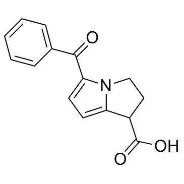
5-benzoyl-2,3-dihydro-1H-pyrrolizine-1-carboxylic acid
|
|
| 别名 |
|
|
| HS Tariff Code |
2934.99.9001
|
|
| 存储方式 |
Powder -20°C 3 years 4°C 2 years In solvent -80°C 6 months -20°C 1 month 注意: 请将本产品存放在密封且受保护的环境中,避免吸湿/受潮。 |
|
| 运输条件 |
Room temperature (This product is stable at ambient temperature for a few days during ordinary shipping and time spent in Customs)
|
| 溶解度 (体外实验) |
|
|||
|---|---|---|---|---|
| 溶解度 (体内实验) |
注意: 如下所列的是一些常用的体内动物实验溶解配方,主要用于溶解难溶或不溶于水的产品(水溶度<1 mg/mL)。 建议您先取少量样品进行尝试,如该配方可行,再根据实验需求增加样品量。
注射用配方
注射用配方1: DMSO : Tween 80: Saline = 10 : 5 : 85 (如: 100 μL DMSO → 50 μL Tween 80 → 850 μL Saline)(IP/IV/IM/SC等) *生理盐水/Saline的制备:将0.9g氯化钠/NaCl溶解在100 mL ddH ₂ O中,得到澄清溶液。 注射用配方 2: DMSO : PEG300 :Tween 80 : Saline = 10 : 40 : 5 : 45 (如: 100 μL DMSO → 400 μL PEG300 → 50 μL Tween 80 → 450 μL Saline) 注射用配方 3: DMSO : Corn oil = 10 : 90 (如: 100 μL DMSO → 900 μL Corn oil) 示例: 以注射用配方 3 (DMSO : Corn oil = 10 : 90) 为例说明, 如果要配制 1 mL 2.5 mg/mL的工作液, 您可以取 100 μL 25 mg/mL 澄清的 DMSO 储备液,加到 900 μL Corn oil/玉米油中, 混合均匀。 View More
注射用配方 4: DMSO : 20% SBE-β-CD in Saline = 10 : 90 [如:100 μL DMSO → 900 μL (20% SBE-β-CD in Saline)] 口服配方
口服配方 1: 悬浮于0.5% CMC Na (羧甲基纤维素钠) 口服配方 2: 悬浮于0.5% Carboxymethyl cellulose (羧甲基纤维素) 示例: 以口服配方 1 (悬浮于 0.5% CMC Na)为例说明, 如果要配制 100 mL 2.5 mg/mL 的工作液, 您可以先取0.5g CMC Na并将其溶解于100mL ddH2O中,得到0.5%CMC-Na澄清溶液;然后将250 mg待测化合物加到100 mL前述 0.5%CMC Na溶液中,得到悬浮液。 View More
口服配方 3: 溶解于 PEG400 (聚乙二醇400) 请根据您的实验动物和给药方式选择适当的溶解配方/方案: 1、请先配制澄清的储备液(如:用DMSO配置50 或 100 mg/mL母液(储备液)); 2、取适量母液,按从左到右的顺序依次添加助溶剂,澄清后再加入下一助溶剂。以 下列配方为例说明 (注意此配方只用于说明,并不一定代表此产品 的实际溶解配方): 10% DMSO → 40% PEG300 → 5% Tween-80 → 45% ddH2O (或 saline); 假设最终工作液的体积为 1 mL, 浓度为5 mg/mL: 取 100 μL 50 mg/mL 的澄清 DMSO 储备液加到 400 μL PEG300 中,混合均匀/澄清;向上述体系中加入50 μL Tween-80,混合均匀/澄清;然后继续加入450 μL ddH2O (或 saline)定容至 1 mL; 3、溶剂前显示的百分比是指该溶剂在最终溶液/工作液中的体积所占比例; 4、 如产品在配制过程中出现沉淀/析出,可通过加热(≤50℃)或超声的方式助溶; 5、为保证最佳实验结果,工作液请现配现用! 6、如不确定怎么将母液配置成体内动物实验的工作液,请查看说明书或联系我们; 7、 以上所有助溶剂都可在 Invivochem.cn网站购买。 |
| 制备储备液 | 1 mg | 5 mg | 10 mg | |
| 1 mM | 3.9174 mL | 19.5871 mL | 39.1742 mL | |
| 5 mM | 0.7835 mL | 3.9174 mL | 7.8348 mL | |
| 10 mM | 0.3917 mL | 1.9587 mL | 3.9174 mL |
1、根据实验需要选择合适的溶剂配制储备液 (母液):对于大多数产品,InvivoChem推荐用DMSO配置母液 (比如:5、10、20mM或者10、20、50 mg/mL浓度),个别水溶性高的产品可直接溶于水。产品在DMSO 、水或其他溶剂中的具体溶解度详见上”溶解度 (体外)”部分;
2、如果您找不到您想要的溶解度信息,或者很难将产品溶解在溶液中,请联系我们;
3、建议使用下列计算器进行相关计算(摩尔浓度计算器、稀释计算器、分子量计算器、重组计算器等);
4、母液配好之后,将其分装到常规用量,并储存在-20°C或-80°C,尽量减少反复冻融循环。
计算结果:
工作液浓度: mg/mL;
DMSO母液配制方法: mg 药物溶于 μL DMSO溶液(母液浓度 mg/mL)。如该浓度超过该批次药物DMSO溶解度,请首先与我们联系。
体内配方配制方法:取 μL DMSO母液,加入 μL PEG300,混匀澄清后加入μL Tween 80,混匀澄清后加入 μL ddH2O,混匀澄清。
(1) 请确保溶液澄清之后,再加入下一种溶剂 (助溶剂) 。可利用涡旋、超声或水浴加热等方法助溶;
(2) 一定要按顺序加入溶剂 (助溶剂) 。
Dosing of Ketorolac in the Emergency Department
CTID: NCT03464461
Phase: Phase 4 Status: Terminated
Date: 2024-11-05
 Butanixin
 COX-2-IN-56
 COX-2-IN-57
 COX-2-IN-58
InvivoChem的所有产品仅用于作科学研究,不面向患者销售
Copyright 2020 InvivoChem LLC | All Rights Reserved 粤ICP备20063088号-1


 COA
COA
 463611831
