| 规格 | 价格 | 库存 | 数量 |
|---|---|---|---|
| 1mg |
|
||
| 2mg |
|
||
| 5mg |
|
||
| 10mg |
|
||
| 25mg |
|
||
| 50mg |
|
||
| 100mg |
|
||
| 250mg |
|
||
| 500mg |
|
||
| Other Sizes |
|
| 靶点 |
MLL1-WDR5 PPI (IC50 = 2.4 nM)
MLL1-WDR5 protein-protein interaction (Ki = ~1.2 nM determined by isothermal titration calorimetry (ITC) for the interaction between recombinant MLL1 SET domain (residues 3749–3969) and WDR5; IC₅₀ = ~5 nM in AlphaScreen binding assay; no significant binding to other WDR5-interacting proteins (e.g., Ash2L) with Ki > 10 μM, confirming selectivity) [1] - MLL1-WDR5 protein-protein interaction (IC₅₀ = ~6 nM in HTRF binding assay for MLL1-WDR5 complex; inhibits MLL1-mediated histone H3K4 methylation in renal tubular cells, with no off-target effects on EZH2 or DOT1L) [2] |
|---|---|
| 体外研究 (In Vitro) |
体外活性:MM-102 作为 MLL1 模拟物,对 WDR5 显示出高结合亲和力,IC50 为 2.9 nM,Ki 为 < 1 nM。在 MLL1-AF9 转导的小鼠细胞中,MM-102 特异性降低两个关键 MLL1 靶基因(HoxA9 和 Meis-1)的表达,这两个基因是 MLL1 介导的白血病发生所必需的。此外,MM-102 可有效、选择性地抑制含有 MLL1 融合蛋白的白血病细胞的细胞生长并诱导细胞凋亡。 激酶测定:HMT 测定在 50 mM HEPES pH 7.8、100 mM NaCl、1.0 mM EDTA 和 5% 甘油中进行。 22℃。每个反应含有 1.5 μCi 的辅助因子 3H-S-腺苷甲硫氨酸。 H3 10 残基肽用作 50 μM 的底物。添加浓度范围为 0.125 至 128 μM 的化合物,并与预组装的 WDR5/RbBP5/ASH2L 复合物一起孵育,每种蛋白质的终浓度为 0.5 μM 2-5 分钟。通过添加最终浓度为 0.5 μM 的 MLL1 蛋白来启动反应,并在准备闪烁计数之前进行 30 分钟。为了对样品进行计数,将反应物点在不同的 P81 滤纸方块上,并通过浸没在新鲜制备的 50 mM 碳酸氢钠缓冲液(pH 9.0)中进行沉淀。清洗和干燥后,将样品在 Ultima Gold 闪烁液中涡旋并计数。作为阴性对照,使用与非相互作用突变体 WDR5D107A 组装的 0.5 μM MLL1/WDR5/RbBP5/ASH2L 复合物进行测定。细胞分析:MM-102 显着抑制 HoxA9 和 Meis-1 的表达,这是 MLL1 融合蛋白介导的白血病发生中的两个关键 MLL1 靶基因。 MM-102 (6.25、25、50 μM) 还特异性抑制含有 MLL1 融合蛋白的白血病细胞的细胞生长并诱导细胞凋亡。 MV4;11、KOPN8 和 K562 细胞在补充有 10% 胎牛血清和 100 U/L 青霉素-链霉素的 RPMI 1640 培养基 (ATCC) 中培养,并在 5% CO2 下于 37°C 孵育。将细胞以每孔 5 × 10⁵ 个 (1 mL) 的密度接种到 12 孔板中进行悬浮,并用载体对照(DMSO,0.2%)或 MM-102 处理 7 天。每 2 天更换一次培养基,并重新供应化合物。 CellTiter-Glo 发光细胞活力测定试剂盒按照制造商的说明使用。首先,每孔加入 100 μL 检测试剂,在定轨摇床上混合 2 分钟以诱导细胞裂解。室温孵育 10 分钟后,在酶标仪上读取发光值
1. 结合亲和力与选择性(白血病方向):MM-102特异性结合MLL1-WDR5相互作用界面。ITC实验显示MM-102与MLL1-WDR5复合物呈1:1结合化学计量比,Ki≈1.2 nM;AlphaScreen实验证实其抑制MLL1-WDR5结合的IC₅₀≈5 nM,而对WDR5-Ash2L(Ki>10 μM)或WDR5-RbBP5(Ki>15 μM)无结合活性[1] 2. 抑制MLL1甲基转移酶活性:MM-102(0.1–10 nM)剂量依赖性抑制重组PRC1样复合物(MLL1-WDR5-Ash2L-RbBP5)中MLL1介导的H3K4三甲基化(H3K4me3)。5 nM剂量下,H3K4me3降低约80%(Western blot);即使在1 μM浓度下,对其他组蛋白甲基转移酶(如EZH2、G9a)也无抑制作用[1] 3. MLL重排白血病细胞抗增殖活性:MM-102抑制MLL重排白血病细胞增殖:MV4-11(MLL-AF4)的IC₅₀≈0.8 μM,RS4;11(MLL-AF4)的IC₅₀≈1.0 μM;非MLL重排白血病细胞(K562)敏感性较低(IC₅₀>10 μM)。1 μM MM-102处理72 h可使MV4-11细胞活力降低约75%(MTT实验)[1] 4. 白血病细胞中MLL靶基因下调:MM-102(0.5–2 μM处理48 h)剂量依赖性降低MV4-11细胞HOXA9和MEIS1启动子区H3K4me3水平(ChIP-qPCR:1 μM时降低约65%);qRT-PCR显示HOXA9 mRNA(-60%)和MEIS1 mRNA(-55%)下调,Western blot证实HOXA9蛋白降低约50%[1] 5. 抑制顺铂诱导的肾小管细胞凋亡:MM-102(1–5 μM)保护HK-2人肾小管细胞免受顺铂(20 μM)诱导的凋亡。Annexin V-FITC/PI染色显示凋亡率从顺铂单独处理组的约45%降至联合处理组(顺铂+3 μM MM-102)的约12%;Western blot检测到切割型caspase-3(-70%)和切割型PARP(-65%)降低[2] 6. 调控肾小管细胞中E-钙粘蛋白和p53表达:MM-102(3 μM处理48 h)逆转顺铂诱导的HK-2细胞E-钙粘蛋白下调(蛋白水平增加2.5倍,Western blot);同时降低顺铂诱导的p53激活:磷酸化p53(Ser15)降低约65%,p53靶促凋亡基因Bax mRNA下调约50%(qRT-PCR)[2] |
| 体内研究 (In Vivo) |
MM-102减轻小鼠顺铂给药后AKI [2]
为了研究MLL1/WDR5在顺铂诱导AKI中的作用,小鼠在顺铂给药前2小时用MLL1/WDR5复合物抑制剂MM102或载药(20 mg/kg,腹腔注射)治疗。然后每天给予MM102,连续三天。注射顺铂后72h采集血液和肾脏组织。血尿素氮(BUN)和血清肌酐(SCr)作为肾功能指标。如图1A所示,顺铂组BUN水平明显高于对照组(6.217±0.374 vs. 2.420±0.470 mmol/L) (***P < 0.001);MM102治疗使顺铂组BUN降低至3.172±0.114 mmol/L (**P < 0.01)。单用顺铂组SCr为68.126±10.217 μmol/L(图1B),高于对照组(10.322±2.135 μmol/L) (**P < 0.01);MM102处理显著降低SCr为20.922±4.016 μmol/L (**P < 0.01);单独使用MM102对BUN或SCr的影响很小。 MM-102可减少细胞凋亡,同时降低p53磷酸化水平,保留E-cadherin在体内的表达[2] IF染色显示,相对于假手术肾脏,暴露于顺铂的肾脏中中性粒细胞明胶酶相关脂钙蛋白(中性粒细胞明胶酶相关脂钙蛋白,AKI的早期生物标志物)增加。给药MM102显著降低了顺铂损伤肾脏中NGAL的表达(图2A, B)。与此一致,tdt介导的dUTP-X镍端标记(TUNEL)染色显示损伤肾脏中凋亡细胞数量增加,MM102在很大程度上抑制了这种反应(图2A, C)。此外,通过免疫印迹分析检测到顺铂给药后肾脏中NGAL表达增加,caspase-3 (C-cas3,一种公认的细胞凋亡标志物)的切割;MM102治疗使这些变化恢复到基础水平。 1. MLL重排白血病异种移植瘤生长抑制:在荷MV4-11(MLL-AF4)皮下异种移植瘤的NOD/SCID小鼠中,MM-102以20 mg/kg剂量每日口服灌胃1次,持续21天。第21天肿瘤体积:处理组约250 mm³ vs 对照组约820 mm³,肿瘤生长抑制率(TGI)≈69%;处死时肿瘤重量:处理组约100 mg vs 对照组约340 mg,减少约71%;肿瘤组织中H3K4me3降低约60%,HOXA9蛋白降低约55%(Western blot)[1] 2. 播散性白血病生存期延长:在MV4-11静脉注射播散性白血病模型中,MM-102(20 mg/kg,口服灌胃,每日1次,持续21天)将中位生存期从对照组22天延长至33天;第38天时,25%的处理组小鼠存活,对照组全部死亡[1] 3. 保护免受顺铂诱导的急性肾损伤(AKI):C57BL/6小鼠在顺铂(20 mg/kg,腹腔注射)前1 h,预先腹腔注射MM-102(10 mg/kg)。顺铂处理后第7天,MM-102降低血清肌酐(从180 μmol/L降至95 μmol/L)和血尿素氮(BUN,从35 mmol/L降至18 mmol/L)(AKI关键标志物);肾组织病理学显示肾小管坏死减少(肾小管损伤评分:联合组1.2 vs 顺铂单独组3.8),TUNEL阳性凋亡细胞减少约70%[2] 4. AKI模型中肾组织分子变化:MM-102处理组小鼠肾组织中E-钙粘蛋白蛋白增加2.3倍,磷酸化p53(-65%)和切割型caspase-3(-70%)降低(Western blot);MLL1靶基因Hoxa9 mRNA下调约50%(qRT-PCR),证实靶点特异性抑制[2] |
| 酶活实验 |
竞争结合试验[1]
采用基于荧光偏振(FP)的竞争结合法测定所有合成化合物的结合亲和力;这个实验的细节已经在前面描述过了。 体外组蛋白甲基转移酶(HMT)测定[1] HMT检测在50 mM HEPES pH 7.8、100 mM NaCl、1.0 mM EDTA和5%甘油中进行,温度为22°C。每个反应含有1.5 μCi的辅助因子3h - s -腺苷蛋氨酸。以h310 -残基肽为底物,深度为50 μM。加入浓度为0.125 ~ 128 μM的化合物,并与预组装好的WDR5/RbBP5/ASH2L复合物一起孵育,每个蛋白的终浓度为0.5 μM,孵育2-5分钟。加入终浓度为0.5 μM的MLL1蛋白开始反应,并允许进行30分钟,然后准备闪烁计数。为了对样品进行计数,将反应在P81滤纸 的单独正方形上进行标记,并将其浸入新鲜配制的pH为9.0的50 mM碳酸氢钠缓冲液中沉淀。洗涤和干燥后,样品在Ultima Gold闪烁液中旋转并计数。作为阴性对照,用0.5 μM MLL1/WDR5/RbBP5/ASH2L复合物与非相互作用突变体WDR5D107A组装进行检测。 1. MLL1-WDR5结合ITC实验:重组人WDR5(20 μM)与MLL1 SET结构域(10 μM)在缓冲液(20 mM Tris-HCl pH 7.5、150 mM NaCl、1 mM DTT)中混合形成MLL1-WDR5复合物;将MM-102(50 μM,同缓冲液)在25°C下滴定至复合物中,记录热量变化,数据拟合1:1结合模型计算Ki[1] 2. MLL1-WDR5抑制AlphaScreen实验:生物素化MLL1肽段(3765–3785位氨基酸,50 nM)与GST标签WDR5(50 nM)在实验缓冲液(50 mM HEPES pH 7.4、100 mM NaCl、0.1% BSA)中,与系列浓度MM-102(0.1 nM–100 nM)室温孵育1 h;加入抗生物素供体微球和抗GST受体微球,检测615 nm荧光,从量效曲线推导IC₅₀[1] 3. MLL1甲基转移酶活性实验:重组MLL1复合物(MLL1-WDR5-Ash2L-RbBP5,各10 nM)与组蛋白H3(1–21)肽段(2 μM)、S-腺苷-L-甲硫氨酸(SAM,10 μM)及MM-102(0.1 nM–10 μM)37°C孵育2 h;用抗H3K4me3抗体通过ELISA检测H3K4me3水平,计算相对于对照组的抑制率,确定IC₅₀[1] 4. MLL1-WDR5结合HTRF实验(肾脏方向):荧光素标记MLL1肽段(50 nM)与WDR5(50 nM)在HTRF缓冲液(50 mM Tris-HCl pH 7.5、100 mM NaCl)中,与MM-102(0.1 nM–100 nM)孵育1 h;加入抗荧光素穴状化合物抗体和抗WDR5 d2抗体,检测时间分辨荧光,从量效曲线计算IC₅₀[2] |
| 细胞实验 |
HOXA9和MEIS-1基因的qRT-PCR分析[1]
用MLL1-AF9致癌基因转染正常小鼠骨髓细胞,获得MLL1-AF9转化的小鼠骨髓细胞,方法由Tan等描述。 MM-102和C-MM-102溶解于DMSO中。转化后的细胞分别用MM-102 (25 μM, 50 μM)、C-MM-102 (50 μM)和Mock (0.2% DMSO)处理,所有样品的终浓度均为0.2% DMSO。用Trizol和RNEASY试剂盒按照前面描述的方法处理96小时后,从MLL1-AF9转导的小鼠骨髓细胞中分离总RNA利用SuperScript III试剂盒随机引物生成cDNA。在SYBR染料存在的情况下,用每种基因特异性引物对HoxA9、Meis1和GAPDH基因进行实时PCR扩增。每个基因转录物的相对定量按照我们之前的工作进行在归一化到内部负载控制(例如,GAPDH或总输入RNA)后,将结果呈现为模拟处理的相对表达。 白血病细胞系细胞生长和凋亡的研究[1] MV4;11, KOPN8和K562细胞是来自密歇根大学Jolanta Grembecka博士的慷慨馈赠。MV4;11、KOPN8和K562细胞在添加10%胎牛血清和100 U/L青霉素-链霉素的RPMI 1640培养基(ATCC)中培养,在37℃、5% CO2下培养。将细胞以5 × 10⁵ /孔(1ml)的密度接种于12孔板中悬浮,用载体对照(DMSO, 0.2%)或MM-102处理7天。每2天更换一次培养基,并补充化合物。 1. 白血病细胞MTT抗增殖实验:MV4-11或RS4;11细胞以3×10³个/孔接种96孔板,过夜培养;加入系列浓度MM-102(0.01 μM–20 μM),37°C、5% CO₂孵育72 h;每孔加MTT试剂(5 mg/mL,10 μL)孵育4 h,加二甲亚砜(100 μL/孔)溶解甲臜,检测570 nm吸光度,非线性回归计算IC₅₀[1] 2. 白血病细胞表观遗传与凋亡标志物Western blot实验:MV4-11细胞用MM-102(0.5–2 μM)处理48 h,提取核蛋白检测H3K4me3/HOXA9,提取总蛋白检测凋亡标志物;样品经SDS-PAGE电泳后转移至PVDF膜,用一抗(抗H3K4me3、抗HOXA9)和HRP偶联二抗孵育,信号相对于GAPDH定量[1] 3. 靶基因启动子区H3K4me3 ChIP-qPCR实验:1 μM MM-102处理48 h的MV4-11细胞用1%甲醛交联,超声破碎染色质,与抗H3K4me3抗体孵育,蛋白A/G珠捕获免疫复合物;纯化DNA后,用HOXA9/MEIS1启动子特异性引物进行qPCR[1] 4. 肾小管细胞凋亡实验:HK-2细胞用MM-102(1–5 μM)预处理2 h,再暴露于顺铂(20 μM)48 h;细胞用Annexin V-FITC和PI室温避光染色15 min,流式细胞术分析,计数凋亡细胞(Annexin V⁺/PI⁻ + Annexin V⁺/PI⁺)[2] 5. 肾小管细胞标志物Western blot实验:HK-2细胞按上述处理后提取总蛋白,膜用抗E-钙粘蛋白、抗磷酸化p53(Ser15)、抗切割型caspase-3和抗GAPDH抗体孵育,ImageJ软件定量条带强度[2] 6. 肾小管细胞基因表达qRT-PCR实验:处理后的HK-2细胞用TRIzol提取总RNA,逆转录为cDNA,用Bax、Hoxa9和GAPDH的特异性引物进行qPCR,2^(-ΔΔCt)法计算相对mRNA水平[2] |
| 动物实验 |
急性肾损伤(AKI)动物模型及治疗[2]
雄性C57BL/6J小鼠(6-8周龄,体重20-25 g)购自杰克逊实验室(Jackson Laboratory)。小鼠随机分为四组:(1)对照组,(2)MM-102组,(3)顺铂组,(4)MM-102联合顺铂组。顺铂以20 mg/kg的剂量腹腔注射。MM-102(15 mg/kg)溶于含10% DMSO和90%玉米油的溶剂中,于顺铂注射前2 h腹腔注射,之后连续3天每日一次。MM-102的剂量参考既往报道。对照组和顺铂组小鼠注射等量的溶剂。对照组和MM-102组小鼠均注射等体积的生理盐水。所有小鼠在顺铂注射后72小时处死。采集血液样本和肾脏组织进行后续分析。所有实验方案均按照美国国立卫生研究院(NIH)《实验动物饲养和使用指南》执行,并经Lifespan动物福利委员会批准。实验动物使用许可编号为5074-19。 1. 白血病皮下异种移植模型:将5×10⁶个MV4-11细胞(PBS:Matrigel = 1:1)皮下注射到6-8周龄雌性NOD/SCID小鼠的右侧腹部。当肿瘤体积达到100-150 mm³时,将小鼠随机分为载体组(n=6)和MM-102组(n=6)。将 MM-102 溶解于 DMSO:PEG400:0.9% 生理盐水 (15:35:50, v/v/v) 混合溶液中,配制成 4 mg/mL 的溶液。小鼠每日灌胃一次,每次灌胃给予 20 mg/kg 的 MM-102,连续 21 天;对照组灌胃给予等体积的溶剂。每 3 天测量一次肿瘤体积(长 × 宽² / 2)和体重。处死小鼠后收集肿瘤组织进行分子分析 [1] 2. 白血病播散性异种移植模型:雌性 NOD/SCID 小鼠经尾静脉注射 2×10⁶ 个 MV4-11 细胞。三天后,将小鼠随机分为对照组(n=8)和 MM-102 组(n=8)。MM-102 的给药方式同上(20 mg/kg,灌胃,每日一次),连续 21 天。对小鼠进行发病率监测(体重减轻>20%,嗜睡),并记录生存时间[1] 3. 顺铂诱导的急性肾损伤(AKI)小鼠模型:将雄性C57BL/6小鼠(8-10周龄)分为3组(每组n=6):对照组、顺铂单药组和顺铂+MM-102组。MM-102溶于DMSO:玉米油(10:90,v/v)混合溶液中,浓度为2 mg/mL。联合用药组小鼠在腹腔注射顺铂(20 mg/kg,腹腔注射)前1小时腹腔注射MM-102(10 mg/kg)。对照组小鼠注射溶剂。顺铂给药后第7天,采集血液样本进行肌酐/尿素氮(BUN)测定,并取肾脏进行组织病理学和Western blot分析[2] |
| 药代性质 (ADME/PK) |
1. 小鼠口服生物利用度:雌性CD-1小鼠分别经口灌胃(20 mg/kg)或静脉注射(5 mg/kg)给予MM-102。分别于给药后0.25、0.5、1、2、4、8和24小时采集血样。采用液相色谱-串联质谱法(LC-MS/MS)测定血浆中MM-102的浓度。口服生物利用度计算为~42%(口服AUC₀₋∞ / 静脉注射AUC₀₋∞ × 静脉注射剂量 / 口服剂量 × 100%)[1]
2. 血浆药代动力学(口服):CD-1小鼠口服20 mg/kg MM-102后,关键参数为:Cₘₐₓ = ~3.1 μM,Tₘₐₓ = ~1.2 h,t₁/₂ = ~3.5 h,AUC₀₋₂₄ₕ = ~10.8 μM·h [1] 3. 组织分布:荷瘤NOD/SCID小鼠(MV4-11异种移植瘤)灌胃给予20 mg/kg MM-102。给药后 1.2 小时 (Tₘₐₓ),收集组织样本并进行 LC-MS/MS 分析。浓度分别为:肿瘤组织 ≈ 2.8 μM,肝脏 ≈ 4.5 μM,脾脏 ≈ 3.8 μM,肺组织 ≈ 2.2 μM,肾脏 ≈ 1.9 μM。肿瘤组织浓度超过了 MV4-11 细胞的体外 IC₅₀ 值 (0.8 μM) [1] 4. 肾组织分布(AKI 模型):C57BL/6 小鼠腹腔注射 10 mg/kg MM-102。给药后 1 小时,肾组织中 MM-102 浓度约为 1.5 μM (LC-MS/MS),足以抑制 MLL1-WDR5 相互作用 [2] |
| 毒性/毒理 (Toxicokinetics/TK) |
1. 小鼠急性毒性:雌性 CD-1 小鼠经口灌胃给予 MM-102,剂量分别为 50、100、150 和 200 mg/kg。在 200 mg/kg 剂量下未观察到死亡或明显的毒性反应(例如体重减轻、嗜睡)。LD₅₀ 测定为 >200 mg/kg [1]
2. 白血病模型慢性毒性:在为期 21 天的灌胃研究中(20 mg/kg),MM-102 治疗的小鼠未出现明显的体重减轻(最大变化:与载体组相比下降 5%)。血清生化指标(ALT、AST、肌酐、尿素)正常,血液学指标(白细胞、红细胞、血小板)未见异常[1] 3. AKI模型毒性:与单独使用顺铂相比,MM-102(10 mg/kg,腹腔注射)联合顺铂治疗的小鼠未出现额外的肝肾毒性。血清ALT/AST水平正常,肾脏组织病理学检查未见MM-102诱导的损伤。未观察到体重或行为变化[2] 4. 血浆蛋白结合率:将MM-102(1 μM)与小鼠血浆在37°C孵育1小时。通过超滤(30 kDa截留分子量)分离未结合的药物,并用LC-MS/MS进行测定。血浆蛋白结合率约为93%[1][2] |
| 参考文献 |
|
| 其他信息 |
混合谱系白血病1 (MLL1) 是一种组蛋白H3赖氨酸4 (H3K4) 甲基转移酶,靶向MLL1酶活性已被提出作为治疗携带MLL1融合蛋白的急性白血病的新型治疗策略。MLL1/WDR5蛋白-蛋白相互作用对MLL1酶活性至关重要。在本研究中,我们基于MLL1的最小结合基序-CO-ARA-NH-,设计了大量靶向MLL1/WDR5相互作用的肽模拟物。我们的研究设计出了高亲和力肽模拟物,这些肽模拟物与WDR5的结合亲和力(Ki < 1 nM)在完全重组的体外H3K4甲基转移酶活性测定中表现出对MLL1活性的强效拮抗作用。两种强效肽模拟物与WDR5复合物的共晶结构测定,揭示了它们与WDR5高亲和力结合的结构基础。其中一种肽模拟物MM-102在转染了MLL1-AF9融合构建体的骨髓细胞中的评估结果表明,该化合物能有效降低HoxA9和Meis-1的表达,这两个基因是MLL1融合蛋白介导的白血病发生过程中的关键靶基因。MM-102还能特异性抑制携带MLL1融合蛋白的白血病细胞的生长并诱导其凋亡。我们的研究首次验证了设计WDR5/MLL1蛋白-蛋白相互作用小分子抑制剂作为治疗携带MLL1融合蛋白的急性白血病的新型治疗方法的概念。[1]混合谱系白血病1 (MLL1) 是一种组蛋白H3赖氨酸4 (H3K4) 甲基转移酶,它与WD重复结构域5 (WDR5) 相互作用,从而调节细胞存活、增殖和衰老。MLL1在急性肾损伤 (AKI) 发病机制中的作用尚不清楚。在本研究中,我们发现顺铂诱导的小鼠AKI肾小管细胞中MLL1、WDR5和三甲基化H3K4 (H3K4me3) 表达上调,同时p53磷酸化水平升高,E-钙黏蛋白表达降低。选择性MLL1/WDR5复合物抑制剂MM102的给药可改善肾功能,减轻肾小管损伤和细胞凋亡,同时抑制MLL1、WDR5和H3K4me3的表达,使p53去磷酸化,并维持E-钙黏蛋白的表达。在培养的小鼠肾近端小管细胞(RPTCs)中,用顺铂处理后,MM102治疗或转染MLL1或WDR5的siRNA均可抑制细胞凋亡和p53磷酸化,同时维持E-钙黏蛋白的表达;用Pifithrin-α抑制p53可降低顺铂诱导的细胞凋亡,但不影响MLL1、WDR5和H3K4me3的表达。有趣的是,沉默E-钙黏蛋白可抵消MM102的细胞保护作用,但对p53磷酸化没有影响。这些研究结果表明,MLL1/WDR5激活p53,进而抑制E-钙黏蛋白,最终导致顺铂诱导的急性肾损伤(AKI)期间的细胞凋亡。进一步研究表明,MM102能有效抑制顺铂诱导的DNA损伤反应(DDR),表现为共济失调毛细血管扩张突变蛋白(ATM)和ATM及Rad-3相关蛋白(ATR)的去磷酸化、检查点激酶1和2(Chk1和Chk2)的去磷酸化、γ-H2AX的抑制以及细胞周期阻滞的抑制(表现为体外和体内p21表达降低以及丝氨酸10位点磷酸化组蛋白H3的减少)。总体而言,我们发现 MLL1 是一种新型的 DNA 损伤反应 (DDR) 调节因子,它通过调控 MLL1/WDR5/ATR/ATM-Chk-p53-E-cadherin 轴驱动顺铂诱导的肾小管上皮细胞 (RPTC) 凋亡和急性肾损伤 (AKI)。靶向 MLL1/WDR5 复合物可能具有治疗 AKI 的潜力。[2] 1. 作用机制:MM-102 是一种 MLL1-WDR5 蛋白-蛋白相互作用的肽模拟抑制剂。它与 WDR5 的“WIN”位点结合,阻断 MLL1 募集到 WDR5,从而破坏 MLL1 甲基转移酶复合物。这抑制了MLL1靶基因(例如HOXA9、MEIS1)上的H3K4me3修饰,从而抑制白血病中的致癌转录以及顺铂诱导的AKI中p53介导的细胞凋亡[1][2]
2. 白血病治疗背景:MLL重排白血病依赖于MLL1-WDR5相互作用来表达癌基因和维持细胞存活。MM-102靶向这种依赖性,在异种移植模型中显示出临床前疗效,支持其开发用于治疗MLL重排白血病[1] 3. AKI治疗潜力:顺铂诱导的AKI涉及MLL1介导的E-钙黏蛋白抑制和p53依赖性细胞凋亡的激活。 MM-102 可抑制 MLL1,恢复 E-钙黏蛋白的表达,并减少肾小管细胞凋亡,代表了一种治疗顺铂诱导肾损伤的新型治疗策略[2] 4. 肽模拟设计:MM-102 基于 MLL1“WIN”基序肽设计,并通过结构修饰(例如环化、疏水取代)来增强其结合亲和力、稳定性和口服生物利用度——这些是其优于线性肽抑制剂的关键优势[1] |
| 分子式 |
C35H49F2N7O4
|
|
|---|---|---|
| 分子量 |
669.8
|
|
| 精确质量 |
669.381
|
|
| 元素分析 |
C, 62.76; H, 7.37; F, 5.67; N, 14.64; O, 9.55
|
|
| CAS号 |
1417329-24-8
|
|
| 相关CAS号 |
MM-102 TFA;1883545-52-5
|
|
| PubChem CID |
54766613
|
|
| 外观&性状 |
White to off-white solid powder
|
|
| LogP |
6.433
|
|
| tPSA |
178.3
|
|
| 氢键供体(HBD)数目 |
6
|
|
| 氢键受体(HBA)数目 |
7
|
|
| 可旋转键数目(RBC) |
16
|
|
| 重原子数目 |
48
|
|
| 分子复杂度/Complexity |
1080
|
|
| 定义原子立体中心数目 |
1
|
|
| SMILES |
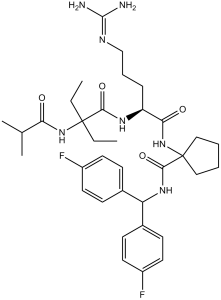
O=C(C1(NC([C@@H](NC(C(NC(C(C)C)=O)(CC)CC)=O)CCCNC(N)=N)=O)CCCC1)NC(C2=CC=C(F)C=C2)C3=CC=C(F)C=C3
|
|
| InChi Key |
RZKSQRIPRKWVBU-MHZLTWQESA-N
|
|
| InChi Code |
InChI=1S/C35H49F2N7O4/c1-5-34(6-2,43-29(45)22(3)4)31(47)41-27(10-9-21-40-33(38)39)30(46)44-35(19-7-8-20-35)32(48)42-28(23-11-15-25(36)16-12-23)24-13-17-26(37)18-14-24/h11-18,22,27-28H,5-10,19-21H2,1-4H3,(H,41,47)(H,42,48)(H,43,45)(H,44,46)(H4,38,39,40)/t27-/m0/s1
|
|
| 化学名 |
N-[Bis(4-fluorophenyl)methyl]-1-[[(2S)-5-(diaminomethylideneamino)-2-[[2-ethyl-2-(2-methylpropanoylamino)butanoyl]amino]pentanoyl]amino]cyclopentane-1-carboxamide
|
|
| 别名 |
|
|
| HS Tariff Code |
2934.99.9001
|
|
| 存储方式 |
Powder -20°C 3 years 4°C 2 years In solvent -80°C 6 months -20°C 1 month 注意: 请将本产品存放在密封且受保护的环境中(例如氮气保护),避免吸湿/受潮和光照。 |
|
| 运输条件 |
Room temperature (This product is stable at ambient temperature for a few days during ordinary shipping and time spent in Customs)
|
| 溶解度 (体外实验) |
|
|||
|---|---|---|---|---|
| 溶解度 (体内实验) |
注意: 如下所列的是一些常用的体内动物实验溶解配方,主要用于溶解难溶或不溶于水的产品(水溶度<1 mg/mL)。 建议您先取少量样品进行尝试,如该配方可行,再根据实验需求增加样品量。
注射用配方
注射用配方1: DMSO : Tween 80: Saline = 10 : 5 : 85 (如: 100 μL DMSO → 50 μL Tween 80 → 850 μL Saline)(IP/IV/IM/SC等) *生理盐水/Saline的制备:将0.9g氯化钠/NaCl溶解在100 mL ddH ₂ O中,得到澄清溶液。 注射用配方 2: DMSO : PEG300 :Tween 80 : Saline = 10 : 40 : 5 : 45 (如: 100 μL DMSO → 400 μL PEG300 → 50 μL Tween 80 → 450 μL Saline) 注射用配方 3: DMSO : Corn oil = 10 : 90 (如: 100 μL DMSO → 900 μL Corn oil) 示例: 以注射用配方 3 (DMSO : Corn oil = 10 : 90) 为例说明, 如果要配制 1 mL 2.5 mg/mL的工作液, 您可以取 100 μL 25 mg/mL 澄清的 DMSO 储备液,加到 900 μL Corn oil/玉米油中, 混合均匀。 View More
注射用配方 4: DMSO : 20% SBE-β-CD in Saline = 10 : 90 [如:100 μL DMSO → 900 μL (20% SBE-β-CD in Saline)] 口服配方
口服配方 1: 悬浮于0.5% CMC Na (羧甲基纤维素钠) 口服配方 2: 悬浮于0.5% Carboxymethyl cellulose (羧甲基纤维素) 示例: 以口服配方 1 (悬浮于 0.5% CMC Na)为例说明, 如果要配制 100 mL 2.5 mg/mL 的工作液, 您可以先取0.5g CMC Na并将其溶解于100mL ddH2O中,得到0.5%CMC-Na澄清溶液;然后将250 mg待测化合物加到100 mL前述 0.5%CMC Na溶液中,得到悬浮液。 View More
口服配方 3: 溶解于 PEG400 (聚乙二醇400) 请根据您的实验动物和给药方式选择适当的溶解配方/方案: 1、请先配制澄清的储备液(如:用DMSO配置50 或 100 mg/mL母液(储备液)); 2、取适量母液,按从左到右的顺序依次添加助溶剂,澄清后再加入下一助溶剂。以 下列配方为例说明 (注意此配方只用于说明,并不一定代表此产品 的实际溶解配方): 10% DMSO → 40% PEG300 → 5% Tween-80 → 45% ddH2O (或 saline); 假设最终工作液的体积为 1 mL, 浓度为5 mg/mL: 取 100 μL 50 mg/mL 的澄清 DMSO 储备液加到 400 μL PEG300 中,混合均匀/澄清;向上述体系中加入50 μL Tween-80,混合均匀/澄清;然后继续加入450 μL ddH2O (或 saline)定容至 1 mL; 3、溶剂前显示的百分比是指该溶剂在最终溶液/工作液中的体积所占比例; 4、 如产品在配制过程中出现沉淀/析出,可通过加热(≤50℃)或超声的方式助溶; 5、为保证最佳实验结果,工作液请现配现用! 6、如不确定怎么将母液配置成体内动物实验的工作液,请查看说明书或联系我们; 7、 以上所有助溶剂都可在 Invivochem.cn网站购买。 |
| 制备储备液 | 1 mg | 5 mg | 10 mg | |
| 1 mM | 1.4930 mL | 7.4649 mL | 14.9298 mL | |
| 5 mM | 0.2986 mL | 1.4930 mL | 2.9860 mL | |
| 10 mM | 0.1493 mL | 0.7465 mL | 1.4930 mL |
1、根据实验需要选择合适的溶剂配制储备液 (母液):对于大多数产品,InvivoChem推荐用DMSO配置母液 (比如:5、10、20mM或者10、20、50 mg/mL浓度),个别水溶性高的产品可直接溶于水。产品在DMSO 、水或其他溶剂中的具体溶解度详见上”溶解度 (体外)”部分;
2、如果您找不到您想要的溶解度信息,或者很难将产品溶解在溶液中,请联系我们;
3、建议使用下列计算器进行相关计算(摩尔浓度计算器、稀释计算器、分子量计算器、重组计算器等);
4、母液配好之后,将其分装到常规用量,并储存在-20°C或-80°C,尽量减少反复冻融循环。
计算结果:
工作液浓度: mg/mL;
DMSO母液配制方法: mg 药物溶于 μL DMSO溶液(母液浓度 mg/mL)。如该浓度超过该批次药物DMSO溶解度,请首先与我们联系。
体内配方配制方法:取 μL DMSO母液,加入 μL PEG300,混匀澄清后加入μL Tween 80,混匀澄清后加入 μL ddH2O,混匀澄清。
(1) 请确保溶液澄清之后,再加入下一种溶剂 (助溶剂) 。可利用涡旋、超声或水浴加热等方法助溶;
(2) 一定要按顺序加入溶剂 (助溶剂) 。
Link: https://www.clinicaltrialsregister.eu/ctr-search/search?query=2008-002703-20
Condition:Locally advanced (stage IIIb with supraclavicular lymph node metastases or malignant pleural or pericardial effusion), metastatic (stage IV) or recurrent non-small cell lung cancer. |
|---|
 BI-9466
 MRK-740-NC
 SKLB-03220
 (Rac)-NSD2-PWWP1-IN-4
InvivoChem的所有产品仅用于作科学研究,不面向患者销售
Copyright 2020 InvivoChem LLC | All Rights Reserved 粤ICP备20063088号-1


 COA
COA
 463611831
