| 规格 | 价格 | 库存 | 数量 |
|---|---|---|---|
| 1mg |
|
||
| 5mg |
|
||
| 10mg |
|
||
| 25mg |
|
||
| Other Sizes |
|

| 靶点 |
Monopolar spindle 1 (MPS1)
|
|---|---|
| 体外研究 (In Vitro) |
设计用于测量MPS1酶活性抑制的体外激酶测定法鉴定出三种得分最高的化合物:Mps-BAY1,一种三唑并吡啶,以及Mps-BAY2a和Mps-BAY2b,两种咪唑并吡嗪(补充图1)。这两类化合物都含有氢键供体/受体氮原子,这在与蛋白激酶的ATP口袋和相关铰链区结合的分子中很常见。Mps-BAY1Mps-BAY2a和Mps-BAY2b抑制人MPS1,IC50范围在1至10 nM之间(补充表1)。当以高浓度(10μM)使用时,Mps-BAY1、Mps-BAY2a和Mps-BAY2b对220种人类激酶显示出有限的抑制作用,与广谱激酶抑制剂瑞香和蒽[1,9-cd]吡唑-6(2H)-酮(SP600125)相比(补充表2)。10,15值得注意的是,Mps-BAY1、Mps-BAY2 a和Mps-BAY2b未能抑制几种已知在有丝分裂中起作用的激酶。Mps-BAY1、Mps-BAY2a和Mps-BAY2b以130nM的IC50抑制SAC的激活,分别为95 nM和670 nM,如在对300 nM诺考达唑有反应的HeLa细胞中评估组蛋白3(H3)磷酸化(在前期/中期发生的翻译后修饰)消失的测定中监测的(数据未显示)。因此,Mps-BAY1、Mps-BAY2a和Mps-BAY2b被培养的细胞有效吸收,并能够达到其分子靶点。根据这一概念,所有这些MPS1抑制剂都降低了绝大多数原代和转化的人类和大鼠细胞的增殖,并对小鼠细胞产生了更高的抗增殖作用(补充表3)。Mps-BAY2a在一组人结肠癌细胞系中引起异质性抗增殖反应,敏感性(IC50)范围为160 nM至>10μM(补充表4)。值得注意的是,CIN和微卫星不稳定性(MIN)均与人类结直肠癌癌症细胞系对Mps-BAY2a的耐药性/敏感性无关(补充表4)。Mps-BAY1、Mps-BAY2a和Mps-BAY2b对人结直肠癌HCT 116(图1和补充图2)和人宫颈癌HeLa细胞(补充图3和4)的细胞周期进展和存活有重大影响,这两种细胞对这些化合物特别敏感(补充表3)。因此,Mps-BAY1、Mps-BAY2a和Mps-BAY2b诱导了细胞周期的剂量和时间依赖性扰动,表现为出现超倍体DNA含量(>4n)的细胞频率增加(图1a-c和补充图2和3),以及死亡细胞(即失去线粒体跨膜电位Δψm的细胞)和细胞尸体(质膜破裂)的逐渐积累(图1d和补充图4)。这些发现确定Mps-BAY1、Mps-BAY2a和Mps-BAY2b是具有强效抗增殖和细胞毒性作用的新型MPS1抑制剂[1]。
Mps-BAY1和Mps-BAY2a[1] 诱导的细胞周期扰动特征 然后,我们研究了MPS1抑制剂对细胞周期进程的精确影响。在施用Mps-BAY1、Mps-BAY2a或Mps-BAY2b后,HCT 116细胞中掺入DNA前体5-乙炔基-2′-脱氧尿苷(EdU,仅在细胞周期的S期被吸收)的比例随着时间的推移而降低,尽管这种抑制作用在SP600125中更为明显(图2a)。值得注意的是,即使在暴露于MPS1抑制剂48小时后,仍有相当一部分细胞复制了它们的DNA。然后,我们对细胞周期蛋白E和B1的水平进行了深入的细胞荧光测定和(荧光)显微镜分析,这两种标记物分别在细胞周期的G1期和G2期积累。针对Mps-BAY1、Mps-BAY2a和Mps-BAY2b(标准剂量:1μM,分别为1μM和3μM),细胞周期蛋白B1+HCT 116细胞的频率降低,尽管这些影响不如SP600125介导的一致(图2b和c)。 |
| 体内研究 (In Vivo) |
最后,研究人员评估了Mps-BAY2b联合紫杉醇在体内对免疫缺陷小鼠中生长的HeLa Matu宫颈癌的治疗潜力。研究人员使用Mps-BAY2b,因为它显示出比Mps-BAY1和Mps-BAY2a更高的体内稳定性(补充表5)。通过免疫组织化学测定,紫杉醇给药24小时后,HeLa Matu细胞衍生的异种移植物显示出比未治疗的肿瘤更高的磷酸化H3水平。经紫杉醇治疗的荷瘤小鼠短时间(1小时)暴露于Mps-BAY2b导致H3磷酸化减少(图8a)。这一发现表明,Mps-BAY2b在体内有效分布,到达异种移植肿瘤并穿透癌症细胞以抑制MPS1。在这种异种移植物模型中,Mps-BAY2b和紫杉醇的组合诱导了比单独干预更高水平的凋亡和更高的巨型单核细胞(核直径>25μm)发生率(图8b)。此外,与载体和紫杉醇或Mps-BAY2b单独给药相比,紫杉醇和Mps-BAY2b联合给药具有更优的抗肿瘤作用(图8c)。总之,这些数据强调了有利地将MPS1抑制剂与MT靶向剂结合的可能性[1]。
|
| 细胞实验 |
细胞荧光测定研究[1]
为了同时定量质膜完整性和Δψm,收集细胞并在37°C下用1μg/ml碘化丙啶和40 nM 3,3′-二己基氧杂碳菁碘化物(DiOC6)染色30分钟。为了评估细胞周期分布,收集细胞,用50μg/ml PI染色,并如前所述通过细胞荧光法进行分析。对于EdU掺入试验,根据制造商的说明,将细胞与10μM EdU在37°C下孵育30分钟,固定、渗透并用荧光染料叠氮化物和PI染色。为了同时测量DNA含量和细胞周期蛋白B1水平,将固定细胞与10 μM 4′,6-二脒基-2-苯基吲哚和先前报道的针对细胞周期蛋白B1的小鼠抗血清。在配备70μm喷嘴的FACSCalibur细胞荧光计或Gallios细胞荧光计上进行细胞荧光采集。 免疫荧光和视频显微镜[1] 根据常规程序进行免疫荧光显微镜检查。使用蔡司Axio Observer捕获58张图像。配备ApoTome系统的Z1显微镜。对于视频显微镜,稳定表达H2B-GFP嵌合体的HCT 116细胞在标准条件下在黑色/透明96孔成像板上生长,并用BD途径855自动活细胞显微镜进行脉冲观察(每13分钟一次,持续72小时)。使用开源软件ImageJ对图像进行分析。细胞命运曲线如前所述。 |
| 动物实验 |
为了定量分析循环中的MPS1抑制剂,将Mps-Bay1、Mps-BAY2a和Mps-BAY2b以可溶性形式经口(po)给予雌性无胸腺nu/nu小鼠(每种化合物和时间点n=2只小鼠)。分别于给药后1、7和24小时采集血清样本,并用冰冷的1:5 (v:v)乙腈/水混合溶液沉淀。采用液相色谱-串联质谱法分析上清液中Mps-BAY1、Mps-BAY2a和Mps-BAY2b的含量。在肿瘤异种移植研究中,使用50日龄、平均体重20-22克的雌性无胸腺nu/nu小鼠,小鼠需经过14天的适应期。取自指数生长期的HeLa-Matu人宫颈癌细胞培养物,重悬于1:1 (v:v)的无血清培养基/Matrigel(BD Biosciences)混合液中,最终浓度为1.5 × 10⁷个细胞/ml。随后,将1.5 × 10⁶个细胞皮下植入腹股沟区域。每周两次测量肿瘤面积(用游标卡尺测量,近似为最长直径与其垂直距离的乘积)和体重。当肿瘤面积达到约21 mm²时,将动物随机分为以下几组(每组8只小鼠):对照组,每周一次口服3:1 (v:v)的聚乙二醇/水(溶剂);Mps-BAY2b组,每周一次口服30 mg/kg的Mps-BAY2b(溶于3:1的聚乙二醇/水中);紫杉醇组,每周一次静脉注射10 mg/kg紫杉醇(溶于1:1:18 (v:v:v) 的Cremophor/乙醇/PBS混合液);Mps-BAY2b联合紫杉醇组,每周一次口服30 mg/kg Mps-BAY2b(溶于3:1的聚乙二醇/水混合液)加10 mg/kg紫杉醇(溶于1:1:18的Cremophor/乙醇/PBS混合液)。当肿瘤面积超过150 mm²时,根据德国动物福利指南对动物实施安乐死。对于免疫组织化学研究,当肿瘤大小达到40-50 mm²时,将动物随机分为以下几组(每组三只小鼠):对照组,口服一次3:1的聚乙二醇/水(溶剂); Mps-BAY2b组,口服一次,剂量为30 mg/kg Mps-BAY2b(溶于3:1聚乙二醇/水);紫杉醇组,腹腔注射一次,剂量为30 mg/kg紫杉醇(溶于1:1:18 Cremophor/乙醇/PBS)。进行苏木精-伊红染色时,当肿瘤大小达到50-80 mm²时,将动物随机分为以下几组(每组4只小鼠):对照组,口服3:1聚乙二醇/水(溶剂),每日两次,连续2天;Mps-BAY2b组,口服30 mg/kg Mps-BAY2b(溶于3:1聚乙二醇/水),每日两次,连续2天;紫杉醇组,静脉注射一次,剂量为8 mg/kg紫杉醇(溶于1:1:18 Cremophor/乙醇/PBS)。 Mps-BAY2b联合紫杉醇组,受试者口服30 mg/kg Mps-BAY2b(溶于3:1聚乙二醇/水溶液),每日两次,连续2天;同时静脉注射10 mg/kg紫杉醇(溶于1:1:18 Cremophor/乙醇/PBS溶液),单次给药。首次给药72小时后,取出肿瘤组织,用4%(w/v)多聚甲醛固定4小时,然后包埋于石蜡中。将10微米厚的组织切片按照标准方案进行苏木精-伊红染色,并按先前描述的方法进行分析。[1]
|
| 参考文献 | |
| 其他信息 |
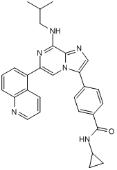
单极纺锤体 1 (MPS1) 是一种有丝分裂激酶,在多种人类癌症中过度表达,它有助于染色体排列到中期板,并有助于纺锤体组装检查点 (SAC) 的执行。在此,我们报道了两种独立结构类别的三种新型 MPS1 抑制剂的鉴定和功能表征,分别为 N-(4-{2-[(2-氰基苯基)氨基][1,2,4]三唑并[1,5-a]吡啶-6-基}苯基)-2-苯基乙酰胺 (Mps-BAY1)(一种三唑并吡啶类化合物)、N-环丙基-4-{8-[(2-甲基丙基)氨基]-6-(喹啉-5-基)咪唑并[1,2-a]吡嗪-3-基}苯甲酰胺 (Mps-BAY2a) 和 N-环丙基-4-{8-(异丁基氨基)咪唑并[1,2-a]吡嗪-3-基}苯甲酰胺 (Mps-BAY2b)(两种咪唑并吡嗪类化合物)。通过选择性地灭活MPS1,这些小分子抑制剂可以抑制癌细胞的增殖,导致其多倍体化和/或死亡。用Mps-BAY1或Mps-BAY2a处理的癌细胞表现出多种有丝分裂紊乱的迹象,包括中期染色体排列效率低下、纺锤体组装检查点(SAC)失活异常以及严重的后期缺陷。对表达组蛋白2B-绿色荧光蛋白的细胞进行显微细胞命运分析表明,MPS1抑制剂能够扰乱有丝分裂的正常时序,因为它们会在中期板错位的情况下诱导细胞过早进入后期。因此,在MPS1抑制剂存在的情况下,细胞要么以双极(但通常是不对称的)方式分裂,要么进入一轮或多轮无效的有丝分裂,分别产生严重的非整倍体和多倍体。在这两种情况下,细胞最终都因有丝分裂灾难诱导的线粒体凋亡途径激活而死亡。值得注意的是,低剂量的MPS1抑制剂和紫杉醇(一种微管毒素)在纺锤体组装检查点(SAC)失活的情况下,协同作用增加了染色体错配和分离错误的发生频率。这导致了大量的多倍体化,随后激活了有丝分裂灾难。体内实验也证实了紫杉醇和MPS1抑制剂之间的协同作用,因为这两种药物的联合使用能有效抑制肿瘤异种移植瘤的生长,并且与单独使用任何一种化合物相比,都表现出更优异的抗肿瘤效果。总之,这些结果表明,MPS1抑制剂可能具有强大的抗癌活性,无论是作为独立的治疗手段,还是与微管靶向药物联合使用。 [1]
本文报道了三种新型高效MPS1抑制剂的鉴定和功能表征,它们分别是三唑并吡啶类化合物Mps-BAY1和咪唑并吡嗪类化合物Mps-BAY2a和Mps-BAY2b。所有这些化合物均能抑制纺锤体组装检查点(SAC)的功能,表现为暴露于MPS1抑制剂的细胞在接触微管毒素后无法维持有丝分裂阻滞。即使在没有SAC激活剂的情况下,这两类MPS1抑制剂也能显著增加由微管-动粒连接错误导致的染色体错位率,并促进细胞过早进入后期(即在正确的赤道板形成之前)。这些结果与之前使用其他 MPS1 特异性抑制剂在 MPS1 耗竭后 1 或在 TTK 条件性敲除后 11 获得的结果一致,证实了这种有丝分裂激酶在 SAC 功能和染色体排列中的核心作用。[1] |
| 分子式 |
C29H28N6O
|
|---|---|
| 分子量 |
476.58
|
| 精确质量 |
476.232
|
| 元素分析 |
C, 73.09; H, 5.92; N, 17.63; O, 3.36
|
| CAS号 |
1382477-96-4
|
| PubChem CID |
57381882
|
| 外观&性状 |
Off-white to light yellow solid powder
|
| 密度 |
1.3±0.1 g/cm3
|
| 折射率 |
1.716
|
| LogP |
5.34
|
| tPSA |
84.2
|
| 氢键供体(HBD)数目 |
2
|
| 氢键受体(HBA)数目 |
5
|
| 可旋转键数目(RBC) |
7
|
| 重原子数目 |
36
|
| 分子复杂度/Complexity |
750
|
| 定义原子立体中心数目 |
0
|
| InChi Key |
MDYKTGNHXNTATG-UHFFFAOYSA-N
|
| InChi Code |
InChI=1S/C29H28N6O/c1-18(2)15-31-27-28-32-16-26(19-8-10-20(11-9-19)29(36)33-21-12-13-21)35(28)17-25(34-27)23-5-3-7-24-22(23)6-4-14-30-24/h3-11,14,16-18,21H,12-13,15H2,1-2H3,(H,31,34)(H,33,36)
|
| 化学名 |
N-cyclopropyl-4-[8-(2-methylpropylamino)-6-quinolin-5-ylimidazo[1,2-a]pyrazin-3-yl]benzamide
|
| 别名 |
Mps BAY 2a; Mps-BAY-2a; 1382477-96-4; CHEMBL3422104; N-cyclopropyl-4-[8-(2-methylpropylamino)-6-quinolin-5-ylimidazo[1,2-a]pyrazin-3-yl]benzamide; N-Cyclopropyl-4-[8-[(2-methylpropyl)amino]-6-(5-quinolinyl)imidazo[1,2-a]pyrazin-3-yl]benzamide; MpsBAY2a
|
| HS Tariff Code |
2934.99.9001
|
| 存储方式 |
Powder -20°C 3 years 4°C 2 years In solvent -80°C 6 months -20°C 1 month |
| 运输条件 |
Room temperature (This product is stable at ambient temperature for a few days during ordinary shipping and time spent in Customs)
|
| 溶解度 (体外实验) |
DMSO : ~10 mg/mL (~20.98 mM)
|
|---|---|
| 溶解度 (体内实验) |
注意: 如下所列的是一些常用的体内动物实验溶解配方,主要用于溶解难溶或不溶于水的产品(水溶度<1 mg/mL)。 建议您先取少量样品进行尝试,如该配方可行,再根据实验需求增加样品量。
注射用配方
注射用配方1: DMSO : Tween 80: Saline = 10 : 5 : 85 (如: 100 μL DMSO → 50 μL Tween 80 → 850 μL Saline)(IP/IV/IM/SC等) *生理盐水/Saline的制备:将0.9g氯化钠/NaCl溶解在100 mL ddH ₂ O中,得到澄清溶液。 注射用配方 2: DMSO : PEG300 :Tween 80 : Saline = 10 : 40 : 5 : 45 (如: 100 μL DMSO → 400 μL PEG300 → 50 μL Tween 80 → 450 μL Saline) 注射用配方 3: DMSO : Corn oil = 10 : 90 (如: 100 μL DMSO → 900 μL Corn oil) 示例: 以注射用配方 3 (DMSO : Corn oil = 10 : 90) 为例说明, 如果要配制 1 mL 2.5 mg/mL的工作液, 您可以取 100 μL 25 mg/mL 澄清的 DMSO 储备液,加到 900 μL Corn oil/玉米油中, 混合均匀。 View More
注射用配方 4: DMSO : 20% SBE-β-CD in Saline = 10 : 90 [如:100 μL DMSO → 900 μL (20% SBE-β-CD in Saline)] 口服配方
口服配方 1: 悬浮于0.5% CMC Na (羧甲基纤维素钠) 口服配方 2: 悬浮于0.5% Carboxymethyl cellulose (羧甲基纤维素) 示例: 以口服配方 1 (悬浮于 0.5% CMC Na)为例说明, 如果要配制 100 mL 2.5 mg/mL 的工作液, 您可以先取0.5g CMC Na并将其溶解于100mL ddH2O中,得到0.5%CMC-Na澄清溶液;然后将250 mg待测化合物加到100 mL前述 0.5%CMC Na溶液中,得到悬浮液。 View More
口服配方 3: 溶解于 PEG400 (聚乙二醇400) 请根据您的实验动物和给药方式选择适当的溶解配方/方案: 1、请先配制澄清的储备液(如:用DMSO配置50 或 100 mg/mL母液(储备液)); 2、取适量母液,按从左到右的顺序依次添加助溶剂,澄清后再加入下一助溶剂。以 下列配方为例说明 (注意此配方只用于说明,并不一定代表此产品 的实际溶解配方): 10% DMSO → 40% PEG300 → 5% Tween-80 → 45% ddH2O (或 saline); 假设最终工作液的体积为 1 mL, 浓度为5 mg/mL: 取 100 μL 50 mg/mL 的澄清 DMSO 储备液加到 400 μL PEG300 中,混合均匀/澄清;向上述体系中加入50 μL Tween-80,混合均匀/澄清;然后继续加入450 μL ddH2O (或 saline)定容至 1 mL; 3、溶剂前显示的百分比是指该溶剂在最终溶液/工作液中的体积所占比例; 4、 如产品在配制过程中出现沉淀/析出,可通过加热(≤50℃)或超声的方式助溶; 5、为保证最佳实验结果,工作液请现配现用! 6、如不确定怎么将母液配置成体内动物实验的工作液,请查看说明书或联系我们; 7、 以上所有助溶剂都可在 Invivochem.cn网站购买。 |
| 制备储备液 | 1 mg | 5 mg | 10 mg | |
| 1 mM | 2.0983 mL | 10.4914 mL | 20.9828 mL | |
| 5 mM | 0.4197 mL | 2.0983 mL | 4.1966 mL | |
| 10 mM | 0.2098 mL | 1.0491 mL | 2.0983 mL |
1、根据实验需要选择合适的溶剂配制储备液 (母液):对于大多数产品,InvivoChem推荐用DMSO配置母液 (比如:5、10、20mM或者10、20、50 mg/mL浓度),个别水溶性高的产品可直接溶于水。产品在DMSO 、水或其他溶剂中的具体溶解度详见上”溶解度 (体外)”部分;
2、如果您找不到您想要的溶解度信息,或者很难将产品溶解在溶液中,请联系我们;
3、建议使用下列计算器进行相关计算(摩尔浓度计算器、稀释计算器、分子量计算器、重组计算器等);
4、母液配好之后,将其分装到常规用量,并储存在-20°C或-80°C,尽量减少反复冻融循环。
计算结果:
工作液浓度: mg/mL;
DMSO母液配制方法: mg 药物溶于 μL DMSO溶液(母液浓度 mg/mL)。如该浓度超过该批次药物DMSO溶解度,请首先与我们联系。
体内配方配制方法:取 μL DMSO母液,加入 μL PEG300,混匀澄清后加入μL Tween 80,混匀澄清后加入 μL ddH2O,混匀澄清。
(1) 请确保溶液澄清之后,再加入下一种溶剂 (助溶剂) 。可利用涡旋、超声或水浴加热等方法助溶;
(2) 一定要按顺序加入溶剂 (助溶剂) 。
InvivoChem的所有产品仅用于作科学研究,不面向患者销售
Copyright 2020 InvivoChem LLC | All Rights Reserved 粤ICP备20063088号-1


 463611831
