| 规格 | 价格 | |
|---|---|---|
| 500mg | ||
| 1g | ||
| Other Sizes |

| 靶点 |
Proteasome
|
|---|---|
| 体外研究 (In Vitro) |
蛋白酶体标记和凝胶内检测[1]
MV151(6)的效价通过荧光底物测定蛋白酶体活性来确定。将EL-4裂解物与浓度增加的MV151孵育,监测底物su - leu - leu - val - tir - amc(乳糜蛋白酶样活性)、Z-Ala-Ala-Arg-AMC(胰蛋白酶样活性)和Z-Leu-Leu-Glu-βNA(肽酰谷氨酰肽水解活性)的裂解情况。在浓度低于1 μM时,MV151似乎比抑制PGPH活性更有效地抑制胰蛋白酶样活性和凝乳胰蛋白酶样活性(图2A)。这可能是由于亚基之间的活性差异,变构效应,小亚基特异性的探针,或非饱和动力学。在浓度为1 μM及更高时,MV151完全抑制上述三种活性。 使用荧光扫描仪探索MV151标记的蛋白酶体亚基的直接凝胶可视化。纯化的人20S蛋白酶体用MV151处理后,活性亚基β1、β2和β5被均匀标记(图2B)。为了确定凝胶内检测的灵敏度,我们直接将凝胶的荧光读数(图2B)与蛋白质的银染色检测蛋白酶体亚基(图2C)进行了比较。凝胶内检测显示非常敏感,因为只需3 ng蛋白酶体就足以检测单个mv151标记的蛋白酶体亚基;这种方法的检测灵敏度至少是银染色的三倍。 接下来,我们比较了组成β1、β2和β5亚基与免疫蛋白酶体β1i、β2i和β5i亚基的标记。为此,我们在人宫颈癌细胞系HeLa(表达组成性蛋白酶体)和小鼠淋巴样细胞系EL-4(表达组成性蛋白酶体和免疫蛋白酶体)的溶出物中标记蛋白酶体,浓度增加MV151。所有活性组成型和诱导型β蛋白酶体亚基被MV151整齐而均匀地标记(图2D和2E)。所有亚基在10 nM MV151浓度下均可检测到,在1 μM MV151浓度下荧光信号达到饱和。在较高浓度的MV151中,高分子量区域的非特异性标记增加。 活细胞功能蛋白酶体抑制[1] 接下来,我们研究了MV151是否能够标记活细胞中的蛋白酶体亚基。增加MV151浓度孵育EL-4和HeLa细胞。在EL-4细胞(图4A)和HeLa细胞(图4B)中观察到所有蛋白酶体亚基的特异性和敏感性标记,尽管比在裂解物中标记亚基需要更高的浓度。β1亚基的标记比裂解物的强度低,而β5标记看起来更明显。细胞裂解物和活细胞中蛋白酶体标记谱的差异此前已被报道;然而,其原因尚不清楚。重要的是,EL-4和HeLa细胞与无活性对照化合物MV152(7)孵育后,没有发现蛋白酶体或任何其他蛋白质的标记(图4A和4B), MV152与MV151几乎相同,但缺乏活性的乙烯基砜战斗部。 |
| 体内研究 (In Vivo) |
小鼠蛋白酶体抑制的监测[1]
在细胞系中获得的结果促使我们研究MV151是否可以用于标记小鼠蛋白酶体。为检测MV151的生物利用度,C57Bl/6小鼠单次腹腔注射MV151 (20 μmol/kg体重),24小时后处死。 荧光显微镜对小鼠组织的分析显示了MV151在体内穿透组织的能力。在肝脏(图5A)和胰腺(图5B)中检测到最高的Bodipy TMR荧光。有趣的是,Bodipy TMR荧光在组织周围较高,表明探针可能通过腹膜腔扩散而不是通过进入血液分布而最有效地到达肝脏。 为了研究蛋白酶体探针的作用,我们利用最近开发的一种转基因小鼠模型来监测泛素-蛋白酶体系统,该模型基于UbG76V-GFP报告基因的普遍表达。我们之前的研究表明,使用蛋白酶体抑制剂epoxomicin和MG262会导致UbG76V-GFP报告基因在受影响的组织中大量积累。这种积累主要在肝脏中发现,在其他组织中浓度较高。在本实验中,给UbG76V-GFP报告小鼠单次腹腔注射MV151 (20 μmol/kg体重)。注射后共24小时,处死小鼠,荧光显微镜下对多个组织进行分析。在肝脏(图5C)和胰腺(图5E)中检测到积累UbG76V-GFP的细胞,在所有检查组织(脾、肠、肾、肝和胰腺)中,胰腺也含有最高的Bodipy TMR荧光。重要的是,所有积累UbG76V-GFP报告基因的细胞都含有非常高的Bodipy TMR荧光。蛋白酶体探针分布在聚集报告基因的细胞的细胞质和细胞核中。与我们在细胞培养实验中观察到的结果相似,小鼠受影响的细胞在靠近细胞核的细胞质中含有MV151的颗粒状积聚。我们证实了UbG76V-GFP在肝脏和胰腺中的积累与MV151的蛋白酶体阻断相一致。用MV151处理动物的肝脏(图5D)和胰腺(图5F)匀浆的SDS-PAGE凝胶内荧光分析显示,蛋白酶体催化亚基被标记为预期的,尽管与体外研究相比,观察到更高的背景标记。(图5D和图5F分别使用图5C和图5E图像中的组织。)脾脏匀浆的SDS-PAGE和凝胶内荧光分析显示了组成型和诱导型蛋白酶体催化亚基的标记(图5G)。 作为最后一组实验,我们监测了MG262在UbG76V-GFP转基因小鼠中的生物分布。我们选择硼酸MG262用于此目的,因为它既有纯化形式的商业可用性,又与药物硼替佐米最相似。分别皮下注射5 μmol/kg或10 μmol/kg体重的硼酸MG262,注射24小时后处死。脾胰溶解,MV151处理。SDS-PAGE分析显示,与未经处理的动物组织裂解物相比,蛋白酶体催化亚基对应的标记带显著减少(图6A,胰腺;图6B,脾脏)。同样组织的荧光显微镜分析(图6A和6B)证实了mg262处理的UbG76V-GFP小鼠中蛋白酶体的浓度依赖性抑制,UbG76V-GFP报告基因积累水平增加。 |
| 酶活实验 |
荧光底物测定蛋白酶体活性[1]
EL-4蛋白裂解物(1 mg/ml)与不同浓度的MV151(6)在37℃下孵育1小时。为了测定蛋白酶体的活性,将10 μg标记的裂解液加入100 μl底物缓冲液中,其中含有20 mM HEPES (pH 8.2)、0.5 mM EDTA、1% DMSO、1 mM ATP和10 μM Z-Ala-Ala-Arg-AMC(色氨酸样)、60 μM su - leu - leu - val - tyr - amc (chymotryptic样)或60 μM Z-Leu-Leu-Glu-βNA (caspase样)。采用Fluostar Optima 96孔板荧光仪(AMC为λex/λem = 355/450 nm, βNA为320/405 nm),在37°C下每分钟测量一次荧光,持续25 min,以每分钟最大荧光增量计算每个样品的比活性。非特异性水解通过1 μM环氧霉素在37℃预孵育1小时来评估,并从每次测量中减去。 <人力资源> 标记蛋白酶体亚基的凝胶内检测[1] 全细胞裂解液在含有50 mM Tris (pH 7.5), 1 mM DTT, 5 mM MgCl2, 250 mM蔗糖,2 mM ATP的裂解缓冲液中制备。用比色Bradford法测定蛋白浓度。标记反应中,10 μg总蛋白裂解物在37℃下孵育1小时,增加MV151的浓度,总反应体积为10 μl。如有需要,使用50 ng纯化20S蛋白酶体。在竞争研究中,将细胞裂解物(10 μg)暴露于已知抑制剂中1小时,然后与MV151 (0.1 μM)在37°C下孵育1小时。为了评估背景标记,用MV151处理热灭活裂解物(10 μg,用1% SDS煮沸3分钟)。反应混合物用含有β-巯基乙醇的Laemmli缓冲液煮沸3分钟,在12.5% SDS-PAGE上分离。使用台风变模成像仪上的Cy3/Tamra设置(λex 532, λem 560)直接在湿凝胶板上进行凝胶内可视化。用1 - 10 μM MV151在37°C的培养基中培养8小时,测定活细胞中的标记谱。细胞裂解,凝胶内检测,如上所述。 |
| 细胞实验 |
显微镜观察[1]< br >
将约0.5 × 10⁶个细胞接种于35mm培养皿中,14mm微孔/ 1.5盖和玻璃底微孔培养皿中,并允许贴壁过夜。在配备Radiance 2100 MP集成激光检测系统和Tsunami多光子激光模块的Nikon Eclipse TE 2000U显微镜上,用60×油浸透镜观察细胞。显微镜控制和数据采集采用LaserSharp 2K软件,图像处理采用Image Pro 3DS 5.1软件。GFP在λex = 488 nm处激发,在500 ~ 530 nm处检测。MV151和MV152在λex = 543 nm处激发,在560 ~ 620 nm处检测到。使用Photoshop软件调整CLSM图像的亮度和对比度。
|
| 动物实验 |
小鼠实验[1]
小鼠按照瑞典动物饲养规程饲养,光照周期为12小时,并自由摄取标准实验室饲料和自来水。成年C57Bl/6和UbG76V-GFP/1小鼠[16],性别和年龄匹配,腹腔注射200 μl,注射液分别为载体(60% DMSO,40% PBS)、MV151(20 μmol/kg体重)或MG262(5或10 μmol/kg体重)。根据我们实验室之前的经验,硼酸类抑制剂比乙烯砜类抑制剂更有效,且体内组织穿透性更好。因此,我们选择20 μmol/kg体重的剂量作为MV151的给药剂量,该剂量在小鼠中未显示明显的毒性。注射后24小时,小鼠经吸入异氟烷(4.4%异氟烷/氧气)麻醉后处死,随后经心脏灌注50 ml PBS以清除污染血液。用于免疫细胞化学分析的组织按先前所述方法处理。简而言之,将12 μm厚的冰冻切片用4%多聚甲醛/PBS固定15分钟,并用PBS洗涤;如前所述,在黑暗中用Hoechst核染料(2 μg/ml,溶于H₂O)染色15分钟,然后用PBS洗涤。将切片封片于含2.5% DABCO的基质中。使用蔡司LSM 510 META系统进行共聚焦显微镜观察。用于凝胶内分析的组织用Heidolph组织匀浆器在300 μl裂解缓冲液中裂解,并按上述方法进行后续处理。 |
| 参考文献 |
[1]. A fluorescent broad-spectrum proteasome inhibitor for labeling proteasomes in vitro and in vivo. Chem Biol. 2006 Nov;13(11):1217-26.
|
| 其他信息 |
蛋白酶体是一种进化上高度保守的重要蛋白酶,参与多种调控系统。本文描述了活性型、荧光标记且可渗透细胞的蛋白酶体抑制剂Bodipy TMR-Ahx(3)L(3)VS (MV151)的合成和表征。该抑制剂特异性靶向活细胞中蛋白酶体和免疫蛋白酶体的所有活性亚基,可实现快速灵敏的凝胶内检测。通过MV151标记,可以轻松测定一系列常用蛋白酶体抑制剂的抑制谱。给小鼠注射MV151后,可实现蛋白酶体的体内标记,且标记结果与受影响组织中蛋白酶体降解的抑制程度相关。该探针可用于多种应用,包括蛋白酶体活性的临床分析、抑制剂亚基特异性的生化分析以及活细胞中蛋白酶体功能和动态的细胞生物学分析。 [1]
总之,我们描述了MV151的合成,并将其表征为一种细胞渗透性广谱蛋白酶体抑制剂。MV151能够对细胞裂解液和活细胞中的蛋白酶体进行广谱分析。Bodipy TMR染料被证明非常适用于凝胶内标记活性亚基的读数,因为它提供了一种直接、灵敏的蛋白酶体分析方法,无需进行蛋白质印迹、放射性标记和凝胶干燥。给小鼠注射MV151后,可以很容易地检测到其浓度,并且与受影响组织中蛋白酶体的抑制程度相关。最后,MV151介导的蛋白酶体标记结合UbG76V-GFP转基因小鼠是监测蛋白酶体抑制剂生物分布的有效策略。[1] 蛋白酶体是维持细胞稳态的关键酶。本文介绍了活性荧光染料MV151的合成与表征,该染料具有细胞渗透性。MV151特异性靶向蛋白酶体,并通过与免疫诱导型和组成型活性β亚基的催化N端苏氨酸残基发生共价且不可逆的结合,展现出广谱活性。其明亮的荧光基团有助于通过凝胶内检测和活细胞荧光显微镜快速灵敏地检测活性蛋白酶体亚基。MV151有望应用于蛋白酶体研究的多个领域:在医学研究中,可用于分析临床相关样本中的活性蛋白酶体组分;在化学和生物化学领域,可用于快速测定新型蛋白酶体抑制剂的效力和亚基特异性。[1] |
| 分子式 |
C59H91BN8O9F2S
|
|---|---|
| 分子量 |
1137.27
|
| 精确质量 |
1136.669
|
| 元素分析 |
C, 62.31; H, 8.07; B, 0.95; F, 3.34; N, 9.85; O, 12.66; S, 2.82
|
| CAS号 |
945611-88-1
|
| 相关CAS号 |
945611-88-1 (MV-151); 945611-89-2 (MV-152)
|
| PubChem CID |
154732122
|
| 外观&性状 |
Typically exists as solid at room temperature
|
| tPSA |
234
|
| 氢键供体(HBD)数目 |
6
|
| 氢键受体(HBA)数目 |
12
|
| 可旋转键数目(RBC) |
36
|
| 重原子数目 |
80
|
| 分子复杂度/Complexity |
2280
|
| 定义原子立体中心数目 |
3
|
| SMILES |
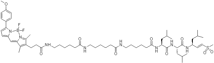
[B-]1(N2C(=C(C(=C2C=C3[N+]1=C(C=C3)C4=CC=C(C=C4)OC)C)CCC(=O)NCCCCCC(=O)NCCCCCC(=O)NCCCCCC(=O)N[C@@H](CC(C)C)C(=O)N[C@@H](CC(C)C)C(=O)N[C@@H](CC(C)C)/C=C/S(=O)(=O)C)C)(F)F
|
| InChi Key |
JNIDQOOVQFMCNR-FWDZYPIVSA-N
|
| InChi Code |
InChI=1S/C59H91BF2N8O9S/c1-40(2)36-46(31-35-80(10,77)78)66-58(75)51(38-42(5)6)68-59(76)50(37-41(3)4)67-57(74)22-16-13-19-33-64-55(72)20-14-11-17-32-63-54(71)21-15-12-18-34-65-56(73)30-28-49-43(7)53-39-47-25-29-52(45-23-26-48(79-9)27-24-45)70(47)60(61,62)69(53)44(49)8/h23-27,29,31,35,39-42,46,50-51H,11-22,28,30,32-34,36-38H2,1-10H3,(H,63,71)(H,64,72)(H,65,73)(H,66,75)(H,67,74)(H,68,76)/b35-31+/t46-,50+,51+/m1/s1
|
| 化学名 |
(2S)-2-[6-[6-[6-[3-[2,2-difluoro-12-(4-methoxyphenyl)-4,6-dimethyl-3-aza-1-azonia-2-boranuidatricyclo[7.3.0.03,7]dodeca-1(12),4,6,8,10-pentaen-5-yl]propanoylamino]hexanoylamino]hexanoylamino]hexanoylamino]-4-methyl-N-[(2S)-4-methyl-1-[[(E,3S)-5-methyl-1-methylsulfonylhex-1-en-3-yl]amino]-1-oxopentan-2-yl]pentanamide
|
| 别名 |
MV151; MV 151; 945611-88-1; (2S)-2-[6-[6-[6-[3-[2,2-difluoro-12-(4-methoxyphenyl)-4,6-dimethyl-3-aza-1-azonia-2-boranuidatricyclo[7.3.0.03,7]dodeca-1(12),4,6,8,10-pentaen-5-yl]propanoylamino]hexanoylamino]hexanoylamino]hexanoylamino]-4-methyl-N-[(2S)-4-methyl-1-[[(E,3S)-5-methyl-1-methylsulfonylhex-1-en-3-yl]amino]-1-oxopentan-2-yl]pentanamide; 6-(3-(5,5-difluoro-7-(4-methoxyphenyl)-1,3-dimethyl-5H-4l4,5l4-dipyrrolo[1,2-c:2',1'-f][1,3,2]diazaborinin-2-yl)propanamido)-N-((4S,7S,10S)-7,10-diisobutyl-2-methyl-4-((E)-2-(methylsulfonyl)vinyl)-6,9,12,19-tetraoxo-5,8,11,18-tetraazatetracosan-24-yl)hexanamide; MV-151; Bodipy TMRAhx(3)L(3)VS.
|
| HS Tariff Code |
2934.99.9001
|
| 存储方式 |
Powder -20°C 3 years 4°C 2 years In solvent -80°C 6 months -20°C 1 month |
| 运输条件 |
Room temperature (This product is stable at ambient temperature for a few days during ordinary shipping and time spent in Customs)
|
| 溶解度 (体外实验) |
May dissolve in DMSO (in most cases), if not, try other solvents such as H2O, Ethanol, or DMF with a minute amount of products to avoid loss of samples
|
|---|---|
| 溶解度 (体内实验) |
注意: 如下所列的是一些常用的体内动物实验溶解配方,主要用于溶解难溶或不溶于水的产品(水溶度<1 mg/mL)。 建议您先取少量样品进行尝试,如该配方可行,再根据实验需求增加样品量。
注射用配方
注射用配方1: DMSO : Tween 80: Saline = 10 : 5 : 85 (如: 100 μL DMSO → 50 μL Tween 80 → 850 μL Saline)(IP/IV/IM/SC等) *生理盐水/Saline的制备:将0.9g氯化钠/NaCl溶解在100 mL ddH ₂ O中,得到澄清溶液。 注射用配方 2: DMSO : PEG300 :Tween 80 : Saline = 10 : 40 : 5 : 45 (如: 100 μL DMSO → 400 μL PEG300 → 50 μL Tween 80 → 450 μL Saline) 注射用配方 3: DMSO : Corn oil = 10 : 90 (如: 100 μL DMSO → 900 μL Corn oil) 示例: 以注射用配方 3 (DMSO : Corn oil = 10 : 90) 为例说明, 如果要配制 1 mL 2.5 mg/mL的工作液, 您可以取 100 μL 25 mg/mL 澄清的 DMSO 储备液,加到 900 μL Corn oil/玉米油中, 混合均匀。 View More
注射用配方 4: DMSO : 20% SBE-β-CD in Saline = 10 : 90 [如:100 μL DMSO → 900 μL (20% SBE-β-CD in Saline)] 口服配方
口服配方 1: 悬浮于0.5% CMC Na (羧甲基纤维素钠) 口服配方 2: 悬浮于0.5% Carboxymethyl cellulose (羧甲基纤维素) 示例: 以口服配方 1 (悬浮于 0.5% CMC Na)为例说明, 如果要配制 100 mL 2.5 mg/mL 的工作液, 您可以先取0.5g CMC Na并将其溶解于100mL ddH2O中,得到0.5%CMC-Na澄清溶液;然后将250 mg待测化合物加到100 mL前述 0.5%CMC Na溶液中,得到悬浮液。 View More
口服配方 3: 溶解于 PEG400 (聚乙二醇400) 请根据您的实验动物和给药方式选择适当的溶解配方/方案: 1、请先配制澄清的储备液(如:用DMSO配置50 或 100 mg/mL母液(储备液)); 2、取适量母液,按从左到右的顺序依次添加助溶剂,澄清后再加入下一助溶剂。以 下列配方为例说明 (注意此配方只用于说明,并不一定代表此产品 的实际溶解配方): 10% DMSO → 40% PEG300 → 5% Tween-80 → 45% ddH2O (或 saline); 假设最终工作液的体积为 1 mL, 浓度为5 mg/mL: 取 100 μL 50 mg/mL 的澄清 DMSO 储备液加到 400 μL PEG300 中,混合均匀/澄清;向上述体系中加入50 μL Tween-80,混合均匀/澄清;然后继续加入450 μL ddH2O (或 saline)定容至 1 mL; 3、溶剂前显示的百分比是指该溶剂在最终溶液/工作液中的体积所占比例; 4、 如产品在配制过程中出现沉淀/析出,可通过加热(≤50℃)或超声的方式助溶; 5、为保证最佳实验结果,工作液请现配现用! 6、如不确定怎么将母液配置成体内动物实验的工作液,请查看说明书或联系我们; 7、 以上所有助溶剂都可在 Invivochem.cn网站购买。 |
| 制备储备液 | 1 mg | 5 mg | 10 mg | |
| 1 mM | 0.8793 mL | 4.3965 mL | 8.7930 mL | |
| 5 mM | 0.1759 mL | 0.8793 mL | 1.7586 mL | |
| 10 mM | 0.0879 mL | 0.4396 mL | 0.8793 mL |
1、根据实验需要选择合适的溶剂配制储备液 (母液):对于大多数产品,InvivoChem推荐用DMSO配置母液 (比如:5、10、20mM或者10、20、50 mg/mL浓度),个别水溶性高的产品可直接溶于水。产品在DMSO 、水或其他溶剂中的具体溶解度详见上”溶解度 (体外)”部分;
2、如果您找不到您想要的溶解度信息,或者很难将产品溶解在溶液中,请联系我们;
3、建议使用下列计算器进行相关计算(摩尔浓度计算器、稀释计算器、分子量计算器、重组计算器等);
4、母液配好之后,将其分装到常规用量,并储存在-20°C或-80°C,尽量减少反复冻融循环。
计算结果:
工作液浓度: mg/mL;
DMSO母液配制方法: mg 药物溶于 μL DMSO溶液(母液浓度 mg/mL)。如该浓度超过该批次药物DMSO溶解度,请首先与我们联系。
体内配方配制方法:取 μL DMSO母液,加入 μL PEG300,混匀澄清后加入μL Tween 80,混匀澄清后加入 μL ddH2O,混匀澄清。
(1) 请确保溶液澄清之后,再加入下一种溶剂 (助溶剂) 。可利用涡旋、超声或水浴加热等方法助溶;
(2) 一定要按顺序加入溶剂 (助溶剂) 。
InvivoChem的所有产品仅用于作科学研究,不面向患者销售
Copyright 2020 InvivoChem LLC | All Rights Reserved 粤ICP备20063088号-1


 COA
COA
 463611831
