| 规格 | 价格 | 库存 | 数量 |
|---|---|---|---|
| 10mg |
|
||
| 25mg |
|
||
| 50mg |
|
||
| 100mg |
|
| 靶点 |
K+/H+ ionophore; NLRP3
|
|---|---|
| 体外研究 (In Vitro) |
通过 caspase 1/GSDMD,尼日利亚霉素(0–4 μg/ml;24 小时)有效引起 TNBC 细胞焦亡 [6]。凭借其集中作用,尼日利亚菌素(0-0.25 μg/ml;24 小时)可抑制 H460 细胞中 Wnt/β-连环蛋白的上调,并表现出抗癌特性 [7]。革兰氏阳性菌对抗生素具有耐药性[4]。
|
| 体内研究 (In Vivo) |
通过每 12 小时腹腔注射 1 mg/kg 尼日利亚霉素,持续三天,可以减少金黄色葡萄球菌 USA300 感染的小鼠 [4]。尼日利亚霉素(皮下注射,每两天一次;0.025 mg/kg)。
体积数据表明,ngericin(4 mg/kg,腹腔注射)表现出肿瘤生长的显着减少,并与化疗剂 DDP 表现出良好的相互作用。在体内,尼日利亚菌素可显着降低 Bmi-1。在尼日利亚素处理下,鼻咽癌细胞的CSC含量和转移潜力部分受到Bmi-1过表达的影响。尼日利亚菌素对鼻咽癌干细胞的抑制可能与Bmi-1的下降有关[3]。 尼日利亚菌素对CSCs在体内表现出选择性毒性[3] 为评价尼日利亚菌素对鼻咽癌细胞的体内作用,将分别皮下接种S18和S26细胞的裸鼠随机分为4个亚组,分别给药、DDP、尼日利亚菌素或DPP和尼日利亚菌素联合给药。在S18组中,尼日利亚菌素显著抑制肿瘤生长,并与化疗药物DDP协同作用,从肿瘤体积来看(P < 0.05或<0.01,图6A)。然而,在S26组中,尼日利亚菌素仅轻微抑制S26异种移植物的生长,而DDP单独或联合尼日利亚菌素均显著抑制肿瘤生长(P < 0.05或<0.01,图6B)。重要的是,与对照组相比,每种药物治疗对小鼠体重的减轻都有影响,从尼日利亚菌素,DDP到两种药物的联合,其严重程度逐渐增加(图6C)。 免疫组化分析显示,尼日利亚菌素处理组S18异种移植物中Bmi-1的表达较低,提示尼日利亚菌素可以下调体内Bmi-1。 此外,我们还评估了尼日利亚菌素治疗后参与Bmi-1通路的两个分子PTEN和Snail的变化。正如预期的那样,与对照组相比,在体内使用尼日利亚菌素后,PTEN蛋白水平升高,蜗牛表达降低。这些结果表明,Bmi-1的下调可能有助于尼日利亚菌素对NPC CSCs的抑制作用。 接下来,我们在4T1异种移植物模型中评估了Nigericin与抗pd -1抗体的协同抗肿瘤作用,该模型包括相对冷的肿瘤,并且由于缺乏T细胞浸润和激活而对免疫检查点抑制剂治疗产生抗性[43,44]。将4T1细胞原位移植到BALB/c小鼠乳腺脂肪垫中。将小鼠随机分为4组,分别给予对照组、尼日利亚菌素(皮下注射)或抗pd -1抗体(腹腔注射)单独和联合治疗。正如预期的那样,单独的PD-1抗体没有明显的抗肿瘤作用,而尼日利亚菌素则有中等的抗肿瘤作用。尼日利亚菌素与抗pd -1抗体联合使用几乎完全抑制肿瘤生长(图6B-D)。在尼日利亚菌素治疗组和联合治疗组中,肿瘤浸润CD4+或CD8+ T细胞的数量一致增加(图6E),提示尼日利亚菌素介导的热亡调节肿瘤微环境,促进T细胞浸润,从而使冷肿瘤转变为热肿瘤。尼日利亚菌素和抗pd -1联合治疗组CD4+或CD8+细胞分泌的TNF-α和IFN-γ水平较高(图6F和图S4C)。根据上述结果,在尼日利亚菌素处理组中,Caspase-1和Caspase-3的裂解量也有所增加(图6F)。此外,我们没有观察到尼日利亚菌素影响免疫细胞和癌细胞中PD-1或PD-L1的表达(图S4D)。这些数据证实了尼日利亚菌素与抗pd -1抗体的协同抗肿瘤作用。 同时,在体内评估了这些治疗的系统性副作用。心、肝、脾、肺、肾组织未见明显组织学毒性(图S5A)。治疗结束时,血液学参数包括白细胞、血红蛋白、天冬氨酸转氨酶、丙氨酸转氨酶、白蛋白和肌酐均在正常范围内(图S5B)。总之,这些结果表明,应用尼日利亚菌素是使tnbc对免疫检查点阻断治疗增敏的有效策略,并且系统副作用可接受。[5] 尼日利亚霉素对USA300感染小鼠模型有效[6] 由于NIG出色的抗菌性能及其在感染动物模型中的疗效尚未得到评价,我们进一步利用金黄色葡萄球菌USA300感染小鼠模型来测试NIG的体内疗效。我们首先使用金黄色葡萄球菌USA300菌株的小鼠深部生物膜感染模型来模拟人类深部慢性感染。每只小鼠大腿注射5 × 10⁷ CFU金黄色葡萄球菌USA300。细菌感染24小时后,小鼠每12小时腹腔注射50 mg/kg VAN或1 mg/kg NIG,连续3天。VAN处理没有显著降低金黄色葡萄球菌USA300的丰度,这表明在该模型中存在对VAN耐受的细菌群体(图3A)。值得注意的是,NIG治疗导致感染大腿的USA300负荷减少了约103倍(图3A)。NIG在体内减少固定细胞和生物膜数量的效果令人鼓舞。 然后,我们使用了金黄色葡萄球菌USA300菌株的创伤感染模型。在小鼠背部皮肤上穿孔创口,随后感染金黄色葡萄球菌USA300。感染后第1天,应用NIG软膏,每日1次,连用9天。结果显示,NIG将伤口中的USA300负荷降低了约104倍(图3B)。同时,NIG治疗10天后伤口大小减少了80%,莫匹罗星(一种已建立的抗葡萄球菌药物)也是如此(图3C, D),这表明NIG和莫匹罗星可能通过大幅减少感染部位的细菌负荷来促进伤口愈合。通过感染皮肤的苏木精和伊红(H&E)染色,usa300感染小鼠的皮肤表皮层被破坏,间质中有大量淋巴细胞浸润,而莫匹罗星或NIG处理小鼠的皮肤结构愈合程度更高,间质中淋巴细胞水平降低(图3E)。值得注意的是,NIG的疗效与莫匹罗星相当,但浓度低8,000倍。 进一步采用小鼠血流感染模型。小鼠尾静脉注射致死剂量USA300 1小时后,每12 h给药(经ip),连续3天。1mg /kg NIG处理显著延长了usa300感染小鼠的存活时间,与5mg /kg VAN处理小鼠的存活时间相当(图3F)。感染72 h后检测心脏、肝脏、脾脏、肺和肾脏中USA300的细菌载量。NIG治疗使主要器官的细菌负担减少了1000到10000倍。1 mg/kg NIG的效果略好于2.5 mg/kg VAN(图3G-K)。这些结果证明了NIG在体内治疗金黄色葡萄球菌感染的有效性。 |
| 酶活实验 |
K(+)、H(+)离子载体和抗生素黑曲霉素已被证明可以引发细胞凋亡,因此被认为可以治疗恶性肿瘤。所涉及的细胞机制包括诱导氧化应激,已知氧化应激会激活红细胞内可渗透钙的非选择性阳离子通道,导致钙进入,增加细胞质钙活性([Ca(2+)]i),随后刺激红细胞凋亡,这是一种自杀性红细胞死亡,其特征是细胞收缩和细胞膜紊乱,磷脂酰丝氨酸易位到红细胞表面。本研究探讨了黑曲霉素是否以及如何诱导红细胞凋亡。根据膜联蛋白V结合、前向散射的细胞体积、Fluo3荧光的[Ca(2+)]i、BCECF荧光的pHi、利用抗体的神经酰胺丰度和DCFDA依赖性荧光的活性氧(ROS)形成来估计细胞表面的磷脂酰丝氨酸暴露量。人红细胞暴露于黑曲霉素48小时后,膜联蛋白-V结合细胞的百分比显著增加(0.1-10 nM),前向散射显著降低(0.1-1 nM)、细胞质pH值显著降低(0.1-1 nM)和Fluo3荧光显著增加(0.1-10 nM)。黑种草素(1 nM)略微但显著地增加了ROS,但没有显著改变神经酰胺的丰度。黑曲霉素对膜联蛋白V结合的影响显著减弱,但去除细胞外钙(2+)不会消除。nigericin诱导的[Ca(2+)]i和膜联蛋白V结合的增加再次被Na(+)/H(+)交换抑制剂cariporide(10μM)显著减弱,但没有被消除。黑种皮素引发红细胞下垂,这是一种与ROS形成平行的效应,部分依赖于Ca(2+)进入的刺激,并涉及cariporide敏感的Na(+)/H(+)交换器[3]。
|
| 细胞实验 |
细胞活力测定 [4]
细胞类型:金黄色葡萄球菌、表皮葡萄球菌、粪肠球菌、屎肠球菌、肺炎链球菌)和无乳链球菌 测试浓度: 0 μg/ml、0.05 μg/ml、0.125 μg/ml、0.25 μg/ml 孵育时间:24 小时 实验结果:表现出对这些临床 MDR 菌株的有效活性,MIC 值范围为 0.004-0.25 mg/ml。 蛋白质印迹分析[6] 细胞类型: MDA-MB-231 和 4T1 细胞 测试浓度: 0 μg/ml ,2 μg/ml,4 μg/ml 孵育时间:24 小时 实验结果:显示 caspase 1 蛋白水平增加和 N-GSDMD 。 细胞活力测定 [7] 细胞类型: H460 细胞 测试浓度: 0.5 μM、1 μM、2.5 μM 孵育持续时间:24 小时 实验结果:经典 Wnt 蛋白(LRP6、Wnt5a/b 和 β-)表达下调连环蛋白)信号通路。 人结直肠癌癌症细胞系HT29和SW480在规定的条件下用nigericin/尼格瑞辛或奥沙利铂处理。进行细胞活力测定和侵袭转移测定,以评估nigericin对CRC细胞的影响。采用球化试验和软琼脂集落形成试验来评估尼格瑞金对经上皮-间质转化(EMT)的CRC细胞的癌症干细胞特性的作用。 结果:与奥沙利铂相比,nigericin对HT29细胞系的毒性更大(IC50,12.92±0.25μmol vs 37.68±0.34μmol)。SW116细胞系也得到了类似的结果(IC50,15.86±0.18μmol vs 41.02±0.23μmol)。Boyden室试验表明,与载体处理组相比,nigericin/尼日利亚霉素处理组中通过聚偏二氟乙烯膜迁移的HT29细胞数量显著减少[11±2个细胞/高倍视野(HPF)vs 19.33±1.52个细胞/HPF,P<0.05)。与对照组相比,nigericin处理组侵入Matrigel涂层膜的HT29细胞数量也减少了(每HPF 6.66±1.52个细胞vs每HPF 14.66±1.51个细胞,P<0.05)。与对照组相比,nigericin还将CD133+细胞的比例从83.57%降低到63.93%(P<0.05)。与对照组相比,nigericin减少了球体的数量(0.14±0.01 vs 0.35±0.01,P<0.05),而奥沙利铂增加了球体的数目(0.75±0.02 vs 0.35±01;P<0.05)。与对照组相比,在标准软琼脂试验中,培养14天后,nigericin在非锚定条件下形成集落的能力也降低了(1.66±0.57 vs 7±1.15,P<0.05),而奥沙利铂组的集落数高于赋形剂处理的对照组(14.33±0.57 vs7±1.15,P<0.05)。我们进一步检测了经nigericin和奥沙利铂处理的细胞中E-钙粘蛋白和波形蛋白的表达。结果表明,与载体对照相比,用nigericin处理的HT29细胞诱导E-钙粘蛋白表达增加,波形蛋白表达减少。相比之下,与载体对照组相比,奥沙利铂下调了HT29细胞中E-cadherin的表达,上调了波形蛋白的表达。 结论:本研究表明,nigericin可以部分逆转细胞侵袭和转移过程中的EMT过程[4]。 |
| 动物实验 |
动物/疾病模型: 原位注射4T1细胞的balb/c(Bagg白化)小鼠[6]
剂量: 0.025 mg/kg 给药途径: 皮下注射 实验结果: 抗PD-1抗体几乎完全抑制了肿瘤生长。 动物/疾病模型: 金黄色葡萄球菌感染的USA300小鼠[4] 剂量: 1 mg/kg 给药途径: 腹腔注射 实验结果: 器官内的细菌载量显著降低至原来的1000-10000倍。 深部小鼠生物膜感染模型 [6] 本研究采用先前报道的深部小鼠生物膜感染模型(Conlon 等,2013),并进行了一些改进。将 100 μL 处于稳定期的金黄色葡萄球菌 USA300(5 × 10⁷ CFU)注射到每只小鼠的大腿。感染后 24 小时开始,每 12 小时腹腔注射 200 μL 尼日利亚菌素(1 mg/kg,溶于 20% (w/v) Kolliphor® HS 15)、万古霉素(50 mg/kg)或溶剂对照,持续 3 天。感染后 4 天处死小鼠,取其感染的大腿组织匀浆、稀释后接种于 MH 琼脂平板上进行菌落计数。 小鼠伤口感染[6] 在每只小鼠背部制造直径为1 cm的圆形伤口。每个伤口接种10 μL浓度为3 × 10⁷ CFU/mL的金黄色葡萄球菌USA300菌株。感染后24小时开始,每天一次,连续9天,在伤口上涂抹尼日利亚菌素(每个伤口0.025 μg)或莫匹罗星(每个伤口200 μg),均用0.1%乙醇稀释。分别在感染后第1、5和10天观察伤口情况。切除伤口组织并研磨,取部分研磨组织用生理盐水稀释后接种于无药琼脂平板上,以测定菌落形成单位(CFU)。另取部分伤口组织进行苏木精-伊红(H&E)染色,用于组织病理学分析。 小鼠血流感染模型[6] 小鼠经静脉注射100 μL浓度为1 × 10⁸ CFU/mL的金黄色葡萄球菌USA300感染。感染后4小时开始,每12小时腹腔注射200 μL尼日利亚菌素(0.5、1 mg/kg,溶于20% (w/v) Kolliphor® HS 15)、万古霉素(5 mg/kg)或溶剂(20% (w/v) Kolliphor® HS 15),持续3天。记录3天治疗期间的生存曲线。为测定USA300的细菌载量,于感染后3天处死小鼠,取其心脏、肝脏、脾脏、肺脏和肾脏组织匀浆、稀释后接种于MH琼脂平板上进行菌落计数。 本研究使用6至8周龄的免疫功能正常的雌性BALB/c小鼠。随后,将5 × 10⁵个4T1细胞原位注射到BALB/c小鼠左侧腹股沟乳腺脂肪垫中。小鼠随机分为四组,并同时接受指定药物治疗。每两天皮下注射一次尼日利亚菌素(2 mg/kg),每周腹腔注射一次抗PD-1抗体(250 μg/只)。每三至四天监测一次肿瘤体积。实验结束时(约4周后),采用二氧化碳安乐死法处死小鼠,并收集肿瘤进行进一步分析。[5] 将S18细胞注射到裸鼠肩胛骨附近。注射9天后,将小鼠随机分为四组,每组六只(对照组、顺铂组、尼日利亚菌素组和顺铂联合尼日利亚菌素组)。连续五天腹腔注射DDP(2.5 mg/kg),每两天腹腔注射尼日利亚菌素(4 mg/kg)。每隔一天用游标卡尺测量肿瘤的长和宽。肿瘤体积采用公式V = 0.5 × (长 × 宽²)计算。每两天记录小鼠的体重。当肿瘤体积达到2000 mm³时,对小鼠实施安乐死。[3] |
| 毒性/毒理 (Toxicokinetics/TK) |
小鼠口服LD50为190 mg/kg,《CRC抗生素化合物手册》,第1卷至第5卷,Berdy, J.,佛罗里达州博卡拉顿,CRC出版社,1980年,5(477),1981年。
小鼠腹腔注射LD50为2500 μg/kg,《抗生素与化疗》,1(594),1951年。 |
| 参考文献 |
|
| 其他信息 |
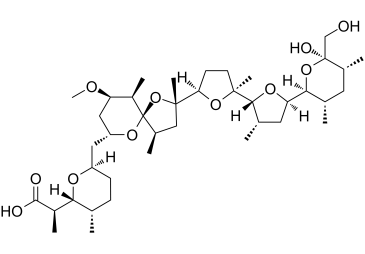
尼日利亚菌素是一种聚醚类抗生素,可影响线粒体中的离子转运和ATP酶活性。它由吸水链霉菌(Streptomyces hygroscopicus)产生。它具有抗菌、抗病毒、钾离子载体和细菌代谢产物等多种功能。
一种聚醚类抗生素,可影响线粒体中的离子转运和ATP酶活性。它由吸水链霉菌(Streptomyces hygroscopicus)产生。(摘自《默克索引》,第11版) 据报道,尼日利亚菌素存在于链霉菌属(Streptomyces)、紫黑链霉菌(Streptomyces violaceus niger)和吸水链霉菌(Streptomyces hygroscopicus)中,并有相关数据。 一种聚醚类抗生素,可影响线粒体中的离子转运和ATP酶活性。它由吸水链霉菌(Streptomyces hygroscopicus)产生。 (摘自《默克索引》,第11版) 肺癌的耐药性与多种因素相关,包括肿瘤异质性以及内在或获得性耐药。干细胞特性增强和癌细胞可塑性提高已被认为是重要的耐药机制;因此,针对不依赖于干细胞表型的癌细胞的治疗方法在肺癌治疗中将更加有效。本文中,我们研究了抗生素尼日利亚菌素在具有不同程度干细胞特性和抗癌药物耐药性的细胞中的抗癌作用,这些细胞的产生源于:(1) 常规培养条件;(2) 长期血清饥饿。这些细胞对紫杉醇、羟基脲、秋水仙碱、奥巴托克拉、沃特曼宁和LY294002等传统抗癌药物具有高度耐药性,在长期血清饥饿条件下生长的细胞的多药耐药表型可能是信号通路广泛重编程的结果;(3) 富含癌干细胞样细胞的肺肿瘤球。我们发现,尼日利亚菌素能有效抑制在常规培养条件下、长期血清饥饿条件下以及肺肿瘤球体中生长的细胞的活力。此外,我们发现尼日利亚菌素能下调Wnt经典信号通路中关键蛋白(如LRP6、Wnt5a/b和β-catenin)的表达,但能促进β-catenin转位至细胞核。Wnt激活剂HLY78以及美国食品药品监督管理局(FDA)批准的药物地高辛及其新型合成类似物MonoD的治疗剂量均能增强尼日利亚菌素的抗肿瘤作用。我们认为,尼日利亚菌素可与其他新型化疗药物联合用于联合治疗,以有效抑制具有不同程度干性的癌症,并可能产生持久的抗癌效果。 [1] K(+),H(+)离子载体和抗生素尼日利亚菌素已被证实能够诱导细胞凋亡,因此被认为可用于治疗恶性肿瘤。其细胞机制包括诱导氧化应激,已知氧化应激可激活红细胞中Ca(2+)通透性非选择性阳离子通道,导致Ca(2+)内流,胞质Ca(2+)活性([Ca(2+)]i)升高,并随后刺激红细胞凋亡,即红细胞自杀性死亡,其特征是细胞收缩和细胞膜磷脂酰丝氨酸易位至红细胞表面。本研究探讨了尼日利亚菌素是否以及如何诱导红细胞凋亡。通过膜联蛋白V结合测定细胞表面磷脂酰丝氨酸的暴露量,通过前向散射测定细胞体积,通过Fluo3荧光测定细胞内钙离子浓度[Ca²⁺]i,通过BCECF荧光测定细胞内pH值,利用抗体测定神经酰胺丰度,并通过DCFDA依赖性荧光测定活性氧(ROS)生成量。人红细胞暴露于尼日利亚菌素48小时后,膜联蛋白V结合细胞的百分比显著增加(0.1-10 nM),前向散射显著降低(0.1-1 nM),胞质pH显著降低(0.1-1 nM),Fluo3荧光显著增加(0.1-10 nM)。1 nM尼日利亚菌素轻微但显著地增加了ROS水平,但对神经酰胺丰度没有显著影响。去除细胞外钙离子后,尼日利亚菌素对膜联蛋白V结合的影响显著减弱,但并未完全消除。尼日利亚菌素诱导的细胞内钙离子浓度[Ca(2+)]i升高和膜联蛋白V结合增加再次被Na(+)/H(+)交换抑制剂卡利泊利(10 μM)显著抑制,但并未完全消除。尼日利亚菌素诱导红细胞凋亡,这一效应伴随着活性氧(ROS)的生成,部分依赖于Ca(2+)内流的刺激,并涉及卡利泊利敏感的Na(+)/H(+)交换器。[2] 鼻咽癌(NPC)在中国南方、北非和阿拉斯加地区较为常见。早期NPC患者的预后良好,而晚期患者预后较差。癌症干细胞(CSCs)被认为与肿瘤的发生、复发和转移相关,NPC预后不良可能源于治疗后残留的CSCs。尽管针对CSCs的治疗在其他癌症中已受到广泛关注,但针对NPC中CSCs的治疗研究仍然不足。本研究以鼻咽癌细胞系中癌症干细胞(CSCs)比例高低不同的细胞系为模型,探讨了抗生素尼日利亚菌素对CSCs的影响。我们发现,尼日利亚菌素能够选择性地靶向CSCs,并在体外和体内均能增强鼻咽癌细胞对常用临床药物顺铂的敏感性。此外,多梳蛋白Bmi-1的下调可能有助于尼日利亚菌素对CSCs的抑制作用。进一步地,利用体外鼻咽癌细胞模型,我们发现尼日利亚菌素能够显著降低细胞的迁移和侵袭能力,而这些能力已知与CSCs密切相关。综上所述,我们的结果表明,尼日利亚菌素能够选择性靶向鼻咽癌干细胞(CSCs),可能是一种有待临床评估的CSCs靶向药物。[3] 目的:评估尼日利亚菌素对结直肠癌的影响并探讨其可能的作用机制。方法:在特定条件下,用尼日利亚菌素或奥沙利铂处理人结直肠癌(CRC)细胞系HT29和SW480。通过细胞活力检测、侵袭和转移实验评估尼日利亚菌素对CRC细胞的影响。采用球体形成实验和软琼脂集落形成实验评估尼日利亚菌素对经历上皮-间质转化(EMT)的结直肠癌细胞的癌干细胞特性的影响。结果:与奥沙利铂相比,尼日利亚菌素对HT29细胞系的毒性更高(IC50分别为12.92 ± 0.25 μmol和37.68 ± 0.34 μmol)。在SW116细胞系中也观察到了类似的结果(IC50分别为15.86 ± 0.18 μmol和41.02 ± 0.23 μmol)。 Boyden小室迁移实验表明,与载体对照组相比,尼日利亚菌素处理组中穿过聚偏二氟乙烯膜的HT29细胞数量显著减少[11 ± 2 个细胞/高倍视野 (HPF) vs 19.33 ± 1.52 个细胞/HPF,P < 0.05]。与对照组相比,尼日利亚菌素处理组中穿过Matrigel包被膜的HT29细胞数量也减少(6.66 ± 1.52 个细胞/HPF vs 14.66 ± 1.52 个细胞/HPF,P < 0.05)。此外,与对照组相比,尼日利亚菌素处理组的CD133+细胞比例也从83.57%降低至63.93%(P < 0.05)。与对照组相比,尼日利亚菌素降低了球体数量(0.14 ± 0.01 vs 0.35 ± 0.01,P < 0.05),而奥沙利铂则增加了球体数量(0.75 ± 0.02 vs 0.35 ± 0.01;P < 0.05)。在标准软琼脂平板培养14天后,与对照组相比,尼日利亚菌素在非锚定条件下形成菌落的能力也降低(1.66 ± 0.57 vs 7 ± 1.15,P < 0.05),而奥沙利铂组的菌落数量高于载体对照组(14.33 ± 0.57 vs 7 ± 1.15,P < 0.05)。我们进一步检测了经尼日利亚菌素和奥沙利铂处理的细胞中E-钙黏蛋白和波形蛋白的表达。结果显示,与载体对照组相比,尼日利亚菌素处理的HT29细胞中E-钙黏蛋白表达增加,波形蛋白表达降低。相反,与载体对照组相比,奥沙利铂处理HT29细胞后,E-钙黏蛋白表达下调,波形蛋白表达上调。结论:本研究表明,尼日利亚菌素可以部分逆转细胞侵袭和转移过程中的EMT过程。[4] 尽管免疫检查点抑制剂改善了晚期三阴性乳腺癌(TNBC)患者的临床预后,但有效率仍然相对较低。尼日利亚菌素是一种源自疏水链霉菌的抗生素。我们发现,尼日利亚菌素通过诱导细胞焦亡和凋亡同时发生,导致TNBC细胞系MDA-MB-231和4T1发生细胞死亡。由于尼日利亚菌素促进了细胞钾离子外流,我们发现它会导致线粒体功能障碍,进而导致线粒体活性氧(ROS)的产生,并激活三阴性乳腺癌(TNBC)细胞中Caspase-1/GSDMD介导的细胞焦亡和Caspase-3介导的细胞凋亡。值得注意的是,尼日利亚菌素诱导的细胞焦亡可通过增强CD4+和CD8+ T细胞的浸润和抗肿瘤作用来放大抗肿瘤免疫反应。此外,尼日利亚菌素与抗PD-1抗体联合用于TNBC治疗时显示出协同治疗效果。我们的研究表明,尼日利亚菌素可能是一种有前景的抗肿瘤药物,尤其是在与免疫检查点抑制剂联合用于晚期TNBC治疗时。[5] 多重耐药菌(MDR)对人类健康构成重大的临床威胁,但抗生素的研发无法满足对有效药物的迫切需求,特别是那些能够杀灭持续性细菌和生物膜的药物。本文报道了尼日利亚菌素对多种临床多重耐药革兰氏阳性菌、持续感染菌和生物膜均表现出强效杀菌活性,且耐药性发生率低。此外,尼日利亚菌素在小鼠深层生物膜、小鼠皮肤和血流感染模型中均显示出良好的体内疗效。对于金黄色葡萄球菌,尼日利亚菌素可破坏ATP生成和电子传递链;细胞死亡与膜结构和通透性的改变有关。获得尼日利亚菌素耐药/耐受突变株需要多次攻毒试验,且未观察到对多种抗菌药物的交叉耐药性,这可能是由于尼日利亚菌素与GraSR双组分调控系统具有独特的作用机制。因此,我们的研究表明,尼日利亚菌素是一种有前景的抗生素候选药物,可用于治疗革兰氏阳性菌引起的慢性或复发性感染。[6] |
| 分子式 |
C40H68O11
|
|---|---|
| 分子量 |
746.94318
|
| 精确质量 |
746.458
|
| 元素分析 |
C, 66.27; H, 9.45; O, 24.28
|
| CAS号 |
28380-24-7
|
| 相关CAS号 |
Nigericin sodium salt;28643-80-3
|
| PubChem CID |
34230
|
| 外观&性状 |
White to off-white solid powder
|
| 密度 |
1.19g/cm3
|
| 沸点 |
779.9ºC at 760mmHg
|
| 熔点 |
245-255ºC
|
| 闪点 |
226.9ºC
|
| 折射率 |
1.543
|
| LogP |
4.37
|
| tPSA |
145.2
|
| 氢键供体(HBD)数目 |
3
|
| 氢键受体(HBA)数目 |
11
|
| 可旋转键数目(RBC) |
9
|
| 重原子数目 |
51
|
| 分子复杂度/Complexity |
1230
|
| 定义原子立体中心数目 |
19
|
| SMILES |
C[C@H]1CC[C@@H](O[C@H]1[C@@H](C)C(=O)O)C[C@@H]2C[C@H]([C@H]([C@@]3(O2)[C@@H](C[C@@](O3)(C)[C@H]4CC[C@@](O4)(C)[C@H]5[C@H](C[C@@H](O5)[C@@H]6[C@H](C[C@H]([C@@](O6)(CO)O)C)C)C)C)C)OC
|
| InChi Key |
MOYOTUKECQMGHE-PDEFJWSRSA-M
|
| InChi Code |
InChI=1S/C40H68O11.Na/c1-21-11-12-28(46-33(21)26(6)36(42)43)17-29-18-30(45-10)27(7)40(48-29)25(5)19-38(9,51-40)32-13-14-37(8,49-32)35-23(3)16-31(47-35)34-22(2)15-24(4)39(44,20-41)50-34/h21-35,41,44H,11-20H2,1-10H3,(H,42,43)/q+1/p-1/t21-,22-,23-,24+,25+,26+,27+,28+,29+,30+,31+,32+,33+,34-,35+,37-,38-,39-,40+/m0./s1
|
| 化学名 |
sodium
(R)-2-((2R,3S,6R)-6-(((2S,4R,5R,7R,9R,10R)-2-((2S,2'R,3'S,5R,5'R)-5'-((2S,3S,5R,6R)-6-hydroxy-6-(hydroxymethyl)-3,5-dimethyltetrahydro-2H-pyran-2-yl)-2,3'-dimethyloctahydro-[2,2'-bifuran]-5-yl)-9-methoxy-2,4,10-trimethyl-1,6-dioxaspiro[4.5]decan-7-yl)methyl)-3-methyltetrahydro-2H-pyran-2-yl)propanoate
|
| 别名 |
Azalomycin M; Antibiotic K178; nigericin; Azalomycin M; 28380-24-7; Helixin C; Antibiotic X-464; Antibiotic K-178; UNII-RRU6GY95IS; RRU6GY95IS; Helexin C; Antibiotic X464; Polyetherin A
|
| HS Tariff Code |
2934.99.9001
|
| 存储方式 |
Powder -20°C 3 years 4°C 2 years In solvent -80°C 6 months -20°C 1 month |
| 运输条件 |
Room temperature (This product is stable at ambient temperature for a few days during ordinary shipping and time spent in Customs)
|
| 溶解度 (体外实验) |
May dissolve in DMSO (in most cases), if not, try other solvents such as H2O, Ethanol, or DMF with a minute amount of products to avoid loss of samples
|
|---|---|
| 溶解度 (体内实验) |
注意: 如下所列的是一些常用的体内动物实验溶解配方,主要用于溶解难溶或不溶于水的产品(水溶度<1 mg/mL)。 建议您先取少量样品进行尝试,如该配方可行,再根据实验需求增加样品量。
注射用配方
注射用配方1: DMSO : Tween 80: Saline = 10 : 5 : 85 (如: 100 μL DMSO → 50 μL Tween 80 → 850 μL Saline)(IP/IV/IM/SC等) *生理盐水/Saline的制备:将0.9g氯化钠/NaCl溶解在100 mL ddH ₂ O中,得到澄清溶液。 注射用配方 2: DMSO : PEG300 :Tween 80 : Saline = 10 : 40 : 5 : 45 (如: 100 μL DMSO → 400 μL PEG300 → 50 μL Tween 80 → 450 μL Saline) 注射用配方 3: DMSO : Corn oil = 10 : 90 (如: 100 μL DMSO → 900 μL Corn oil) 示例: 以注射用配方 3 (DMSO : Corn oil = 10 : 90) 为例说明, 如果要配制 1 mL 2.5 mg/mL的工作液, 您可以取 100 μL 25 mg/mL 澄清的 DMSO 储备液,加到 900 μL Corn oil/玉米油中, 混合均匀。 View More
注射用配方 4: DMSO : 20% SBE-β-CD in Saline = 10 : 90 [如:100 μL DMSO → 900 μL (20% SBE-β-CD in Saline)] 口服配方
口服配方 1: 悬浮于0.5% CMC Na (羧甲基纤维素钠) 口服配方 2: 悬浮于0.5% Carboxymethyl cellulose (羧甲基纤维素) 示例: 以口服配方 1 (悬浮于 0.5% CMC Na)为例说明, 如果要配制 100 mL 2.5 mg/mL 的工作液, 您可以先取0.5g CMC Na并将其溶解于100mL ddH2O中,得到0.5%CMC-Na澄清溶液;然后将250 mg待测化合物加到100 mL前述 0.5%CMC Na溶液中,得到悬浮液。 View More
口服配方 3: 溶解于 PEG400 (聚乙二醇400) 请根据您的实验动物和给药方式选择适当的溶解配方/方案: 1、请先配制澄清的储备液(如:用DMSO配置50 或 100 mg/mL母液(储备液)); 2、取适量母液,按从左到右的顺序依次添加助溶剂,澄清后再加入下一助溶剂。以 下列配方为例说明 (注意此配方只用于说明,并不一定代表此产品 的实际溶解配方): 10% DMSO → 40% PEG300 → 5% Tween-80 → 45% ddH2O (或 saline); 假设最终工作液的体积为 1 mL, 浓度为5 mg/mL: 取 100 μL 50 mg/mL 的澄清 DMSO 储备液加到 400 μL PEG300 中,混合均匀/澄清;向上述体系中加入50 μL Tween-80,混合均匀/澄清;然后继续加入450 μL ddH2O (或 saline)定容至 1 mL; 3、溶剂前显示的百分比是指该溶剂在最终溶液/工作液中的体积所占比例; 4、 如产品在配制过程中出现沉淀/析出,可通过加热(≤50℃)或超声的方式助溶; 5、为保证最佳实验结果,工作液请现配现用! 6、如不确定怎么将母液配置成体内动物实验的工作液,请查看说明书或联系我们; 7、 以上所有助溶剂都可在 Invivochem.cn网站购买。 |
| 制备储备液 | 1 mg | 5 mg | 10 mg | |
| 1 mM | 1.3388 mL | 6.6940 mL | 13.3880 mL | |
| 5 mM | 0.2678 mL | 1.3388 mL | 2.6776 mL | |
| 10 mM | 0.1339 mL | 0.6694 mL | 1.3388 mL |
1、根据实验需要选择合适的溶剂配制储备液 (母液):对于大多数产品,InvivoChem推荐用DMSO配置母液 (比如:5、10、20mM或者10、20、50 mg/mL浓度),个别水溶性高的产品可直接溶于水。产品在DMSO 、水或其他溶剂中的具体溶解度详见上”溶解度 (体外)”部分;
2、如果您找不到您想要的溶解度信息,或者很难将产品溶解在溶液中,请联系我们;
3、建议使用下列计算器进行相关计算(摩尔浓度计算器、稀释计算器、分子量计算器、重组计算器等);
4、母液配好之后,将其分装到常规用量,并储存在-20°C或-80°C,尽量减少反复冻融循环。
计算结果:
工作液浓度: mg/mL;
DMSO母液配制方法: mg 药物溶于 μL DMSO溶液(母液浓度 mg/mL)。如该浓度超过该批次药物DMSO溶解度,请首先与我们联系。
体内配方配制方法:取 μL DMSO母液,加入 μL PEG300,混匀澄清后加入μL Tween 80,混匀澄清后加入 μL ddH2O,混匀澄清。
(1) 请确保溶液澄清之后,再加入下一种溶剂 (助溶剂) 。可利用涡旋、超声或水浴加热等方法助溶;
(2) 一定要按顺序加入溶剂 (助溶剂) 。
InvivoChem的所有产品仅用于作科学研究,不面向患者销售
Copyright 2020 InvivoChem LLC | All Rights Reserved 粤ICP备20063088号-1


 COA
COA
 463611831
