| 规格 | 价格 | 库存 | 数量 |
|---|---|---|---|
| 5mg |
|
||
| 10mg |
|
||
| 25mg |
|
||
| 50mg |
|
||
| 100mg |
|
||
| 250mg |
|
||
| 500mg |
|
||
| Other Sizes |
|
| 靶点 |
TOPK (IC50 = 2.6 nM)
|
|---|---|
| 体外研究 (In Vitro) |
体外活性:OTS514 强烈抑制 TOPK 阳性癌细胞的生长。它还对卵巢癌细胞系显示出显着的生长抑制作用,IC50 值为 3.0 至 46 nM。 OTS514对5种肾癌细胞系VMRC-RCW、Caki-1、Caki-2、769-P和786-O具有生长抑制作用,其中TOPK高表达。 IC50 值范围为 19.9 至 44.1 nM 激酶测定:OTS514 是一种新型高效 TOPK(T-LAK 细胞源性蛋白激酶)抑制剂,IC50 值为 2.6 nM。细胞测定:细胞在补充有20%胎牛血清和1×StemSpan CC100的RPMI中培养。用 OTS514(20 或 40 nM)或 OTS964(100 或 200 nM)处理细胞 48 小时。收集的细胞用PBS洗涤并重悬于100ml PBS中,随后用CD41a抗体在室温下染色20分钟。最后,再次用PBS洗涤细胞,然后通过流式细胞术分析CD41a染色。使用抗STAT5抗体通过蛋白质印迹检查STAT5的表达。
|
| 体内研究 (In Vivo) |
对人类肺癌细胞的小鼠异种移植研究证明了 OTS514 的体内功效,但发现该化合物也会引起严重的造血毒性 [红细胞 (RBC) 和白细胞 (WBC) 减少,并伴有血小板显着增加。与赋形剂对照相比,口服 OTS514 显着延长了 ES-2 腹部播散异种移植模型的总生存期(P < 0.001)。
|
| 酶活实验 |
如前所述,通过蛋白质印迹检测TOPK的表达和组蛋白H3(Ser10)的磷酸化。用于蛋白质印迹的其他抗体如下:c-Src(1:1000)、Fyn(1:1000”)和Lyn(1:1000“)。使用细胞计数试剂盒-8通过比色测定法测量体外细胞存活率。将细胞(100μl)以产生连续线性生长的密度(A549,1×103个细胞;LU-99,2×10³个电池;DU4475,4×103个手机;MDA-MB-231,3×10³个电脑;T47D,3×104个手机;Daudi,5×103个汽车;UM-UC-3,1×10⁵个手机;HCT-116,1×10⁴个汽车;MKN1,2×104个电脑;MKN45,4×105个电脑;HepG2,4×107个电脑;MIAPaca-2,2×10⁷个手机;22Rv1,6×103个;HT29,3×105个汽车)铺在96孔板中。在37°C下暴露于化合物72小时之前,让细胞粘附过夜。用分光光度计在450nm波长下读取板。所有检测均一式三份。在测量IC50值后,我们计算z分数以产生P值。在IC50值(nM)的对数转换(基数10)后,计算13个TOPK阳性细胞系的IC50对数值的平均值和SD。OTS514的平均值和标准差分别为0.76和0.23,OTS964分别为1.53和0.26。然后,根据HT29 IC50值,OTS514和OTS964的z得分分别为6.44和3.62[1]。
|
| 细胞实验 |
人造血干细胞的体外分化[1]
从健康供体的生长因子动员外周血中纯化CD34+HSC,然后在添加了20%胎牛血清和1×StemSpan CC100的RPMI中培养细胞。用OTS514(20或40 nM)或OTS964(100或200 nM)处理细胞48小时。收集的细胞用磷酸盐缓冲盐水(PBS)洗涤,重新悬浮在100μl PBS中,然后在室温下用CD41a抗体染色20分钟。最后,再次用PBS洗涤细胞,然后在BD FACSCalibur上通过流式细胞术分析CD41a染色。用抗STAT5抗体通过蛋白质印迹检测STAT5的表达。 微阵列分析[2] 5×10⁵ H929细胞用0.015%DMSO、15 nMOTS514、15µM来那度胺(LEN)或5 nM卡非佐米(CFZ)处理24小时。此外,还进行了每种活性药物组合(OTS514/LEN、OTS514/CFZ、OTS514/LEN/CFZ和LEN/CFZ)。使用Qiagen RNeasy迷你试剂盒提取来自三个独立实验(共24个样本)的RNA,并在芝加哥大学功能基因组学核心设施的两个人类HT12v4珠阵列上进行分析。使用分子特征数据库v6.1.27中的标志基因集对分位数标准化、背景减除的数据进行基因集富集分析(GSEA),28通过使用独创性途径分析生成上游调控因子分析。 细胞在补充有1×StemSpan CC100和20%胎牛血清的RPMI中培养。将细胞暴露于 OTS964(100 或 200 nM)或 OTS514(20 或 40 nM)48 小时。 PBS 洗涤并重悬于 100 毫升 PBS 后,使用 CD41a 抗体在室温下对收集的细胞染色 20 分钟。最终,细胞再进行一次 PBS 洗涤,然后进行流式细胞术分析 CD41a 染色。使用抗 STAT5 抗体,使用蛋白质印迹来测量 STAT5 表达。 |
| 动物实验 |
Female BALB/cSLC-nu/nu mice bearing a xenograft model of A549 cells[1]
1, 2.5, and 5 mg/kg Intravenously treated; once every day for 2 weeks In vivo xenograft study[1] A549 (1 × 10⁷ cells) or LU-99 cells (5 × 10⁶ or 1 × 10⁷ cells) were injected subcutaneously in the left flank of female BALB/cSLC-nu/nu mice. When A549 xenografts had reached an average volume of 200 mm3 or when LU-99 xenografts had reached an average volume of 150 or 200 mm3, animals were randomized into groups of six mice. The starting tumor volume of 150 mm3 was used for LU-99 xenografts when tumors were monitored for a longer time period (>14 days), because LU-99 cells grew very rapidly, and thus the starting volume of 200 mm3 prevented longer observation considering animal ethics (for example, 200 mm3 of inoculated LU-99 tumor reached an average tumor volume of about 1100 mm3, whereas A549 tumor reached about 490 mm3 on day 15). For intravenous administration, compounds were formulated in 5% glucose and injected into the tail vein. For oral administration, compounds (e.g. OTS514) were prepared in a vehicle of 0.5% methylcellulose and given by oral gavage at the indicated dose and schedule. An administration volume of 10 ml/kg of body weight was used for both administration routes. Concentrations were indicated in the main text and figures. Tumor volumes were determined using a caliper. The results were converted to tumor volume (mm3) by the formula length × width2 × 1/2. The weight of the mice was determined as an indicator of tolerability on the same days. The animal experiments were conducted at KAC Co. Ltd. for A549 xenograft or at OncoTherapy Science Inc. for LU-99 xenograft, in accordance with the Institutional Guidelines for the Care and Use of Laboratory Animals of each site. TGI was calculated according to the formula [1 − (T − T0)/(C − C0)] × 100, where T and T0 are the mean tumor volumes at day 15 or 22 and day 1, respectively, for the experimental group, and C and C0 are those for the vehicle control group. WBCs were counted with Sysmex XT-1800iV Analyzer (Sysmex Corporation) at KAC Co. Ltd. or with a cell counting chamber. Blood was collected in a blood collection tube with EDTA to prevent coagulation and to perform the blood cell count. |
| 参考文献 | |
| 其他信息 |
TOPK (T-lymphokine-activated killer cell-originated protein kinase) is highly and frequently transactivated in various cancer tissues, including lung and triple-negative breast cancers, and plays an indispensable role in the mitosis of cancer cells. We report the development of a potent TOPK inhibitor, OTS964 {(R)-9-(4-(1-(dimethylamino)propan-2-yl)phenyl)-8-hydroxy-6-methylthieno[2,3-c]quinolin-4(5H)-one}, which inhibits TOPK kinase activity with high affinity and selectivity. Similar to the knockdown effect of TOPK small interfering RNAs (siRNAs), this inhibitor causes a cytokinesis defect and the subsequent apoptosis of cancer cells in vitro as well as in xenograft models of human lung cancer. Although administration of the free compound induced hematopoietic adverse reactions (leukocytopenia associated with thrombocytosis), the drug delivered in a liposomal formulation effectively caused complete regression of transplanted tumors without showing any adverse reactions in mice. Our results suggest that the inhibition of TOPK activity may be a viable therapeutic option for the treatment of various human cancers.[1]
Multiple myeloma (MM) continues to be considered incurable, necessitating new drug discovery. The mitotic kinase T-LAK cell-originated protein kinase/PDZ-binding kinase (TOPK/PBK) is associated with proliferation of tumor cells, maintenance of cancer stem cells, and poor patient prognosis in many cancers. In this report, we demonstrate potent anti-myeloma effects of the TOPK inhibitor OTS514 for the first time. OTS514 induces cell cycle arrest and apoptosis at nanomolar concentrations in a series of human myeloma cell lines (HMCL) and prevents outgrowth of a putative CD138+ stem cell population from MM patient-derived peripheral blood mononuclear cells. In bone marrow cells from MM patients, OTS514 treatment exhibited preferential killing of the malignant CD138+ plasma cells compared with the CD138- compartment. In an aggressive mouse xenograft model, OTS964 given orally at 100 mg/kg 5 days per week was well tolerated and reduced tumor size by 48%-81% compared to control depending on the initial graft size. FOXO3 and its transcriptional targets CDKN1A (p21) and CDKN1B (p27) were elevated and apoptosis was induced with OTS514 treatment of HMCLs. TOPK inhibition also induced loss of FOXM1 and disrupted AKT, p38 MAPK, and NF-κB signaling. The effects of OTS514 were independent of p53 mutation or deletion status. Combination treatment of HMCLs with OTS514 and lenalidomide produced synergistic effects, providing a rationale for the evaluation of TOPK inhibition in existing myeloma treatment regimens.[2] |
| 分子式 |
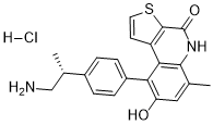
C21H21CLN2O2S
|
|
|---|---|---|
| 分子量 |
400.9216
|
|
| 精确质量 |
364.12
|
|
| 元素分析 |
C, 62.91; H, 5.28; Cl, 8.84; N, 6.99; O, 7.98; S, 8.00
|
|
| CAS号 |
2319647-76-0
|
|
| 相关CAS号 |
OTS514;1338540-63-8; 2319647-76-0 (HCl); 1338544-87-8 (HBr); 1338545-92-8 (S-isomer HCl); 1338541-25-5 (s-isomer);
|
|
| PubChem CID |
92044487
|
|
| 外观&性状 |
Solid powder
|
|
| tPSA |
104
|
|
| 氢键供体(HBD)数目 |
4
|
|
| 氢键受体(HBA)数目 |
4
|
|
| 可旋转键数目(RBC) |
3
|
|
| 重原子数目 |
27
|
|
| 分子复杂度/Complexity |
522
|
|
| 定义原子立体中心数目 |
1
|
|
| SMILES |
CC1=CC(=C(C2=C1NC(=O)C3=C2C=CS3)C4=CC=C(C=C4)[C@@H](C)CN)O.Cl
|
|
| InChi Key |
YCRRQRJUNVBPBW-UHFFFAOYSA-N
|
|
| InChi Code |
InChI=1S/C21H18N2O2S.ClH/c1-11-9-16(24)17(14-5-3-13(4-6-14)12(2)10-22)18-15-7-8-26-20(15)21(25)23-19(11)18;/h3-9,12H,10,22H2,1-2H3;1H
|
|
| 化学名 |
9-[4-(1-aminopropan-2-yl)phenyl]-6-methylthieno[2,3-c]quinoline-4,8-dione;hydrochloride
|
|
| 别名 |
|
|
| HS Tariff Code |
2934.99.9001
|
|
| 存储方式 |
Powder -20°C 3 years 4°C 2 years In solvent -80°C 6 months -20°C 1 month |
|
| 运输条件 |
Room temperature (This product is stable at ambient temperature for a few days during ordinary shipping and time spent in Customs)
|
| 溶解度 (体外实验) |
|
|||
|---|---|---|---|---|
| 溶解度 (体内实验) |
注意: 如下所列的是一些常用的体内动物实验溶解配方,主要用于溶解难溶或不溶于水的产品(水溶度<1 mg/mL)。 建议您先取少量样品进行尝试,如该配方可行,再根据实验需求增加样品量。
注射用配方
注射用配方1: DMSO : Tween 80: Saline = 10 : 5 : 85 (如: 100 μL DMSO → 50 μL Tween 80 → 850 μL Saline)(IP/IV/IM/SC等) *生理盐水/Saline的制备:将0.9g氯化钠/NaCl溶解在100 mL ddH ₂ O中,得到澄清溶液。 注射用配方 2: DMSO : PEG300 :Tween 80 : Saline = 10 : 40 : 5 : 45 (如: 100 μL DMSO → 400 μL PEG300 → 50 μL Tween 80 → 450 μL Saline) 注射用配方 3: DMSO : Corn oil = 10 : 90 (如: 100 μL DMSO → 900 μL Corn oil) 示例: 以注射用配方 3 (DMSO : Corn oil = 10 : 90) 为例说明, 如果要配制 1 mL 2.5 mg/mL的工作液, 您可以取 100 μL 25 mg/mL 澄清的 DMSO 储备液,加到 900 μL Corn oil/玉米油中, 混合均匀。 View More
注射用配方 4: DMSO : 20% SBE-β-CD in Saline = 10 : 90 [如:100 μL DMSO → 900 μL (20% SBE-β-CD in Saline)] 口服配方
口服配方 1: 悬浮于0.5% CMC Na (羧甲基纤维素钠) 口服配方 2: 悬浮于0.5% Carboxymethyl cellulose (羧甲基纤维素) 示例: 以口服配方 1 (悬浮于 0.5% CMC Na)为例说明, 如果要配制 100 mL 2.5 mg/mL 的工作液, 您可以先取0.5g CMC Na并将其溶解于100mL ddH2O中,得到0.5%CMC-Na澄清溶液;然后将250 mg待测化合物加到100 mL前述 0.5%CMC Na溶液中,得到悬浮液。 View More
口服配方 3: 溶解于 PEG400 (聚乙二醇400) 请根据您的实验动物和给药方式选择适当的溶解配方/方案: 1、请先配制澄清的储备液(如:用DMSO配置50 或 100 mg/mL母液(储备液)); 2、取适量母液,按从左到右的顺序依次添加助溶剂,澄清后再加入下一助溶剂。以 下列配方为例说明 (注意此配方只用于说明,并不一定代表此产品 的实际溶解配方): 10% DMSO → 40% PEG300 → 5% Tween-80 → 45% ddH2O (或 saline); 假设最终工作液的体积为 1 mL, 浓度为5 mg/mL: 取 100 μL 50 mg/mL 的澄清 DMSO 储备液加到 400 μL PEG300 中,混合均匀/澄清;向上述体系中加入50 μL Tween-80,混合均匀/澄清;然后继续加入450 μL ddH2O (或 saline)定容至 1 mL; 3、溶剂前显示的百分比是指该溶剂在最终溶液/工作液中的体积所占比例; 4、 如产品在配制过程中出现沉淀/析出,可通过加热(≤50℃)或超声的方式助溶; 5、为保证最佳实验结果,工作液请现配现用! 6、如不确定怎么将母液配置成体内动物实验的工作液,请查看说明书或联系我们; 7、 以上所有助溶剂都可在 Invivochem.cn网站购买。 |
| 制备储备液 | 1 mg | 5 mg | 10 mg | |
| 1 mM | 2.4943 mL | 12.4713 mL | 24.9426 mL | |
| 5 mM | 0.4989 mL | 2.4943 mL | 4.9885 mL | |
| 10 mM | 0.2494 mL | 1.2471 mL | 2.4943 mL |
1、根据实验需要选择合适的溶剂配制储备液 (母液):对于大多数产品,InvivoChem推荐用DMSO配置母液 (比如:5、10、20mM或者10、20、50 mg/mL浓度),个别水溶性高的产品可直接溶于水。产品在DMSO 、水或其他溶剂中的具体溶解度详见上”溶解度 (体外)”部分;
2、如果您找不到您想要的溶解度信息,或者很难将产品溶解在溶液中,请联系我们;
3、建议使用下列计算器进行相关计算(摩尔浓度计算器、稀释计算器、分子量计算器、重组计算器等);
4、母液配好之后,将其分装到常规用量,并储存在-20°C或-80°C,尽量减少反复冻融循环。
计算结果:
工作液浓度: mg/mL;
DMSO母液配制方法: mg 药物溶于 μL DMSO溶液(母液浓度 mg/mL)。如该浓度超过该批次药物DMSO溶解度,请首先与我们联系。
体内配方配制方法:取 μL DMSO母液,加入 μL PEG300,混匀澄清后加入μL Tween 80,混匀澄清后加入 μL ddH2O,混匀澄清。
(1) 请确保溶液澄清之后,再加入下一种溶剂 (助溶剂) 。可利用涡旋、超声或水浴加热等方法助溶;
(2) 一定要按顺序加入溶剂 (助溶剂) 。
 Growth-inhibitory and cytotoxic effects of OTS514 for ovarian cancer cells freshly-isolated from patients.Clin Cancer Res.2016 Dec 15;22(24):6110-6117. |
|---|
In vivoefficacy of OTS514 in ES-2 ovarian cancer peritoneal dissemination xenograft model.Clin Cancer Res.2016 Dec 15;22(24):6110-6117. |
 TOPK expression levels, IC50values to TOPK inhibitors and suppression of FOXM1 in ovarian cancer cell lines.Clin Cancer Res.2016 Dec 15;22(24):6110-6117. |