| 规格 | 价格 | ||
|---|---|---|---|
| 500mg | |||
| 1g | |||
| Other Sizes |

| 靶点 |
Tripeptide
|
|---|---|
| 体外研究 (In Vitro) |
铜三肽(1 nM;0-96 小时)醋酸盐影响受辐射成纤维细胞的群体倍增时间,使其接近对照组 [1]。与正常对照组相比,铜三肽(1 nM;0-120 小时)醋酸盐显著增加了 24 小时受辐射成纤维细胞中碱性成纤维细胞生长因子的产生 [1]。
辐照后的成纤维细胞在无血清培养基中存活并复制。暴露于GHK-Cu的正常和辐照成纤维细胞的种群倍增速度比未处理的对照组快。GHK-Cu处理的成纤维细胞的生长速率与未处理的对照组接近,并且在GHK-Cu暴露后早期产生的碱性成纤维细胞生长因子和血管内皮生长因子明显多于未处理的对照组。 结论:受辐照的成纤维细胞在无血清培养基中存活和复制,建立了体外评价生长因子生成的理想模型。铜三肽加速正常和受辐照的成纤维细胞的生长,使受辐照的成纤维细胞接近正常对照的群体倍增时间。GHK-Cu-处理的成纤维细胞早期增加碱性成纤维细胞生长因子和血管内皮生长因子的产生可能促进伤口愈合。[1] |
| 细胞实验 |
实验在细胞的第一或第二代进行。实验时,用磷酸盐缓冲的生理盐水洗涤成纤维细胞,用0.05%胰蛋白酶将融合细胞从烧瓶壁上释放出来。胰酶大豆抑制剂(GIBCO)以1:1的比例灭活胰蛋白酶。通过台盼蓝染色排除法测定细胞培养活力,并使用血细胞计和相差显微镜进行重复细胞计数。然后使用市售无血清培养基,以5 × 10³(正常)和3 × 10³(辐照)细胞/孔的密度在无菌96孔板的每孔中接种细胞。该培养基已被证明能维持真皮成纤维细胞生长至少7天,存活率高于90%。
在0小时时,在治疗组中加入无血清培养基中的GHK-Cu溶液(1 × 10⁻⁹ M),在未处理的对照组中加入等体积的无血清普通培养基。将每个细胞系中未经处理的细胞作为对照。细胞计数使用细胞增殖试验系统,在启动24、48、72和96小时后使用试剂4-[3-(4-碘苯基)-2-(4-硝基苯基)- 2h -5-四氮唑]-1,3-苯二磺酸盐(WST-1)进行细胞计数,以生成生长曲线。WST-1测定法是一种定量测定细胞增殖和细胞活力的比色法,基于线粒体脱氢酶在活细胞中切割四氮唑盐WST-1。它是一种非放射性的替代氚胸腺嘧啶掺入试验。使用自动车牌阅读器读取化验结果。用市售软件分析光密度。细胞计数通过与标准曲线进行比较来确定,标准曲线是根据每种细胞类型和培养基计算的已知细胞数量得出的。 每隔24小时,从测试井中收集三份无细胞上清液。样品保存在- 80°C的微离心管中,用于随后的生长因子测定。采用固相酶联免疫吸附法每隔24小时检测各组bFGF、TGF-β1和VEGF的表达。我们从对数最佳拟合曲线中计算细胞群体加倍时间(PDT)。[1] |
| 参考文献 | |
| 其他信息 |
Prezatide is a tripeptide consisting of glycine, histidine, and lysine which readily forms a complex with copper ions. Prezatide is used in cosmetic products for the skin and hair. It is known to aid wound healing and its potential applications in chronic obstructive pulmonary disease and metastatic colon cancer are currently being investigated.
Drug Indication Commonly used in cosmetic products for the skin and hair. Mechanism of Action Prezatide in complex with copper increases the synthesis and deposition of type I collagen and glycosaminoglycan. It also increases the expression of matrix metalloproteinase-2 as well as tissue inhibitor of matrix metalloproteinases-1 and -2, suggesting that it plays a role in the modulation of tissue remodeling. It is thought that prezatide's antioxidant activity is due to its ability to supply copper for superoxide dismutase and its anti inflammatory ability due to the blockage the of iron (Fe2+) release during injury. Prezatide also increases angiogenesis to injury sites. The precise mechanisms of these effects are unknown. It is also unknown whether prezatide's effects are due to the action of the tripeptide itself or its ability to localize and transport copper. Prezatide is known to be bound by heparin and heparin sulfate Pharmacodynamics Prezatide in complex with copper improve skin elasticity, density, and firmness, reduces fine lines and wrinkles, reduces photodamage, increases keratinocyte proliferation. Prezatide also displays anti-oxidant and angiogenic effects and appears to modulate tissue remodeling in injury. Objective: To evaluate the effects of copper tripeptide (GHK-Cu) on the growth and autocrine production of basic fibroblast growth factor, transforming growth factor beta1, and vascular endothelial growth factor by normal and irradiated fibroblasts in a serum-free in vitro environment. Methods: Primary human dermal fibroblast cell lines were established after explantation from intraoperative specimens obtained from patients who had undergone radiation therapy for head and neck cancer. Normal and irradiated fibroblasts were propagated in serum- and growth factor-free media. Treatment groups were exposed to GHK-Cu (1 x 10(-9) mol/L). We measured cell counts and production of basic fibroblast growth factor, transforming growth factor beta1, and vascular endothelial growth factor.[1] Several interesting findings are demonstrated. First, survival and growth of irradiated fibroblasts was demonstrated within the serum-free media. To our knowledge, our laboratory is the first to document this phenomenon using irradiated human fibroblasts. Our laboratory has already demonstrated survival and growth of normal, fetal, and keloid fibroblasts in this serum-free environment. Serum-free cell culture is essential when measuring changes in the growth factor milieu and is now a viable model for future studies involving irradiated fibroblasts. Second, the data established differences in the baseline production of growth factors between normal and irradiated fibroblasts in a head-to-head model. Production of bFGF by normal fibroblasts was significantly increased when compared with that of irradiated fibroblasts at all but 1 time point (72 hours). Production of TGF-β1 by normal fibroblasts was significantly increased when compared with that of irradiated fibroblasts at the 24-hour mark. Finally, production of VEGF by normal fibroblasts was significantly increased when compared with that of irradiated fibroblasts at 24 and 48 hours. It is reasonable to suppose that these differences play an influential role in the differing wound-healing properties of these wounds clinically. Third, the data show that modulation of the environment with GHK-Cu is associated with changes in the growth factor milieu. The GHK-Cu–treated irradiated fibroblasts showed significantly greater production of bFGF than controls at 24 and 72 hours. In fact, GHK-Cu–treated irradiated fibroblasts produced significantly more bFGF than normal controls at the 24-hour interval. Furthermore, GHK-Cu–treated irradiated fibroblasts produced significantly more VEGF than normal controls at the 24-hour interval. This finding is of importance given the known benefit of an early presence of these growth factors in the healing wound. Finally, the data show that modulation of the environment with GHK-Cu is associated with a dramatic increase in fibroblast PDT. This was demonstrated in the normal and irradiated cell lines. One striking finding is that population growth in GHK-Cu–treated irradiated fibroblasts assumed that of normal controls. The clinical implications of this are not yet known. However, given the important role of fibroblasts in wound healing, one might hypothesize that more fibroblasts in an irradiated wound bed would lead to a generalized improvement in wound healing. |
| 分子式 |
C16H26CUN6O6
|
|---|---|
| 分子量 |
461.96
|
| 精确质量 |
461.12098
|
| 元素分析 |
C, 41.60; H, 5.67; Cu, 13.76; N, 18.19; O, 20.78
|
| CAS号 |
300801-03-0
|
| 相关CAS号 |
Copper tripeptide;89030-95-5
|
| 外观&性状 |
Typically exists as solids at room temperature
|
| LogP |
0
|
| SMILES |
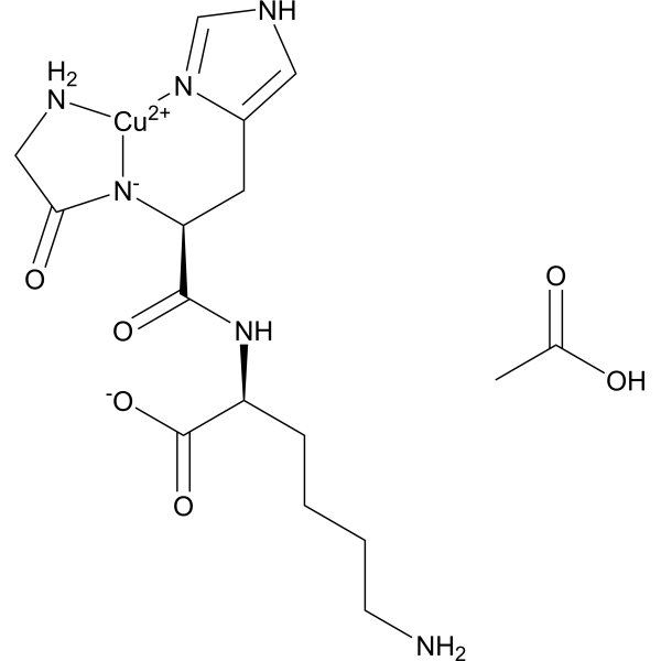
[Cu+2].O=C([C@H](CC1=CN=CN1)NC(CN)=O)N[C@H](C(=O)[O-])CCCCN.O=C([C@H](CC1=CN=CN1)NC(CN)=O)N[C@H](C(=O)[O-])CCCCN
|
| 别名 |
GHK-Cu acetate; GHK Cu acetate; 300801-03-0; copper;acetic acid;(2S)-6-amino-2-[[(2S)-2-(2-aminoacetyl)azanidyl-3-(1H-imidazol-4-yl)propanoyl]amino]hexanoate; GHK-Cu; GHK-Cu acetate, Gly-His-Lys-Cu(II)
|
| HS Tariff Code |
2934.99.9001
|
| 存储方式 |
Powder -20°C 3 years 4°C 2 years In solvent -80°C 6 months -20°C 1 month |
| 运输条件 |
Room temperature (This product is stable at ambient temperature for a few days during ordinary shipping and time spent in Customs)
|
| 溶解度 (体外实验) |
May dissolve in DMSO (in most cases), if not, try other solvents such as H2O, Ethanol, or DMF with a minute amount of products to avoid loss of samples
|
|---|---|
| 溶解度 (体内实验) |
注意: 如下所列的是一些常用的体内动物实验溶解配方,主要用于溶解难溶或不溶于水的产品(水溶度<1 mg/mL)。 建议您先取少量样品进行尝试,如该配方可行,再根据实验需求增加样品量。
注射用配方
注射用配方1: DMSO : Tween 80: Saline = 10 : 5 : 85 (如: 100 μL DMSO → 50 μL Tween 80 → 850 μL Saline)(IP/IV/IM/SC等) *生理盐水/Saline的制备:将0.9g氯化钠/NaCl溶解在100 mL ddH ₂ O中,得到澄清溶液。 注射用配方 2: DMSO : PEG300 :Tween 80 : Saline = 10 : 40 : 5 : 45 (如: 100 μL DMSO → 400 μL PEG300 → 50 μL Tween 80 → 450 μL Saline) 注射用配方 3: DMSO : Corn oil = 10 : 90 (如: 100 μL DMSO → 900 μL Corn oil) 示例: 以注射用配方 3 (DMSO : Corn oil = 10 : 90) 为例说明, 如果要配制 1 mL 2.5 mg/mL的工作液, 您可以取 100 μL 25 mg/mL 澄清的 DMSO 储备液,加到 900 μL Corn oil/玉米油中, 混合均匀。 View More
注射用配方 4: DMSO : 20% SBE-β-CD in Saline = 10 : 90 [如:100 μL DMSO → 900 μL (20% SBE-β-CD in Saline)] 口服配方
口服配方 1: 悬浮于0.5% CMC Na (羧甲基纤维素钠) 口服配方 2: 悬浮于0.5% Carboxymethyl cellulose (羧甲基纤维素) 示例: 以口服配方 1 (悬浮于 0.5% CMC Na)为例说明, 如果要配制 100 mL 2.5 mg/mL 的工作液, 您可以先取0.5g CMC Na并将其溶解于100mL ddH2O中,得到0.5%CMC-Na澄清溶液;然后将250 mg待测化合物加到100 mL前述 0.5%CMC Na溶液中,得到悬浮液。 View More
口服配方 3: 溶解于 PEG400 (聚乙二醇400) 请根据您的实验动物和给药方式选择适当的溶解配方/方案: 1、请先配制澄清的储备液(如:用DMSO配置50 或 100 mg/mL母液(储备液)); 2、取适量母液,按从左到右的顺序依次添加助溶剂,澄清后再加入下一助溶剂。以 下列配方为例说明 (注意此配方只用于说明,并不一定代表此产品 的实际溶解配方): 10% DMSO → 40% PEG300 → 5% Tween-80 → 45% ddH2O (或 saline); 假设最终工作液的体积为 1 mL, 浓度为5 mg/mL: 取 100 μL 50 mg/mL 的澄清 DMSO 储备液加到 400 μL PEG300 中,混合均匀/澄清;向上述体系中加入50 μL Tween-80,混合均匀/澄清;然后继续加入450 μL ddH2O (或 saline)定容至 1 mL; 3、溶剂前显示的百分比是指该溶剂在最终溶液/工作液中的体积所占比例; 4、 如产品在配制过程中出现沉淀/析出,可通过加热(≤50℃)或超声的方式助溶; 5、为保证最佳实验结果,工作液请现配现用! 6、如不确定怎么将母液配置成体内动物实验的工作液,请查看说明书或联系我们; 7、 以上所有助溶剂都可在 Invivochem.cn网站购买。 |
| 制备储备液 | 1 mg | 5 mg | 10 mg | |
| 1 mM | 2.1647 mL | 10.8234 mL | 21.6469 mL | |
| 5 mM | 0.4329 mL | 2.1647 mL | 4.3294 mL | |
| 10 mM | 0.2165 mL | 1.0823 mL | 2.1647 mL |
1、根据实验需要选择合适的溶剂配制储备液 (母液):对于大多数产品,InvivoChem推荐用DMSO配置母液 (比如:5、10、20mM或者10、20、50 mg/mL浓度),个别水溶性高的产品可直接溶于水。产品在DMSO 、水或其他溶剂中的具体溶解度详见上”溶解度 (体外)”部分;
2、如果您找不到您想要的溶解度信息,或者很难将产品溶解在溶液中,请联系我们;
3、建议使用下列计算器进行相关计算(摩尔浓度计算器、稀释计算器、分子量计算器、重组计算器等);
4、母液配好之后,将其分装到常规用量,并储存在-20°C或-80°C,尽量减少反复冻融循环。
计算结果:
工作液浓度: mg/mL;
DMSO母液配制方法: mg 药物溶于 μL DMSO溶液(母液浓度 mg/mL)。如该浓度超过该批次药物DMSO溶解度,请首先与我们联系。
体内配方配制方法:取 μL DMSO母液,加入 μL PEG300,混匀澄清后加入μL Tween 80,混匀澄清后加入 μL ddH2O,混匀澄清。
(1) 请确保溶液澄清之后,再加入下一种溶剂 (助溶剂) 。可利用涡旋、超声或水浴加热等方法助溶;
(2) 一定要按顺序加入溶剂 (助溶剂) 。
InvivoChem的所有产品仅用于作科学研究,不面向患者销售
Copyright 2020 InvivoChem LLC | All Rights Reserved 粤ICP备20063088号-1


 463611831
